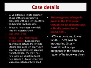
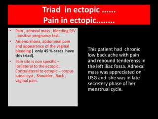
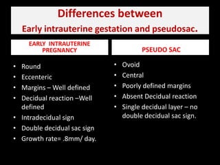
OVARIAN ECTOPIC PREGNANCY.5% To 1% Of Ectopic PregnanciesA woman who is capable of conceiving is capable of having a pregnancy  in a location other than the uterine cavity . COMPLEX  EXOPHYTIC OVARIAN LUTEAL  CYST VERSUS OVARIAN ECTOPIC.Follow up – cyst has rapid change  Complex fluid in the POD supports ectopic as possibility .MERCURY IMAGING INSTITUTE SCO 172-173 SEC 9C  CHANDIGARHMERCURY IMAGING CENTRE SCO 16-17 SEC 20D CHANDIGARH
Case details27 yr old female in late secretery phase of the menstrual cycle presented with pain left illiacfossa and chronic  low back ache.Rebound tenderness in the left iliac fossaapprecaiated.USG –TVS - ? PID Repeat – USG –Transvesicle                        ? Tubal ectopic (Adnexal mass appreciated betwen the left side uterine cornu and left ovary . Left ovary could not be seen separate from the lesion. The mass has  heterogenous  chaotic arterial flow around it . Probe tenderness was appreciated on the lesion.)Heteroegnousechogenic  areas in the POD were thought to be adynamic gut – Finally proved to be Blood clots .hCG was done and it was >2000 . There was no intrauterine G sac – Possibility of ectopic pregnancy in the ampullary region of he tube was given.
 Trans vesicle USGEndometrial lining is homogenous with no decidual cyst ,  pseudogestational sac . Mass is appreciated in the left adnexal region between the corunal end and left ovary .
 Trans vesicle USGLeft adnexal mass is well defined with  an anechoic central crescent shaped area s/o  ? G sac in the extra uterine location.
 Trans vesicle USGRt ovary is normal. Central endometrium is normal.
 Trans vesicle USGMass  is seen medial to the left ovary and  on further sagittal views  mass can not be discretely separated from left ovary.
Follow up.....................Laproscopic  removal of the adnexal mass done . Moderate amount of haemoperitoneum present .Either side tubes were normal.Initial histopathologicalassesement – Corpus luteal cyst .Follow up  beta HCG VALUES DROP DOWN.Possibility of  ? Tubal abortion with ruptured corpus luteal cyst is kept.Further  sections through the ovarian tissue revealed chorionic villiDiagnosis of ovarian ectopic was confirmed.
Ectopic pregnancySALPHINGITIS  TREATMENTTUBAL SCARRING , TUBOPLASTY.ASSISTIVE REPRODUCTIVE  TECHNIQUESOVULATION INDUCTION.All the above mentioned  situations raise the  possibility of ectopic gestation.Some factsBeta Hcg ( International reference ) – Value of more than 2000 – usually the sac is seen in the normal pregnancy.In normal   intrauterine Pregnancy  the  doubling time of beta Hcg is 2 days .Intrauterine sac should grow at  a rate of .8mm / day.Ectopic pregnancy has lower S PROGESTERONE LEVEL  when compared with the normal intrauterine gestation.
Triad  in ectopic ......                                       Pain in ectopic........Pain , adnexal mass , bleeding P/V , positive pregnancy test.Amenorrhoea, abdominal pain and appearance of the vaginal bleeding (  only 45 % cases  have this triad).Pain site is non specific – Ipsilateral to the ectopic , Contralateral to ectopic – corpus luteal cyst , Shoulder , Back , vaginal pain.     This patient had  chronic low back ache with pain  and rebound tenderenss in the left iliac fossa. Adnexal mass was appreciated on USG and  she was in late secretery phase of her menstrual cycle.
Signs in early intrauterine gestation.INTRA DECIDUAL SIGN Small anechoic focus with  echogenic rim around it   with location eccenteric to the endometrial stripe  is suggestive of  early intrauterine gestation .                ( seen  4.5 wks of gestation)                     ( Size as small as 2.2mm)D/D –                                              Decidual cysts, Pseudogestational sac , Small endometrial fluid collection.DOUBLE DECIDUAL SAC SIGNSeen later than the intradecidual sign.     Yolk sac and embroy if appreciated in the  SAC  - increase the confidence level of the intrauterine gestation.Choroinivvilli , endometrial fluid , deciduavera - All three constitue the double decidual sac sign.
Abnormal intrauterine G sac                                                    versus                                                           Pseudogestational sac Combine – Trans vaginal  with trans abdominal – Vaginal probe has better resolution because of the proximity to area of interest but limited field of view  is a limitation hence transabdominalshould be combined with trans vaginal .Look above and below the ovaries . Look between the ovaries and uterus Keep in mind haematosalphinx.  Appreciating  an intrauterine sac of size approximately 13mm   with no secondary yolk sac   raises the suspicion of the abnormal intrauterine gestation.  Post D&C if presence of the chorionic villiis demonstrated – Nothing to worry . If No chorionic villi demonstrated – still chances of ectopic are high and  Serial monitoring with beta Hcg has to be done.
Diagnostic criteria of ectopic pregnancy                                                           ( in patient with positive pregnancy test no intrauterine pregnancy.)Extrauterine sac with Yolk sac/embryo Adnexal ringComplex adnexal mass separate from the ovary.Fluid ( Moderate or large amount of the fluid / complex fluid- echogenic fluid ).Decidual cyst.     This patient had an adnexal mass with minimal amount of fluid  and no intrauterine G sac/  endometrailfuid collection.     The echogenic contents in the POD were assumed to be adynamic gut which were blood clots due to haemoperitoneum.
Differences between Early intrauterine gestation and pseudosac.EARLY  INTRAUTERINE PREGNANCY  PSEUDO SAC OvoidCentralPoorly defined marginsAbsent Decidual reactionSingle decidual layer – no double decidual sac sign.Round EccentericMargins – Well definedDecidual reaction –Well defined Intradecidual signDouble decidual sac signGrowth rate= .8mm/ day.
Endometrium in ectopic pregnancyEndometrium in ectopic pregnancy – Thick , thin Decidual cyst may or may not be present at endometrial/ myometrial junction.D/D  of haematosalphinxRetrograde flow of the blood in spontaneous abortion.Pedunculated fibroidExophytic  corpus luteal cyst Tubo-ovarian abscessTubal cyst Adjacent bowel
ASSESS PERITROPHOBLASTIC FLOWCorpus luteal cyst Implantation site of normal pregnancyExtrautreine ectopic pregnancy All these will have  low impedence  high diastolic flow component . Hence in situation where corpus luteal cyst has to be differentiated from the  ectopic pregnancy – extra –ovarian location is very important.  Colordoppler helps to assess  viability of the tissue – No flow in an ectopic  supports dead non viable tissue and  hence instead of surgical  - more conservative  plans can be thought of.
Brief about  cervical and interstitial ectopicCERVICAL ECTOPICDIFFERENTIATE FROM CERVICAL ABORTION.Sac in abortion changes it’s shape quite fastSac in ectopic is round to hour glass in shape Peritrophoblsatic flow is appreciated in the cervix in case of ectopic gestation and not in the cervical abortions.Profuse bleeding  in cervical ectopicINTERSTITIAL PREGNANCY Pregnancy with less than 5mm myometrial cover at one or more places and stationed in the vicnity of the fundus / cornual region is taken as interstitial pregnancy.
Non invasive approachTVS  with Beta  Hcg – Non invasive preffered  test .Combine TVS with Trans abdominal USG.Doppler suggestion of no flow in the ectopic pregnancy suggests non viable / dead ectopic and conservative approach  can be  thought in this situation.
Lesson learntDifferentiate exophytic corpus luteal cyst  from the  ectopic pregnancy ( be sure about intraovarian / extra ovarian location)Echogenic complex signal in the POD – Think of haem products , Beta Hcg-  take international reference levels into considerationsTrophoblsatic flow – Low impedence and high diatolic component in an extrauterine / extra ovarian mass supports ectopic as diagnosis.Decidual cysts , small endometrial fluid collections – these observations should be given  importance in similar way as other direct positive signs.

Ovarian ectopic pregnancy

  • 1.
    OVARIAN ECTOPIC PREGNANCY.5%To 1% Of Ectopic PregnanciesA woman who is capable of conceiving is capable of having a pregnancy in a location other than the uterine cavity . COMPLEX EXOPHYTIC OVARIAN LUTEAL CYST VERSUS OVARIAN ECTOPIC.Follow up – cyst has rapid change Complex fluid in the POD supports ectopic as possibility .MERCURY IMAGING INSTITUTE SCO 172-173 SEC 9C CHANDIGARHMERCURY IMAGING CENTRE SCO 16-17 SEC 20D CHANDIGARH
  • 2.
    Case details27 yrold female in late secretery phase of the menstrual cycle presented with pain left illiacfossa and chronic low back ache.Rebound tenderness in the left iliac fossaapprecaiated.USG –TVS - ? PID Repeat – USG –Transvesicle ? Tubal ectopic (Adnexal mass appreciated betwen the left side uterine cornu and left ovary . Left ovary could not be seen separate from the lesion. The mass has heterogenous chaotic arterial flow around it . Probe tenderness was appreciated on the lesion.)Heteroegnousechogenic areas in the POD were thought to be adynamic gut – Finally proved to be Blood clots .hCG was done and it was >2000 . There was no intrauterine G sac – Possibility of ectopic pregnancy in the ampullary region of he tube was given.
  • 3.
    Trans vesicleUSGEndometrial lining is homogenous with no decidual cyst , pseudogestational sac . Mass is appreciated in the left adnexal region between the corunal end and left ovary .
  • 4.
    Trans vesicleUSGLeft adnexal mass is well defined with an anechoic central crescent shaped area s/o ? G sac in the extra uterine location.
  • 5.
    Trans vesicleUSGRt ovary is normal. Central endometrium is normal.
  • 6.
    Trans vesicleUSGMass is seen medial to the left ovary and on further sagittal views mass can not be discretely separated from left ovary.
  • 7.
    Follow up.....................Laproscopic removal of the adnexal mass done . Moderate amount of haemoperitoneum present .Either side tubes were normal.Initial histopathologicalassesement – Corpus luteal cyst .Follow up beta HCG VALUES DROP DOWN.Possibility of ? Tubal abortion with ruptured corpus luteal cyst is kept.Further sections through the ovarian tissue revealed chorionic villiDiagnosis of ovarian ectopic was confirmed.
  • 8.
    Ectopic pregnancySALPHINGITIS TREATMENTTUBAL SCARRING , TUBOPLASTY.ASSISTIVE REPRODUCTIVE TECHNIQUESOVULATION INDUCTION.All the above mentioned situations raise the possibility of ectopic gestation.Some factsBeta Hcg ( International reference ) – Value of more than 2000 – usually the sac is seen in the normal pregnancy.In normal intrauterine Pregnancy the doubling time of beta Hcg is 2 days .Intrauterine sac should grow at a rate of .8mm / day.Ectopic pregnancy has lower S PROGESTERONE LEVEL when compared with the normal intrauterine gestation.
  • 9.
    Triad inectopic ...... Pain in ectopic........Pain , adnexal mass , bleeding P/V , positive pregnancy test.Amenorrhoea, abdominal pain and appearance of the vaginal bleeding ( only 45 % cases have this triad).Pain site is non specific – Ipsilateral to the ectopic , Contralateral to ectopic – corpus luteal cyst , Shoulder , Back , vaginal pain. This patient had chronic low back ache with pain and rebound tenderenss in the left iliac fossa. Adnexal mass was appreciated on USG and she was in late secretery phase of her menstrual cycle.
  • 10.
    Signs in earlyintrauterine gestation.INTRA DECIDUAL SIGN Small anechoic focus with echogenic rim around it with location eccenteric to the endometrial stripe is suggestive of early intrauterine gestation . ( seen 4.5 wks of gestation) ( Size as small as 2.2mm)D/D – Decidual cysts, Pseudogestational sac , Small endometrial fluid collection.DOUBLE DECIDUAL SAC SIGNSeen later than the intradecidual sign. Yolk sac and embroy if appreciated in the SAC - increase the confidence level of the intrauterine gestation.Choroinivvilli , endometrial fluid , deciduavera - All three constitue the double decidual sac sign.
  • 11.
    Abnormal intrauterine Gsac versus Pseudogestational sac Combine – Trans vaginal with trans abdominal – Vaginal probe has better resolution because of the proximity to area of interest but limited field of view is a limitation hence transabdominalshould be combined with trans vaginal .Look above and below the ovaries . Look between the ovaries and uterus Keep in mind haematosalphinx. Appreciating an intrauterine sac of size approximately 13mm with no secondary yolk sac raises the suspicion of the abnormal intrauterine gestation. Post D&C if presence of the chorionic villiis demonstrated – Nothing to worry . If No chorionic villi demonstrated – still chances of ectopic are high and Serial monitoring with beta Hcg has to be done.
  • 12.
    Diagnostic criteria ofectopic pregnancy ( in patient with positive pregnancy test no intrauterine pregnancy.)Extrauterine sac with Yolk sac/embryo Adnexal ringComplex adnexal mass separate from the ovary.Fluid ( Moderate or large amount of the fluid / complex fluid- echogenic fluid ).Decidual cyst. This patient had an adnexal mass with minimal amount of fluid and no intrauterine G sac/ endometrailfuid collection. The echogenic contents in the POD were assumed to be adynamic gut which were blood clots due to haemoperitoneum.
  • 13.
    Differences between Earlyintrauterine gestation and pseudosac.EARLY INTRAUTERINE PREGNANCY PSEUDO SAC OvoidCentralPoorly defined marginsAbsent Decidual reactionSingle decidual layer – no double decidual sac sign.Round EccentericMargins – Well definedDecidual reaction –Well defined Intradecidual signDouble decidual sac signGrowth rate= .8mm/ day.
  • 14.
    Endometrium in ectopicpregnancyEndometrium in ectopic pregnancy – Thick , thin Decidual cyst may or may not be present at endometrial/ myometrial junction.D/D of haematosalphinxRetrograde flow of the blood in spontaneous abortion.Pedunculated fibroidExophytic corpus luteal cyst Tubo-ovarian abscessTubal cyst Adjacent bowel
  • 15.
    ASSESS PERITROPHOBLASTIC FLOWCorpusluteal cyst Implantation site of normal pregnancyExtrautreine ectopic pregnancy All these will have low impedence high diastolic flow component . Hence in situation where corpus luteal cyst has to be differentiated from the ectopic pregnancy – extra –ovarian location is very important. Colordoppler helps to assess viability of the tissue – No flow in an ectopic supports dead non viable tissue and hence instead of surgical - more conservative plans can be thought of.
  • 16.
    Brief about cervical and interstitial ectopicCERVICAL ECTOPICDIFFERENTIATE FROM CERVICAL ABORTION.Sac in abortion changes it’s shape quite fastSac in ectopic is round to hour glass in shape Peritrophoblsatic flow is appreciated in the cervix in case of ectopic gestation and not in the cervical abortions.Profuse bleeding in cervical ectopicINTERSTITIAL PREGNANCY Pregnancy with less than 5mm myometrial cover at one or more places and stationed in the vicnity of the fundus / cornual region is taken as interstitial pregnancy.
  • 17.
    Non invasive approachTVS with Beta Hcg – Non invasive preffered test .Combine TVS with Trans abdominal USG.Doppler suggestion of no flow in the ectopic pregnancy suggests non viable / dead ectopic and conservative approach can be thought in this situation.
  • 18.
    Lesson learntDifferentiate exophyticcorpus luteal cyst from the ectopic pregnancy ( be sure about intraovarian / extra ovarian location)Echogenic complex signal in the POD – Think of haem products , Beta Hcg- take international reference levels into considerationsTrophoblsatic flow – Low impedence and high diatolic component in an extrauterine / extra ovarian mass supports ectopic as diagnosis.Decidual cysts , small endometrial fluid collections – these observations should be given importance in similar way as other direct positive signs.