Downloaded 222 times


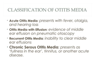


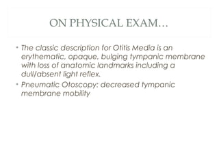


Otitis media is a middle ear infection that is most common in young children. It peaks between 6-12 months of age and 50% of children will have at least one episode by age 1. Common causes are Streptococcus pneumoniae, Haemophilus influenzae, and Moraxella catarrhalis. Risk factors include upper respiratory infections, allergies, craniofacial abnormalities, and passive smoking. Symptoms include fever, ear pain, hearing loss, and irritability. Diagnosis is made through pneumatic otoscopy or tympanometry. Treatment typically involves amoxicillin or other antibiotics for 10-14 days. Complications can include hearing loss, mastoiditis,


