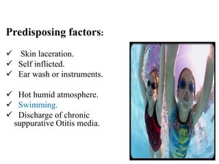
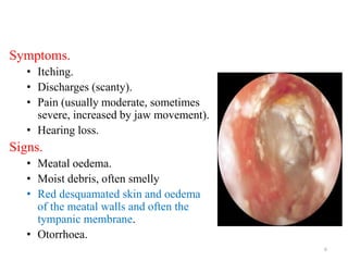
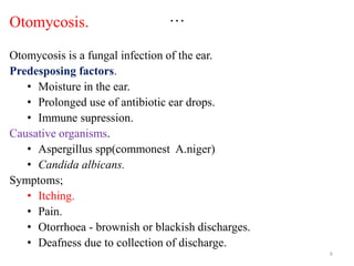
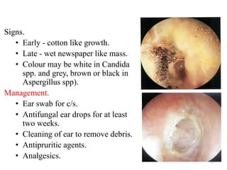
OTITIS EXTERNA
Dr Masoud
1
Otitis externa
• Otitis externa is an inflammation of the external
auditory meatus (EAM).
• can be divided according to the cause as follows;
a. Infectious otitis eterna
• Circumscript otits externa ( furuncle).
• Diffuse otitis externa.
• Malignant otitis externa.
b. Reactive otitis externa
. Eczematous otitis externa.
. Seborrhoeic otitis externa.
2
Circumscript otitis externa.
• This infection affects the outer one third of the ear
canal.
• Normally presents with painful boils and
furuncles(due to infection of hair follicle).
• When boils ripe burst and the ear discharges pus.
Causes:
• Staphylococcus aureus, Pseudomonas
aeruginosa.
Symptoms & signs:
• Pain - out of proportion to the visible lesion.
• Swelling - hyperemic skin.
• Hearing impairment - due to meatal occlusion by
the furuncle.
Treatment.
• Antibiotics, Analgesics depending with severity,
Incision and drainage.
3
Diffuse otitis externa.
• An inflammation of the entire external ear
canal.
• Results to oedema and blockage of the canal(
commonly referred to as "swimmer's ear“).
• The causative organisms are initially fungi
(Aspegillus fumigatus, Aspegillus nigra and
Candida albicans).
• May complicate by mixed bacterial
superinfection like Staphylococcus aureus and
Pseudomonas aeuruginosa. 4
Predisposing factors:
 Skin laceration.
 Self inflicted.
 Ear wash or instruments.
 Hot humid atmosphere.
 Swimming.
 Discharge of chronic
suppurative Otitis media.
5
Symptoms.
• Itching.
• Discharges (scanty).
• Pain (usually moderate, sometimes
severe, increased by jaw movement).
• Hearing loss.
Signs.
• Meatal oedema.
• Moist debris, often smelly
• Red desquamated skin and oedema
of the meatal walls and often the
tympanic membrane.
• Otorrhoea.
6
Management of diffuse otitis externa.
• Ear swab - identification of the offending microorganism.
• Magnesium sulphate pack to reduce edema.
• Aural toilet - to clear the debris.
• Ear drops - antibiotic and steroid.
• Antifungal ear drops.
Prevention of recurrence.
• Prevent water entering the ears.
• Silicone rubber earplugs.
• Avoidance of scratching and poking the ears.
7
…
Otomycosis.
Otomycosis is a fungal infection of the ear.
Predesposing factors.
• Moisture in the ear.
• Prolonged use of antibiotic ear drops.
• Immune supression.
Causative organisms.
• Aspergillus spp(commonest A.niger)
• Candida albicans.
Symptoms;
• Itching.
• Pain.
• Otorrhoea - brownish or blackish discharges.
• Deafness due to collection of discharge.
8
Signs.
• Early - cotton like growth.
• Late - wet newspaper like mass.
• Colour may be white in Candida
spp. and grey, brown or black in
Aspergillus spp).
Management.
• Ear swab for c/s.
• Antifungal ear drops for at least
two weeks.
• Cleaning of ear to remove debris.
• Antipruritic agents.
• Analgesics.
9
Malignant otitis externa.
• Is a progressive necrotising infection.
• starts in the external ear.
• involves tissues of the base of skull, temporal
bone and cranial nerves.
• Primarily occurs in immune suppressed people
eg. Elderly, diabetics, AIDS.
• Causative organism Pseudomonas aeruginosa.
10
Clinical features.
• Severe otalgia
• Granulation tissue protruding through the floor of
ear canal wall at the bone cartilaginous junction
• Extension to involve bone structures of temporal
bone, base of skull and intracranium.
• Cranial nerve VII paralysis.
• Can involve other cranial nerves at jugular foramen.
• Intracranial spread present with headache, fever
neck stiffness and altered level of consciousness.
11
Investigation.
• Swab for culture and
sensitivity.
• CT scan-skull and the brain.
Treatment.
• Aggressive medical treatment.
• Antipseudomonas antibiotics
eg Ciprofloxacillin
intravenous for six weeks.
• Surgical debridment and
dressing.
12
Complications of malignant otitis externa.
• Osteomyelitis of the temporal bone and skull
base.
• Facial nerve paralysis at stylomastoid foramen.
• Last 4 cranial nerves paralysis at the jugular
foramen.
• Meningitis.
• Brain abscess.
• Septicaemia.
13
Reactive otitis externa.
Eczematous otitis externa.
Is an allergic dermatitis of the external auditory
meatus.
Clinical features.
• Irritation and oedema of the canal.
• Weeping eczema with crusting occurs in chronic
cases.
• Secondary infection may lead to acute otitis
externa.
• Canal stenosis due to oedema and fibrosis.
• Fissuring and scalling.
14
Treatment
• Topical steroids.
• Antibiotics locally
and systematically.
• Antihistamines.
Squamous debris covering
the skin of the external
auditory canal can be
noted.
15
Seborrheic dermatitis.
• A chronic inflammatory skin disease.
• Unknown etiology with a predilection for areas of the skin
rich with sebaceous glands.
• Affection of the ear is often distributed along the concha,
scaphoid region, EAC, and postauricular crease.
• Cause has been associated with Pityrosporum ovale and
Malassezia furfur
Clinical Findings.
• Greasy scales overlying erythematous.
• Often pruritic plaques.
• The distribution often involves the scalp, forehead, eyebrows,
glabella, and nasolabial folds.
• Scaling of the scalp is common.
• Superimposed infection and edema may also occur.
16
• Differential Diagnosis.
Seborrheic dermatitis may be confused
with atopic or psoriatic dermatitis, and
scaling within the EAC may be
confused with external otitis or
otomycosis.
• Treatment:
• steroid with antibiotic ointment or
drops.
• Aural toilet.
• Ketoconazole shampoo.
17
THANKS
18

Otitis externa on human ear Pathophysiology

  • 1.
  • 2.
    Otitis externa • Otitisexterna is an inflammation of the external auditory meatus (EAM). • can be divided according to the cause as follows; a. Infectious otitis eterna • Circumscript otits externa ( furuncle). • Diffuse otitis externa. • Malignant otitis externa. b. Reactive otitis externa . Eczematous otitis externa. . Seborrhoeic otitis externa. 2
  • 3.
    Circumscript otitis externa. •This infection affects the outer one third of the ear canal. • Normally presents with painful boils and furuncles(due to infection of hair follicle). • When boils ripe burst and the ear discharges pus. Causes: • Staphylococcus aureus, Pseudomonas aeruginosa. Symptoms & signs: • Pain - out of proportion to the visible lesion. • Swelling - hyperemic skin. • Hearing impairment - due to meatal occlusion by the furuncle. Treatment. • Antibiotics, Analgesics depending with severity, Incision and drainage. 3
  • 4.
    Diffuse otitis externa. •An inflammation of the entire external ear canal. • Results to oedema and blockage of the canal( commonly referred to as "swimmer's ear“). • The causative organisms are initially fungi (Aspegillus fumigatus, Aspegillus nigra and Candida albicans). • May complicate by mixed bacterial superinfection like Staphylococcus aureus and Pseudomonas aeuruginosa. 4
  • 5.
    Predisposing factors:  Skinlaceration.  Self inflicted.  Ear wash or instruments.  Hot humid atmosphere.  Swimming.  Discharge of chronic suppurative Otitis media. 5
  • 6.
    Symptoms. • Itching. • Discharges(scanty). • Pain (usually moderate, sometimes severe, increased by jaw movement). • Hearing loss. Signs. • Meatal oedema. • Moist debris, often smelly • Red desquamated skin and oedema of the meatal walls and often the tympanic membrane. • Otorrhoea. 6
  • 7.
    Management of diffuseotitis externa. • Ear swab - identification of the offending microorganism. • Magnesium sulphate pack to reduce edema. • Aural toilet - to clear the debris. • Ear drops - antibiotic and steroid. • Antifungal ear drops. Prevention of recurrence. • Prevent water entering the ears. • Silicone rubber earplugs. • Avoidance of scratching and poking the ears. 7
  • 8.
    … Otomycosis. Otomycosis is afungal infection of the ear. Predesposing factors. • Moisture in the ear. • Prolonged use of antibiotic ear drops. • Immune supression. Causative organisms. • Aspergillus spp(commonest A.niger) • Candida albicans. Symptoms; • Itching. • Pain. • Otorrhoea - brownish or blackish discharges. • Deafness due to collection of discharge. 8
  • 9.
    Signs. • Early -cotton like growth. • Late - wet newspaper like mass. • Colour may be white in Candida spp. and grey, brown or black in Aspergillus spp). Management. • Ear swab for c/s. • Antifungal ear drops for at least two weeks. • Cleaning of ear to remove debris. • Antipruritic agents. • Analgesics. 9
  • 10.
    Malignant otitis externa. •Is a progressive necrotising infection. • starts in the external ear. • involves tissues of the base of skull, temporal bone and cranial nerves. • Primarily occurs in immune suppressed people eg. Elderly, diabetics, AIDS. • Causative organism Pseudomonas aeruginosa. 10
  • 11.
    Clinical features. • Severeotalgia • Granulation tissue protruding through the floor of ear canal wall at the bone cartilaginous junction • Extension to involve bone structures of temporal bone, base of skull and intracranium. • Cranial nerve VII paralysis. • Can involve other cranial nerves at jugular foramen. • Intracranial spread present with headache, fever neck stiffness and altered level of consciousness. 11
  • 12.
    Investigation. • Swab forculture and sensitivity. • CT scan-skull and the brain. Treatment. • Aggressive medical treatment. • Antipseudomonas antibiotics eg Ciprofloxacillin intravenous for six weeks. • Surgical debridment and dressing. 12
  • 13.
    Complications of malignantotitis externa. • Osteomyelitis of the temporal bone and skull base. • Facial nerve paralysis at stylomastoid foramen. • Last 4 cranial nerves paralysis at the jugular foramen. • Meningitis. • Brain abscess. • Septicaemia. 13
  • 14.
    Reactive otitis externa. Eczematousotitis externa. Is an allergic dermatitis of the external auditory meatus. Clinical features. • Irritation and oedema of the canal. • Weeping eczema with crusting occurs in chronic cases. • Secondary infection may lead to acute otitis externa. • Canal stenosis due to oedema and fibrosis. • Fissuring and scalling. 14
  • 15.
    Treatment • Topical steroids. •Antibiotics locally and systematically. • Antihistamines. Squamous debris covering the skin of the external auditory canal can be noted. 15
  • 16.
    Seborrheic dermatitis. • Achronic inflammatory skin disease. • Unknown etiology with a predilection for areas of the skin rich with sebaceous glands. • Affection of the ear is often distributed along the concha, scaphoid region, EAC, and postauricular crease. • Cause has been associated with Pityrosporum ovale and Malassezia furfur Clinical Findings. • Greasy scales overlying erythematous. • Often pruritic plaques. • The distribution often involves the scalp, forehead, eyebrows, glabella, and nasolabial folds. • Scaling of the scalp is common. • Superimposed infection and edema may also occur. 16
  • 17.
    • Differential Diagnosis. Seborrheicdermatitis may be confused with atopic or psoriatic dermatitis, and scaling within the EAC may be confused with external otitis or otomycosis. • Treatment: • steroid with antibiotic ointment or drops. • Aural toilet. • Ketoconazole shampoo. 17
  • 18.