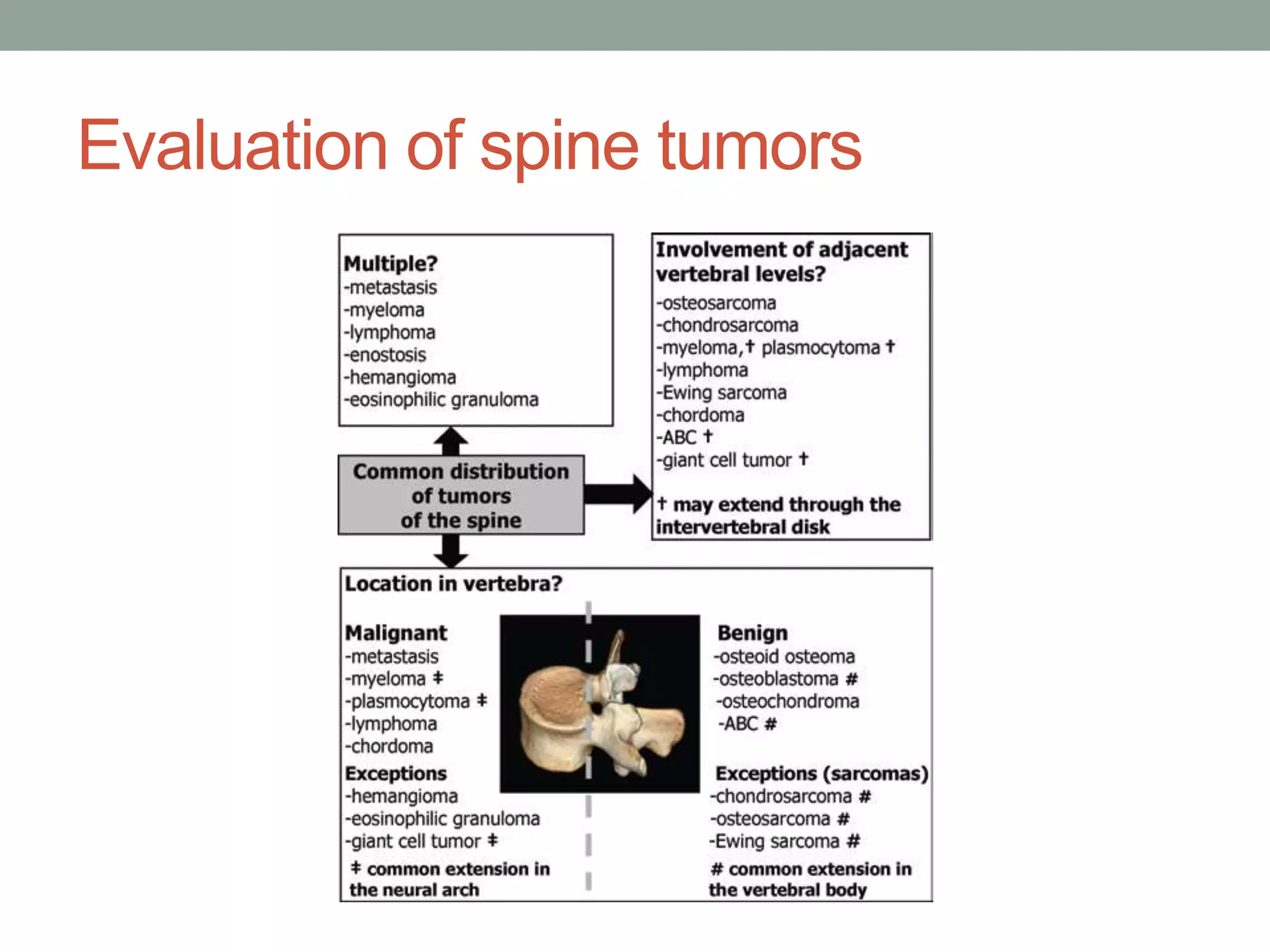
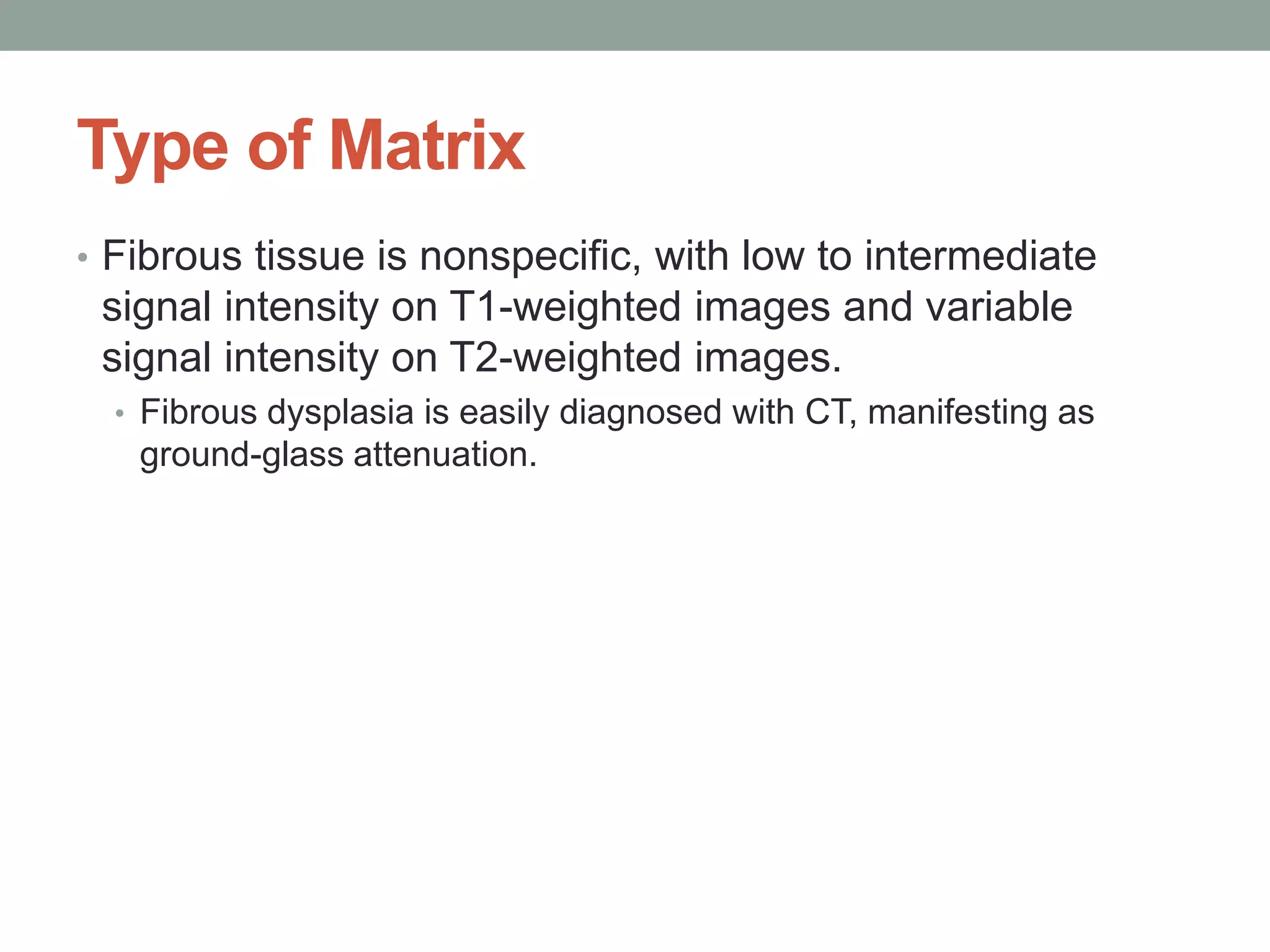
PRIMARY BONE
TUMORS OF THE
SPINE
Ramin Sadeghi, MD
Associate professor of nuclear medicine
Mashhad University of Medical Sciences
• Some tumors have a predilection for specific age groups.
• In patients under 30 years of age, tumors of the spine are fairly
uncommon and are generally benign except for Ewing sarcoma
and osteosarcoma.
• In patients over 30 years of age, most tumors are malignant except
for vertebral hemangiomas and bone islands. Metastases are the
most common lesions.
Evaluation of spine tumors
Topographic Features
• Some tumors have a distinct predilection for the ends of
the spinal column.
• Chordoma is the most common primary distal tumor of the upper
cervical spine. It should be differentiated from pseudotumoral
lesions of the foramen magnum such as calcium pyrophosphate
dihydrate deposits, synovial pannus, and craniovertebral junction
tuberculosis.
• Giant cell tumors commonly involve the sacrum. The sacrum, as a
site of hematopoietic or red marrow in the adult, is also a common
site of metastatic disease as well as of hematologic malignancies
including plasmocytoma, myeloma, lymphoma, and Ewing
sarcoma.
Type of Matrix
• As in the peripheral skeleton, the type of tumor matrix can
help the radiologist diagnose bone- or cartilage-forming
tumors and fibrous dysplasia.
• Osteoblastic tumors can display amorphous ossifications
at radiography or CT. The matrix most often appears
amorphous or cloudlike because it is less dense than
normal bone and lacks an organized trabecular pattern.
The amount and degree of matrix mineralization is widely
variable; thus, the radiographic appearance of
osteoblastic tumors may range from densely blastic to
nearly completely lytic.
Type of Matrix
• Dense osteoblastic lesions display a low T1-T2 signal intensity
pattern at MR imaging.
• The differential diagnosis of osteoblastic tumors includes osteoblastic
metastasis, bone island, lymphoma, and osteosarcoma.
• Osteoblastic tumors should be differentiated from reactive bone
sclerosis adjacent to osteoid osteoma and osteoblastoma.
• Cartilage-forming tumors typically exhibit punctate commalike
or annular calcifications at radiography and CT. These
calcifications appear as low-signal-intensity foci at MR imaging.
Cartilage lobulations display high signal intensity on T2- and
short inversion time inversion-recovery– weighted images
owing to the high water content of the hyaline cartilage. A
pattern of enhancing rings and arcs is seen in cartilaginous
tumors on postcontrast images.
• The differential diagnosis of cartilage-forming tumors includes
osteochondroma, chondroblastoma and chondrosarcoma, ABC, and
chordoma.
Type of Matrix
• Fibrous tissue is nonspecific, with low to intermediate
signal intensity on T1-weighted images and variable
signal intensity on T2-weighted images.
• Fibrous dysplasia is easily diagnosed with CT, manifesting as
ground-glass attenuation.
Other Differential Features
• Fluid-fluid levels were initially described at CT. MR
imaging is now the most sensitive method for detecting
fluid-fluid levels and may also help differentiate liquid from
hemorrhage.
• The imaging finding of prominent fluid-filled hemorrhagic spaces in
a vertebral lesion is suggestive of ABC but has also been described
in telangiectatic osteosarcoma. Moreover, the high prevalence of a
secondary ABC should indicate the possibility of finding fluid-fluid
levels in a coexisting tumor.
• Vertical striations or a “honeycomb” pattern are highly
suggestive of vertebral hemangioma.
• Fat content within lesions can be found in vertebral
hemangioma, fibrous dysplasia, Paget disease, and
Schmörl node.
Margins and Limits
• Benign tumors usually exhibit geographic bone
destruction and sclerotic margins without soft tissue
extension (except ABC, aggressive hemangioma, and
eosinophilic granuloma).
• Conversely, malignant tumors usually exhibit poorly
defined margins, permeative bone destruction, and a soft-
tissue mass. Marrow and, more particularly, soft-tissue
edema, is frequently seen adjacent to primary bone
tumors.
Bone-forming Tumors
Bone Island
• Bone islands, or enostoses, are asymptomatic lesions
that are discovered incidentally in patients of all ages.
They are not true neoplasms and represent dense
compact bone within spongiosa (developmental
abnormality).
• Radiography and CT demonstrate round osteoblastic
lesions with “brush border” at their periphery.
• Enostoses have low signal intensity at T1- and T2-
weighted MR imaging. They may show activity at bone
scintigraphy in less than 10% of cases, and most lesions
remain stable. Enostoses vary in size, and some giant
enostoses have been reported in the literature. The
primary alternative in the differential diagnosis is
osteoblastic metastasis.
Enostosis of the sacrum in a 70-year-old man with a history of
prostate cancer. Coronal reformatted CT image shows a densely
sclerotic lesion with radiating spiculations just beneath the cortex
• A 52-year-old lady who was
suffering from chronic low
back pain
CTscanandMRIofthoracolumbarspineA,B:oncoronalandsagittalviewsofCTscantherewasascleroticlesion
intheT11vertebralbody.C,D:SagittalviewsofMRIfromthoracolumbarspinerevealedasinglewell-
circumscribedhyposignallesioninthebodyofT11onbothT1-andT2-weightedsequences.BothCTscanand
MRIcharacteristicsofthelesionwerecompatiblewithanenostosis.
Osteoid Osteoma
• Osteoid osteoma is a benign osteoblastic lesion
characterized by a nidus of osteoid tissue or even
mineralized immature bone, often surrounded by sclerotic
reactive bone.
• The majority of osteoid osteomas occur in the 2nd and 3rd decades
of life, with a male predilection of 2–3:1.
• The majority of spinal osteoid osteomas are located in the neural
arch. The lumbar spine is most commonly affected, followed by the
cervical, thoracic, and sacral segments. Patients with spinal osteoid
osteoma classically present with painful scoliosis. Aspirin usually
provides pain relief.
Osteoid Osteoma
• At radiography, the complex anatomy of the spine makes the detection
and localization of a radiolucent nidus obscured by reactive sclerosis
much more difficult than that of a nidus located in a long bone.
• Bone scintigraphy is almost invariably positive and has been advocated for
localizing the vertebral level in patients with clinically suspected osteoid osteoma.
• Subsequent targeted CT is generally regarded as the preferred cross-sectional
technique for the demonstration and precise localization of the nidus.
• Osteoid osteoma characteristically manifests as a low-attenuation nidus with central
mineralization and varying degrees of perinidal sclerosis.
• The nidus of osteoid osteoma can have a very heterogeneous, variable appearance
at MR imaging, making detection and characterization difficult. Most tumors have
low to intermediate signal intensity on T1-weighted images and variable signal
intensity on T2-weighted images, possibly depending on the vascularity of the tumor
and the presence of calcification.
• MR imaging is sensitive in detecting nonspecific changes in the bone marrow and
soft tissues, which may have a misleading aggressive appearance. Edema in the
pedicle and lamina extending anteriorly to involve one- to two-thirds of the
posterolateral vertebral body with sparing of the intervertebral disk space should
raise suspicion for the diagnosis. It is also possible to see edema in the neural arch
at a level adjacent to that which harbors the nidus.
Sagittal reformatted CT shows a focal lytic lesion (nidus) with
central density, suspicious of calcification in the right T8 pedicle,
with very minimal sclerotic reaction seen surrounding the nidus.
SagittalT1WimageofthethoracicspineshowsafocalareaofhypointensityintheposterioraspectoftherightsideofT8vertebralbodywith
involvementoftherightpedicle.Additionallynotedisanextensionoftheabnormalsignalintotherightparaspinalsofttissues.
AxialT2WimageofthethoracicspineshowsafocalareaofhyperintensityintheposterioraspectoftherightsideofT8vertebralbodywith
involvementoftherightpedicle.
Sagittalpost-contrastimageofthethoracicspineshowsanabnormalhomogeneousenhancementintheposterioraspectoftherightsideofT8
vertebralbodywithinvolvementoftherightpedicle.
Osteoid osteoma of the lamina at theT8 level in a 34-year-old man with painful
scoliosis. (a) CT scan shows the nidus (arrowhead) with a small area of
calcification and no perinidal sclerosis.
Osteoblastoma
• It tends toward progression and may be locally
aggressive, whereas osteoid osteoma tends toward
regression.
• Osteoblastoma accounts for 1% of all primary bone
tumors, with a male-female ratio of 2:1. Between 32% and
46% of osteoblastomas involve the spine.
• Ninety percent of osteoblastomas manifest in the 2nd and
3rd decades of life. Osteoblastomas originate in the
neural arch and often extend into the vertebral body.
Spinal osteoblastomas appear with more or less equal
frequency in the cervical, thoracic, and lumbar segments.
Osteoblastoma
• At bone scintigraphy, osteoblastoma demonstrates
marked radionuclide uptake.
• Reactive sclerosis adjacent to the lesion is common at
conventional radiography. The radiologic features of
osteoblastoma are a lesion diameter greater than 2 cm,
osseous expansion, soft-tissue components, and
multifocal matrix mineralization.
• Tumors that are mainly lytic at CT with little evidence of
matrix mineralization are hypointense at T1-weighted MR
imaging and hyperintense at T2-weighted imaging.
Lesions showing little matrix mineralization at CT but
diffuse osteoid production at histologic analysis have low
signal intensity at T2-weighted MR imaging
Case 1
Osteoblastoma of the sacrum in a 37-year-old man with sacral
pain. CT scan shows a hypoattenuating area with central
calcifications and minimal surrounding sclerosis.
Aggressiveosteoblastomaof the L5 vertebralbodyin a 34-year-old woman with lowbackpainand
neurologicsymptoms. (a) CTscanshows a destructiveexpansile lesionwith osteoidmatrix extending
intothe spinalcanal.(b)AxialT2-weightedMR imageshows a masswith lowsignal intensity. (c)Axial
contrast-enhanced fat-saturatedT1-weightedMR imageshowsmarked enhancementof the lesion.
Osteoblastomaof the L2 posteriorarch ina 15-year-old girlwith painfulscoliosis.(a) CTscanshows
diffuseosteoidmatrix. (b) Sagittalcontrast-enhancedfat-saturatedT1-weightedMR imageshows
marked enhancementof the lesion(*) and severeinflammationof the surroundingsoft tissueinvolving
the adjacentvertebrallevels(arrowheads)and the posteriorarch of L1 (arrow) (“flare phenomenon”).
Osteosarcoma
• Only 4% of all osteosarcomas involve the spine. Peak
prevalence occurs during the 4th decade of life. Radiation
therapy and Paget disease are usually implicated in cases
of secondary osteosarcoma in elderly patients.
• The thoracic and lumbar segments are involved with
equal frequency, followed by the sacrum and the cervical
column.
• In 79% of cases, the tumor arises in the posterior
elements with partial vertebral body involvement.
• Involvement of two vertebral levels is seen in 17% of
cases
• Conventional osteosarcoma is a high-grade malignant
osteoblastic lesion with varying amounts of osteoid production,
cartilage, or fibrous tissue. In 80% of cases, CT demonstrates
matrix mineralization.
• Rarely, tumors with marked mineralization originating in the vertebral
body may manifest as an “ivory vertebra” (sclerosing osteoblastic
osteosarcoma). A purely lytic pattern is also seen in various subtypes
such as telangiectatic osteosarcoma (predominant cystic architecture
simulating ABC).
• MR imaging signal intensity characteristics are usually
nonspecific. Fluid-fluid levels have been described in
association with telangiectatic osteosarcoma.
• As opposed to ABCs, telangiectatic osteosarcomas with prominent
fluid-filled hemorrhagic spaces are characterized by thick, solid
nodular tissue surrounding the cystic spaces, matrix mineralization,
and a more aggressive growth pattern
Osteoblasticosteosarcomaof the sacrum in a 20-year-old man with sacral pain,
neurologic symptoms, and a palpable mass. (a) CTscan shows permeative bone
destruction and osteoid matrix. (b)Axial T2-weighted MR image shows a slightly
hypointense lesion with cystic foci (arrowheads). (c)Axial contrast-enhanced fat-saturated
T1-weighted MR image shows enhancement of the lesion with cystic foci (arrowheads).
Cartilage-forming Tumors
Osteochondroma
• Osteochondroma represents the most common bone tumor and is a
developmental lesion rather than a true neoplasm
• Most osteochondromas are solitary and sporadic lesions, although some
are multiple, usually with an autosomal dominant inheritance. Although
only 1.3%–4.1% of solitary osteochondromas originate in the spine,
approximately 9% of patients with multiple osteochondromas have spinal
lesions.
• Solitary lesions affect males more frequently than females (1.9:1 ratio),
and the average age at diagnosis is 33 years. Osteochondromas can
arise from any part of the vertebral column, but the cervical spine is
commonly involved, with a predilection for the atlantoaxial area, followed
by the thoracic spine and the lumbar spine. In cases of multiple
exostoses, the thoracolumbar spine is more commonly involved. Most
spinal lesions occur near the tip of spinous or transverse processes.
They can also develop in the vertebral body, a pedicle, or, more rarely,
the articular facet.
• Radiation-induced osteochondromas occur within or at the periphery of
the radiation field and are usually solitary. The prevalence following
irradiation for childhood malignancy is approximately 12%.
Osteochondroma
• Because of overlapping of osseous structures of the spine,
conventional radiography is often insufficient.
• CT is the modality of choice for demonstrating the diagnostic
appearance of marrow and cortical continuity with the
underlying vertebra.
• At MR imaging, the lesion manifests with a peripheral rim of
low signal intensity corresponding to the cortical bone and a
central area of fat signal intensity corresponding to the central
cancellous bone. A thin cartilaginous cap may be present,
especially in children. With age, cartilage tends to thin and
disappears at numerous points on the surface of an
osteochondroma.
• However, a thick (>1-cm) cartilaginous cap in an adult patient should
raise suspicion for an exostotic chondrosarcoma
Osteochondroma of the cervical spine in a 30-year-old man with
cervicobrachial neuralgia. (a) Sagittal reformatted CT image
shows cortical continuity of the osteochondroma with the C6
posterior arch
Case 2
Chondroblastoma
• Chondroblastoma is a benign cartilaginous tumor with a
predilection for the growing skeleton
• Only 1.4% of all chondroblastomas originate in a vertebra.
Most patients present during the 3rd decade of life. The
tumor involves the vertebral body and posterior elements.
• The tumor shows aggressive features at imaging, with
bone destruction and a soft-tissue mass but no
surrounding bone edema. In other cases, CT may
demonstrate a geographic lesion with sclerotic borders.
• Most lesions have hypointense areas on T2-weighted MR images.
Low signal intensity on T2-weighted images is associated with
immature chondroid matrix, hypercellularity, calcifications, and
hemosiderin at histologic analysis
ChondroblastomaofL3ina23-year-oldwomanwitha6-monthhistoryofpainfullumbarscoliosis.(a)Sagittalreformatted
CTimagerevealsalyticlesionoftheL3vertebralbodywithscleroticmargins.Thelesioninvolvestherightpedicleand
articularprocessandextendsintotheadjacentforamina.(b)OnasagittalT2-weightedMRimage,thelesion
(arrowheads)appearsisointenserelativetothenormalvertebralbodiesandisdifficulttovisualize.(c)Axialcontrast-
enhancedfat-saturatedT1-weightedMRimageshowsmarkedenhancementofthelesion
Chondrosarcoma
• Chondrosarcoma is the second most common
nonlymphoproliferative primary malignant tumor of the
spine in adults
• Peak prevalence occurs between 30 and 70 years of age.
Men are affected two to four times more frequently than
women. The thoracic and lumbar spine are most
frequently affected, with the sacrum being affected only
rarely. Chondrosarcoma originates in the vertebral body
(15% of cases), posterior element (40%), or both (45%) at
presentation
Chondrosarcoma
• Chondrosarcomas of the spine usually manifest as a large,
calcified mass with bone destruction. True ossification may also
be present, which sometimes corresponds to residual
osteochondroma in cases of secondary chondrosarcoma.
• Chondroid matrix mineralization is better demonstrated with
CT.
• Calcified matrix is detected as areas of signal void at MR
imaging.
• The nonmineralized portion of the tumor has low attenuation on CT
scans, low to intermediate signal intensity on T1-weighted MR images,
and very high signal intensity on T2-weighted images due to the high
water content of hyaline cartilage. An enhancement pattern of rings
and arcs at gadolinium-enhanced MR imaging reflects the lobulated
growth pattern of these cartilaginous tumors.
• Extension through the intervertebral disk has been reported in
35% of cases
Chondrosarcoma of the L4 vertebral body in a 41-year-old man with low back pain. (a) CT
scan shows a large mass arising from the vertebral body with cortical disruption and ring
and arc calcifications.(b)Axial T2-weighted MR image shows a high-signal-intensity
lobulated mass with linear striations.
Hematopoietic, Reticuloendothelial,
and Lymphatic Tumors
Eosinophilic Granuloma
• Langerhans cell histiocytosis, previously termed histiocytosis X,
includes conditions that range from the usually solitary and curable
eosinophilic granuloma, to the disseminated process that produces
Schüller-Christian syndrome, to the disseminated and rapidly fatal
variety known as Letterer-Siwe disease. These three seemingly
dissimilar conditions are united by common pathologic features
• It usually occurs in children, with a peak prevalence between 5 and
10 years of age.
• Eighty percent of cases occur before the age of 30 years.
• In multifocal involvement (10%–20% of cases), osseous lesions
appear simultaneously or within 1–2 years.
• Vertebral involvement is seen in only 7.8%–25% of cases of
Langerhans cell histiocytosis.
• Patients with spinal lesions usually have pain, which subsides rapidly
after bed rest. Although almost all vertebral lesions involve collapse of
the vertebral body,
Eosinophilic Granuloma
• The radiographic characteristics of a typical spinal lesion
consist of complete or incomplete collapse of the vertebral
body; absence of an osteolytic area; preservation of
pedicles, posterior elements, and adjacent disk spaces;
absence of adjacent paravertebral soft-tissue shadow;
and increased opacity in the collapsed body (vertebra
plana)
osinophilic granuloma in a 12-year-old boy. Radiograph shows
increased opacity in an incompletely collapsed vertebral body
(arrowhead).
Plasmocytoma
• Plasmocytoma represents focal proliferation of malignant
plasma cells without diffuse bone marrow involvement. These
lesions are considered to represent the early stages of multiple
myeloma.
• Solitary plasmocytoma is an uncommon tumor that occurs in
3%–7% of patients with plasma cell neoplasms. Seventy
percent of patients are over 60 years old.
• The vertebral body is the most common site of involvement by
plasmocytoma due to its rich red marrow content, but the tumor
frequently extends to the pedicles.
• Plasmocytoma usually manifests with a single collapsed
vertebra.
• A monoclonal seric immunoglobulin is present at a low seric
level in 40% of cases.
Plasmocytoma
• In two-thirds of cases, the radiographic appearance is characteristic, with
a mixed, predominantly lytic pattern.
• The tumor preferentially replaces the cancellous bone, whereas the cortical bone is
partly preserved or even sclerotic, resulting in a hollow vertebral body or pedicle.
• The cortical thickening in the arrangement of plasmocytoma appears to be unique to
this tumor and results in a “minibrain” appearance on axial images.
• In one-third of cases, the radiographic appearance is less characteristic,
with a multicystic “soap bubble” appearance simulating a hemangioma,
or perhaps a purely lytic appearance.
• Solitary sclerotic plasmocytoma is extremely rare and can be found in
association with polyneuropathy.
• Like many expansile tumors involving the axial skeleton, plasmocytoma
has low signal intensity on T1-weighted MR images, high signal intensity
on T2-weighted images, and homogeneous marked enhancement on
postcontrast T1-weighted images.
• Focal endplate fractures are well described in patients with
plasmocytoma
• Involvement of the intervertebral disk and adjacent vertebrae can be
used to help differentiate plasmocytoma from metastasis
Plasmocytoma of C6 in a 36-year-old man with cervical spinal pain and neurologic
symptoms. (a) Lateral radiograph shows a multicystic-appearing lesion (arrowhead)
involving the vertebral body and the neural arch at C6. (b) CTscan shows the minibrain
sign created by a lytic vertebral lesion with cortical preservation.
Axial contrast-enhanced fat-saturated T1-weighted MR image
shows homogeneous enhancement of the lesion with epidural
extension and spinal cord compression.
Lymphoma
• Primary lymphoma of bone is a rare extranodal
manifestation of non-Hodgkin lymphoma, accounting for
only about 1%–3% of all lymphomas.
• Primary bone lymphomas are mainly diffuse large B-cell
lymphomas. Peak prevalence occurs in the 5th–7th
decades, with a strong male predilection (up to an 8:1
male-female ratio).
• Spinal involvement in lymphomas results much more
frequently from late metastatic dissemination of Hodgkin
disease and non-Hodgkin lymphoma. Spinal involvement
may manifest as paraspinal, vertebral, and epidural
involvement, either in isolation or in combination
• Vertebral lesions may have a sclerotic, lytic, or mixed
appearance. The sclerotic (ivory vertebra) and mixed patterns
are more common in Hodgkin disease. The appearance of
vertebral lymphoma at CT and MR imaging is usually
nonspecific.
• Bone scintigraphy shows increased radionuclide uptake in
nearly all patients.
• Nevertheless, a focus of bone marrow replacement and a
surrounding soft-tissue mass without large areas of cortical
bone destruction suggest lymphoma.
• Lymphoma is caused by tumor spread from the medullary cavity along
the small vascular channels that run through the cortex. It can be seen
with other small round cell tumors such as Ewing sarcoma. Contiguous
vertebral involvement has also been reported
B-celllymphoma inan 80-year-oldwoman withlowbackpainand caudaequinasyndrome. (a) Sagittal
T2-weightedMR imageshowsa low-signal-intensityspinallesion spreadingovermultiplelevels(L4–
S3)and extendingintothe epiduraland prevertebralspaces,but withoutextensionthrough the
intervertebral disk.(b) Sagittalcontrast-enhancedfat-saturatedT1-weightedMR imageshows
heterogeneousenhancementof the lesion.
Ewing Sarcoma
• Although the vertebral column is frequently involved in
preterminal metastatic Ewing sarcoma, primary vertebral Ewing
sarcoma is quite rare, with a reported prevalence of 3.5%–15%
• Ewing sarcoma is an undifferentiated high-grade proliferation of
uniform small round cells. Necrosis is common.
• Primary vertebral Ewing sarcoma is usually seen in the 2nd
decade of life (mean age, 19.3 years), with a slight male
predilection (62% vs 38% of cases)
• The sacrum is the most frequently involved site (55.2% of
cases), followed by the lumbar spine (25%). The cervical spine
is the least frequently affected site (3.2% of cases).
• In the non-sacral spine, the majority (60%) of lesions originate in the
posterior elements with extension into the vertebral body. The ala is
the most frequently affected sacral site (69% of cases). More than one
segment is involved in 8% of cases. The disk spaces are usually
preserved
• Lesions may be lytic, sclerotic, or mixed with associated
vertebral compression. Nearly all tumors (93%) are lytic
and morphologically aggressive. A purely sclerotic pattern
is rare and might correspond to necrotic and reactive
bone formation. Other unusual imaging findings include
vertebra plana, ivory vertebra, and pseudohemangioma
• Invasion of the spinal canal is common (91% of cases),
often with a large mass. The paraspinal component is
often larger than the intraosseous lesion
Vertebral Hemangioma
• Spinal hemangiomas are common and frequently
multiple. The prevalence of hemangiomas seems to
increase with age and is greatest after middle age, with a
slight female predilection. Most hemangiomas are seen in
the thoracic and lumbar spine. They are usually confined
to the vertebral body, although they may occasionally
extend into the posterior elements.
• Most spinal hemangiomas are asymptomatic.
Occasionally, vertebral hemangiomas may increase in
size and compress the spinal cord and nerve roots.
Compressive vertebral hemangiomas can occur in
patients of any age, with a peak prevalence in young
adults, and preferentially occur in the thoracic spine
• Hemangiomas in vertebrae cause rarefaction with exaggerated vertical striations
or a coarse honeycomb appearance.
• CT shows the pattern as multiple dots (polka-dot appearance) representing a
cross-section of reinforced trabeculae.
• At scintigraphy, the appearance of osseous hemangiomas ranges from
photopenia to a moderate increase in radiotracer uptake.
• The presence of high signal intensity on T1- and T2-weighted MR images is
related to the amount of adipocytes or vessels and interstitial edema, respectively.
Thick, low-signal-intensity vertical struts may be seen within hemangiomas.
• Fatty vertebral hemangiomas may represent inactive forms of this lesion whereas low signal
intensity at MR imaging may indicate a more active lesion with the potential to compress the
spinal cord.
• The radiographic and CT appearances of compressive vertebral hemangiomas
can be misleading, with irregular trabeculae and lytic areas; poorly defined,
expanded cortex; and soft-tissue expansion
• Mottled high signal intensity on T1-weighted MR images can be expected in only
about 50% of compressive vertebral hemangiomas and signal voids are the most
useful additional MR imaging sign in lesions that are hypointense on T1-weighted
images. The differential diagnosis of compressive hemangiomas with a coarse
reticular pattern and extradural extension includes rare hemangioblastoma,
lymphangioma, and Ewing sarcoma
Hemangiomaof theT9vertebralbody inan 81-year-oldwoman withmultipleosteoporoticcompression
fractures (T10–T12). (a) SagittalT1-weightedMR imageshows fat signalintensitythroughouttheT9
vertebralbody(arrowhead), with linearlow-signal-intensitystriationsdue to thickened
trabeculae.(b)AxialT2-weightedMR imageshowsa well-circumscribedfatty lesionwith coarsevertical
trabeculae(polkadots).
Aggressive hemangioma in a 53-year-old man.Axial contrast-enhanced fat-
saturatedT1-weighted MR image shows an avidly enhanced lesion with coarse
vertical trabeculae and extraosseous extension into the epidural space
Another case
Another case
Chordoma
• Chordoma is a rare malignant neoplasm arising from the
remnants of the primitive notochord
• Next to lymphoproliferative tumors, chordomas are the most
common primary malignant neoplasm of the spine in adults
• Chordomas generally occur in late middle age, with a peak
prevalence in the 5th–6th decades
• Spinal chordomas have a 2:1 male-female ratio. Chordomas
most commonly arise in the sacrococcygeal region (50% of
cases), followed by the spheno-occipital region (35%) and the
vertebral bodies (15%). Sacrococcygeal tumors usually start in
the lower sacrum and coccyx
• Spinal chordomas arise more frequently in the cervical spine
than in the thoracic and lumbar regions
• The most common site of involvement in the mobile spine is the
vertebral body with sparing of the posterior elements
Chordoma
• The most suggestive manifestation is a destructive lesion of a vertebral body
associated with a soft-tissue mass with a “collar button” or “mushroom”
appearance and a “dumbbell” shape, spanning several segments and sparing the
disks
• Areas of amorphous calcifications are noted in 40% of chordomas of the mobile
spine and in up to 90% of sacrococcygeal lesions.
• Most chordomas are iso- or hypointense relative to muscle on T1-weighted MR
images. The focal areas of hemorrhage and high protein content of the myxoid
and mucinous collections that may be seen in chordomas account for the high
signal intensity on T1-weighted images
• On T2-weighted images, most chordomas have a high signal intensity that reflects
their high water content
• The fibrous septa that divide the gelatinous components of the tumor are seen as
areas of low signal intensity on T2-weighted images.
• The presence of hemosiderin also accounts for the low signal intensity seen on
T2-weighted images. This MR imaging feature has been reported in 72% of
sacrococcygeal chordomas but is rare in spinal chordomas
• After the injection of gadolinium-based contrast material, most tumors
demonstrate moderate heterogeneous enhancement, but ring and arc
enhancement and peripheral enhancement have also been described
Sacrococcygeal chordoma in a 76-year-old man. CT
scan shows a midline soft-tissue mass with amorphous
calcifications.
Chordomaof the cervicalspineina 50-year-old womanwith neurologicsymptoms. (a) Sagittalcontrast-
enhancedT1-weightedMR imageshows a heterogeneouslyenhancedlesioninvadingthe C5 and C6
vertebralbodiesand nearlysparingthe C5–C6intervertebraldisk.(b) SagittalT2-weightedMR image
shows the lesionwith highsignalintensityand invadingthe C6 vertebralbody and epiduralspace.Note
the mushroomlikeappearanceof the lesion.
Another case
Another case
Aneurysmal Bone Cyst
• ABC is a benign bone lesion of unknown origin. It is a relatively rare
lesion that represents 1.4%–2.3% of primary bone tumors. The spine
is involved in 3%–20% of cases
• In 29%–35% of cases, a preexisting lesion can be identified. The
most common of these is giant cell tumor, which accounts for 19%–
39% of cases in which a preexisting lesion is found
• Other common precursor lesions include osteoblastoma, angioma, and
chondroblastoma, whereas uncommon precursor lesions include fibrous
dysplasia, fibrous histiocytoma, eosinophilic granuloma, osteosarcoma, and
even metastatic carcinoma
• ABC usually occurs between the ages of 5 and 20 years but can
manifest at any age. There may be a slight female predilection.
• The cervical spine is affected in 22% of cases, the thoracic spine in 34%, the
lumbar spine in 31%, and the sacrum in 13%.
• Spinal involvement is typically in the posterior elements, although
extension into the vertebral body is common (75% of cases)
• Spinal ABC may extend into the adjacent vertebrae or intervertebral
disk, the ribs, and the paravertebral soft tissue
Aneurysmal Bone Cyst
• The natural history of ABC has been described as
evolving through four radiologic stages: initial, active,
stabilization, and healing
• In the initial phase, the lesion is characterized by a well-defined
area of osteolysis. This is followed by a growth phase, in which the
lesion has a purely lytic pattern and sometimes ill-defined margins.
Later, during the stabilization phase, the characteristic soap bubble
appearance develops as a result of maturation of the bony shell.
CT and MR imaging typically show a well-defined lesion with
internal septation
Aneurysmal Bone Cyst
• Fluid-fluid levels within ABCs are indicative of hemorrhage with
sedimentation and are better demonstrated with MR imaging.
• On T1-weighted images, they may have increased signal intensity due to
methemoglobin in either the dependent or nondependent component.
• Gadolinium-based contrast material injection demonstrates smooth
enhancement of the internal septa within ABCs.
• The lesion margins show a low-signal-intensity rim on MR images that is
thought to be caused by an intact, thickened periosteal membrane
• Other areas usually show high signal intensity on T2-weighted images.
The presence of a solid component with diffuse contrast enhancement
should raise suspicion for secondary ABC, although it might be
encountered in the solid variant of ABC
• The predominant bone scintigraphic pattern in ABC is moderate to
intense radiotracer accumulation at the periphery of the lesion with little
activity at its center (“doughnut sign”), a finding that is evident in about
64% of cases. However, this scintigraphic pattern lacks specificity, since
it is also found in giant cell tumor, chondrosarcoma, and telangiectatic
osteosarcoma
ABC of the lumbar spine (L3) in a 34-year-old woman with low back pain. (a) CT
scan shows a sharply demarcated and multiloculated lesion with peripheral
calcifications and fluid-fluid levels due to layering of blood products.(b) Coronal
contrast-enhancedT1-weighted MR image shows enhanced septa between
cysts. (c) On a coronalT2-weighted MR image, the signal intensity of cysts
varies with stage of blood degradation
Another case
Giant Cell Tumor
• This tumor occurs in skeletally mature patients in the 2nd–4th
decades of life, more frequently in females
• Seven percent of giant cell tumors occur in the spine. The sacrum is
affected in 90% of such cases. The tumor is usually located in the
upper sacrum and frequently lateralized in a sacral wing
• Extension to the iliac wing through the sacroiliac joint is possible.
Above the sacrum, the lumbar, thoracic, and cervical spine (in
decreasing order of frequency) may be affected
• The tumor usually predominates in the vertebral body, with frequent
involvement of the posterior arch. The tumor is limited to the vertebral
body and pedicles in only 21% of cases
• Extraosseous involvement of the soft tissues is seen in 79% of cases.
Giant cell tumors of the thoracic spine can sometimes simulate
posterior mediastinal neoplasms
• Intervertebral disk invasion and extension into an adjacent vertebra is
possible
• Radiography typically shows a lytic lesion with cortical
expansion A purely osteolytic pattern is also possible. CT
demonstrates absence of mineralization and the lack of a
sclerotic rim at the margins of the tumor
• Bone scintigraphy shows increased radiotracer uptake in
all patients.
• The tumor usually has low to intermediate signal intensity
on T1-weighted MR images. Areas of high signal intensity
can suggest relatively recent hemorrhage. More
specifically, most giant cell tumors of the spine have low
to intermediate signal intensity on T2-weighted images.
This appearance seems to be caused by hemosiderin
deposition and high collagen content
Giant cell tumor of the upper sacrum in a 33-year-old woman.
Coronal reformatted CT image shows a well-defined lytic lesion of
the right upper part of the sacrum with extension through the right
sacroiliac joint and absence of a sclerotic rim.
Another case
Fibrous Dysplasia
• It occurs in both children and adults and has an equal gender
distribution. Vertebral involvement is rare but occurs more
frequently with polyostotic disease. The lesion is frequently
asymptomatic, but pain and fractures may be part of the clinical
spectrum.
• Radiography and CT usually show a mildly expansile lesion
with a “blown-out” cortical shell or a lytic lesion with a sclerotic
rim. “Ground-glass” matrix is common and characteristic.
• Lesions may also contain islands of cartilage. Fibrous
dysplasia has variable MR imaging characteristics—typically,
intermediate to low signal intensity on T1-weighted images and
variable signal intensity on T2-weighted images.
• Fibrous dysplasia shows a mild to marked increase in
radionuclide uptake at bone scintigraphy.
•
Fibrous dysplasia of the lumbar spine (L4) in a 45-year-old
woman with low back pain. (a) CT scan shows a mildly expansile
lytic lesion with a sclerotic rim involving the vertebral body and the
neural arch. The lesion also demonstrates ground-glass matrix
(arrowhead).
Differential Diagnosis of Primary
Spinal Tumors
Metastatic Disease
• Metastases are the most common vertebral tumors. Osteolytic
metastases occur more frequently than osteoblastic
metastases. Some metastases have a mixed pattern, with
areas of osteolysis and areas of sclerosis. Typically,
metastases are multiple and of variable size with cortical
disruption (osteolytic lesions). Vertebral compression fracture
and epidural tumor are common in metastases. Some slow-
growing metastases may mimic a primary bone tumor with
mineralization and sclerotic margins.
• Osteolytic metastases are most often caused by carcinoma of the
lung, breast, thyroid, kidney, and colon and (in childhood)
neuroblastoma.
• Osteoblastic metastases are most commonly caused by prostate
carcinoma in elderly men and by breast cancer in women. Other
osteoblastic metastases are caused by lymphoma, carcinoid tumors,
mucinous adenocarcinoma of the gastrointestinal tract, pancreatic
adenocarcinoma, bladder carcinoma, neuroblastoma, and (in
childhood) medulloblastoma.
Paget Disease
• Paget disease is a chronic metabolic disorder of abnormal
bone remodeling in the adult skeleton. It is rare in patients less
than 40 years old.
• Paget disease occurs more frequently in Caucasians of
Northern European descent and is rare in Asians and African-
Americans.
• In the spine, the vertebra is expanded. The typical “picture
frame” vertebra shows a coarse and sclerotic peripheral
trabecular pattern and central osteopenia. Other patterns of
pagetic vertebrae include ivory vertebra and isolated posterior
arch involvement.
• The pagetic bone marrow contains fatty areas with a
heterogeneous distribution. Bone scintigraphy demonstrates
the extent of Paget disease. Sarcomatous transformation of the
lesion is rare (<1% of cases).
Brown Tumor
• Brown tumor, a classic manifestation of
hyperparathyroidism, can arise in the spine, causing
neurologic compromise related to both collapse of
osteolytic vertebral lesions and tumor growth within the
spinal canal
• At histologic analysis, brown tumor cannot be
differentiated from giant cell tumor, so that the diagnosis
relies chiefly on the presence of hyperparathyroidism
• When located in bone, brown tumor undergoes
remineralization after parathyroidectomy
Schmorl Node
• Schmörl nodes represent vertical disk prolapses through
areas of weakness in the vertebral end-plate. Schmörl
nodes are often multiple and occur predominantly in the
thoracolumbar spine. Recently formed Schmörl nodes can
be painful and may be indistinguishable from
inflammatory or tumoral disease in terms of signal
intensity. The identification of endplate defects or intra-
nuclear cleft bending of the disk at either CT or MR
imaging is helpful in making the correct diagnosis of acute
Schmörl nodes
Primary bone tumors of the spine

Primary bone tumors of the spine

  • 1.
    PRIMARY BONE TUMORS OFTHE SPINE Ramin Sadeghi, MD Associate professor of nuclear medicine Mashhad University of Medical Sciences
  • 6.
    • Some tumorshave a predilection for specific age groups. • In patients under 30 years of age, tumors of the spine are fairly uncommon and are generally benign except for Ewing sarcoma and osteosarcoma. • In patients over 30 years of age, most tumors are malignant except for vertebral hemangiomas and bone islands. Metastases are the most common lesions.
  • 7.
  • 8.
    Topographic Features • Sometumors have a distinct predilection for the ends of the spinal column. • Chordoma is the most common primary distal tumor of the upper cervical spine. It should be differentiated from pseudotumoral lesions of the foramen magnum such as calcium pyrophosphate dihydrate deposits, synovial pannus, and craniovertebral junction tuberculosis. • Giant cell tumors commonly involve the sacrum. The sacrum, as a site of hematopoietic or red marrow in the adult, is also a common site of metastatic disease as well as of hematologic malignancies including plasmocytoma, myeloma, lymphoma, and Ewing sarcoma.
  • 9.
    Type of Matrix •As in the peripheral skeleton, the type of tumor matrix can help the radiologist diagnose bone- or cartilage-forming tumors and fibrous dysplasia. • Osteoblastic tumors can display amorphous ossifications at radiography or CT. The matrix most often appears amorphous or cloudlike because it is less dense than normal bone and lacks an organized trabecular pattern. The amount and degree of matrix mineralization is widely variable; thus, the radiographic appearance of osteoblastic tumors may range from densely blastic to nearly completely lytic.
  • 10.
    Type of Matrix •Dense osteoblastic lesions display a low T1-T2 signal intensity pattern at MR imaging. • The differential diagnosis of osteoblastic tumors includes osteoblastic metastasis, bone island, lymphoma, and osteosarcoma. • Osteoblastic tumors should be differentiated from reactive bone sclerosis adjacent to osteoid osteoma and osteoblastoma. • Cartilage-forming tumors typically exhibit punctate commalike or annular calcifications at radiography and CT. These calcifications appear as low-signal-intensity foci at MR imaging. Cartilage lobulations display high signal intensity on T2- and short inversion time inversion-recovery– weighted images owing to the high water content of the hyaline cartilage. A pattern of enhancing rings and arcs is seen in cartilaginous tumors on postcontrast images. • The differential diagnosis of cartilage-forming tumors includes osteochondroma, chondroblastoma and chondrosarcoma, ABC, and chordoma.
  • 11.
    Type of Matrix •Fibrous tissue is nonspecific, with low to intermediate signal intensity on T1-weighted images and variable signal intensity on T2-weighted images. • Fibrous dysplasia is easily diagnosed with CT, manifesting as ground-glass attenuation.
  • 12.
    Other Differential Features •Fluid-fluid levels were initially described at CT. MR imaging is now the most sensitive method for detecting fluid-fluid levels and may also help differentiate liquid from hemorrhage. • The imaging finding of prominent fluid-filled hemorrhagic spaces in a vertebral lesion is suggestive of ABC but has also been described in telangiectatic osteosarcoma. Moreover, the high prevalence of a secondary ABC should indicate the possibility of finding fluid-fluid levels in a coexisting tumor. • Vertical striations or a “honeycomb” pattern are highly suggestive of vertebral hemangioma. • Fat content within lesions can be found in vertebral hemangioma, fibrous dysplasia, Paget disease, and Schmörl node.
  • 13.
    Margins and Limits •Benign tumors usually exhibit geographic bone destruction and sclerotic margins without soft tissue extension (except ABC, aggressive hemangioma, and eosinophilic granuloma). • Conversely, malignant tumors usually exhibit poorly defined margins, permeative bone destruction, and a soft- tissue mass. Marrow and, more particularly, soft-tissue edema, is frequently seen adjacent to primary bone tumors.
  • 14.
  • 15.
    Bone Island • Boneislands, or enostoses, are asymptomatic lesions that are discovered incidentally in patients of all ages. They are not true neoplasms and represent dense compact bone within spongiosa (developmental abnormality). • Radiography and CT demonstrate round osteoblastic lesions with “brush border” at their periphery. • Enostoses have low signal intensity at T1- and T2- weighted MR imaging. They may show activity at bone scintigraphy in less than 10% of cases, and most lesions remain stable. Enostoses vary in size, and some giant enostoses have been reported in the literature. The primary alternative in the differential diagnosis is osteoblastic metastasis.
  • 16.
    Enostosis of thesacrum in a 70-year-old man with a history of prostate cancer. Coronal reformatted CT image shows a densely sclerotic lesion with radiating spiculations just beneath the cortex
  • 17.
    • A 52-year-oldlady who was suffering from chronic low back pain
  • 18.
  • 19.
    Osteoid Osteoma • Osteoidosteoma is a benign osteoblastic lesion characterized by a nidus of osteoid tissue or even mineralized immature bone, often surrounded by sclerotic reactive bone. • The majority of osteoid osteomas occur in the 2nd and 3rd decades of life, with a male predilection of 2–3:1. • The majority of spinal osteoid osteomas are located in the neural arch. The lumbar spine is most commonly affected, followed by the cervical, thoracic, and sacral segments. Patients with spinal osteoid osteoma classically present with painful scoliosis. Aspirin usually provides pain relief.
  • 20.
    Osteoid Osteoma • Atradiography, the complex anatomy of the spine makes the detection and localization of a radiolucent nidus obscured by reactive sclerosis much more difficult than that of a nidus located in a long bone. • Bone scintigraphy is almost invariably positive and has been advocated for localizing the vertebral level in patients with clinically suspected osteoid osteoma. • Subsequent targeted CT is generally regarded as the preferred cross-sectional technique for the demonstration and precise localization of the nidus. • Osteoid osteoma characteristically manifests as a low-attenuation nidus with central mineralization and varying degrees of perinidal sclerosis. • The nidus of osteoid osteoma can have a very heterogeneous, variable appearance at MR imaging, making detection and characterization difficult. Most tumors have low to intermediate signal intensity on T1-weighted images and variable signal intensity on T2-weighted images, possibly depending on the vascularity of the tumor and the presence of calcification. • MR imaging is sensitive in detecting nonspecific changes in the bone marrow and soft tissues, which may have a misleading aggressive appearance. Edema in the pedicle and lamina extending anteriorly to involve one- to two-thirds of the posterolateral vertebral body with sparing of the intervertebral disk space should raise suspicion for the diagnosis. It is also possible to see edema in the neural arch at a level adjacent to that which harbors the nidus.
  • 21.
    Sagittal reformatted CTshows a focal lytic lesion (nidus) with central density, suspicious of calcification in the right T8 pedicle, with very minimal sclerotic reaction seen surrounding the nidus.
  • 22.
  • 23.
    Osteoid osteoma ofthe lamina at theT8 level in a 34-year-old man with painful scoliosis. (a) CT scan shows the nidus (arrowhead) with a small area of calcification and no perinidal sclerosis.
  • 24.
    Osteoblastoma • It tendstoward progression and may be locally aggressive, whereas osteoid osteoma tends toward regression. • Osteoblastoma accounts for 1% of all primary bone tumors, with a male-female ratio of 2:1. Between 32% and 46% of osteoblastomas involve the spine. • Ninety percent of osteoblastomas manifest in the 2nd and 3rd decades of life. Osteoblastomas originate in the neural arch and often extend into the vertebral body. Spinal osteoblastomas appear with more or less equal frequency in the cervical, thoracic, and lumbar segments.
  • 25.
    Osteoblastoma • At bonescintigraphy, osteoblastoma demonstrates marked radionuclide uptake. • Reactive sclerosis adjacent to the lesion is common at conventional radiography. The radiologic features of osteoblastoma are a lesion diameter greater than 2 cm, osseous expansion, soft-tissue components, and multifocal matrix mineralization. • Tumors that are mainly lytic at CT with little evidence of matrix mineralization are hypointense at T1-weighted MR imaging and hyperintense at T2-weighted imaging. Lesions showing little matrix mineralization at CT but diffuse osteoid production at histologic analysis have low signal intensity at T2-weighted MR imaging
  • 26.
  • 27.
    Osteoblastoma of thesacrum in a 37-year-old man with sacral pain. CT scan shows a hypoattenuating area with central calcifications and minimal surrounding sclerosis.
  • 28.
    Aggressiveosteoblastomaof the L5vertebralbodyin a 34-year-old woman with lowbackpainand neurologicsymptoms. (a) CTscanshows a destructiveexpansile lesionwith osteoidmatrix extending intothe spinalcanal.(b)AxialT2-weightedMR imageshows a masswith lowsignal intensity. (c)Axial contrast-enhanced fat-saturatedT1-weightedMR imageshowsmarked enhancementof the lesion.
  • 29.
    Osteoblastomaof the L2posteriorarch ina 15-year-old girlwith painfulscoliosis.(a) CTscanshows diffuseosteoidmatrix. (b) Sagittalcontrast-enhancedfat-saturatedT1-weightedMR imageshows marked enhancementof the lesion(*) and severeinflammationof the surroundingsoft tissueinvolving the adjacentvertebrallevels(arrowheads)and the posteriorarch of L1 (arrow) (“flare phenomenon”).
  • 30.
    Osteosarcoma • Only 4%of all osteosarcomas involve the spine. Peak prevalence occurs during the 4th decade of life. Radiation therapy and Paget disease are usually implicated in cases of secondary osteosarcoma in elderly patients. • The thoracic and lumbar segments are involved with equal frequency, followed by the sacrum and the cervical column. • In 79% of cases, the tumor arises in the posterior elements with partial vertebral body involvement. • Involvement of two vertebral levels is seen in 17% of cases
  • 31.
    • Conventional osteosarcomais a high-grade malignant osteoblastic lesion with varying amounts of osteoid production, cartilage, or fibrous tissue. In 80% of cases, CT demonstrates matrix mineralization. • Rarely, tumors with marked mineralization originating in the vertebral body may manifest as an “ivory vertebra” (sclerosing osteoblastic osteosarcoma). A purely lytic pattern is also seen in various subtypes such as telangiectatic osteosarcoma (predominant cystic architecture simulating ABC). • MR imaging signal intensity characteristics are usually nonspecific. Fluid-fluid levels have been described in association with telangiectatic osteosarcoma. • As opposed to ABCs, telangiectatic osteosarcomas with prominent fluid-filled hemorrhagic spaces are characterized by thick, solid nodular tissue surrounding the cystic spaces, matrix mineralization, and a more aggressive growth pattern
  • 32.
    Osteoblasticosteosarcomaof the sacrumin a 20-year-old man with sacral pain, neurologic symptoms, and a palpable mass. (a) CTscan shows permeative bone destruction and osteoid matrix. (b)Axial T2-weighted MR image shows a slightly hypointense lesion with cystic foci (arrowheads). (c)Axial contrast-enhanced fat-saturated T1-weighted MR image shows enhancement of the lesion with cystic foci (arrowheads).
  • 33.
  • 34.
    Osteochondroma • Osteochondroma representsthe most common bone tumor and is a developmental lesion rather than a true neoplasm • Most osteochondromas are solitary and sporadic lesions, although some are multiple, usually with an autosomal dominant inheritance. Although only 1.3%–4.1% of solitary osteochondromas originate in the spine, approximately 9% of patients with multiple osteochondromas have spinal lesions. • Solitary lesions affect males more frequently than females (1.9:1 ratio), and the average age at diagnosis is 33 years. Osteochondromas can arise from any part of the vertebral column, but the cervical spine is commonly involved, with a predilection for the atlantoaxial area, followed by the thoracic spine and the lumbar spine. In cases of multiple exostoses, the thoracolumbar spine is more commonly involved. Most spinal lesions occur near the tip of spinous or transverse processes. They can also develop in the vertebral body, a pedicle, or, more rarely, the articular facet. • Radiation-induced osteochondromas occur within or at the periphery of the radiation field and are usually solitary. The prevalence following irradiation for childhood malignancy is approximately 12%.
  • 35.
    Osteochondroma • Because ofoverlapping of osseous structures of the spine, conventional radiography is often insufficient. • CT is the modality of choice for demonstrating the diagnostic appearance of marrow and cortical continuity with the underlying vertebra. • At MR imaging, the lesion manifests with a peripheral rim of low signal intensity corresponding to the cortical bone and a central area of fat signal intensity corresponding to the central cancellous bone. A thin cartilaginous cap may be present, especially in children. With age, cartilage tends to thin and disappears at numerous points on the surface of an osteochondroma. • However, a thick (>1-cm) cartilaginous cap in an adult patient should raise suspicion for an exostotic chondrosarcoma
  • 36.
    Osteochondroma of thecervical spine in a 30-year-old man with cervicobrachial neuralgia. (a) Sagittal reformatted CT image shows cortical continuity of the osteochondroma with the C6 posterior arch
  • 37.
  • 38.
    Chondroblastoma • Chondroblastoma isa benign cartilaginous tumor with a predilection for the growing skeleton • Only 1.4% of all chondroblastomas originate in a vertebra. Most patients present during the 3rd decade of life. The tumor involves the vertebral body and posterior elements. • The tumor shows aggressive features at imaging, with bone destruction and a soft-tissue mass but no surrounding bone edema. In other cases, CT may demonstrate a geographic lesion with sclerotic borders. • Most lesions have hypointense areas on T2-weighted MR images. Low signal intensity on T2-weighted images is associated with immature chondroid matrix, hypercellularity, calcifications, and hemosiderin at histologic analysis
  • 39.
  • 40.
    Chondrosarcoma • Chondrosarcoma isthe second most common nonlymphoproliferative primary malignant tumor of the spine in adults • Peak prevalence occurs between 30 and 70 years of age. Men are affected two to four times more frequently than women. The thoracic and lumbar spine are most frequently affected, with the sacrum being affected only rarely. Chondrosarcoma originates in the vertebral body (15% of cases), posterior element (40%), or both (45%) at presentation
  • 41.
    Chondrosarcoma • Chondrosarcomas ofthe spine usually manifest as a large, calcified mass with bone destruction. True ossification may also be present, which sometimes corresponds to residual osteochondroma in cases of secondary chondrosarcoma. • Chondroid matrix mineralization is better demonstrated with CT. • Calcified matrix is detected as areas of signal void at MR imaging. • The nonmineralized portion of the tumor has low attenuation on CT scans, low to intermediate signal intensity on T1-weighted MR images, and very high signal intensity on T2-weighted images due to the high water content of hyaline cartilage. An enhancement pattern of rings and arcs at gadolinium-enhanced MR imaging reflects the lobulated growth pattern of these cartilaginous tumors. • Extension through the intervertebral disk has been reported in 35% of cases
  • 42.
    Chondrosarcoma of theL4 vertebral body in a 41-year-old man with low back pain. (a) CT scan shows a large mass arising from the vertebral body with cortical disruption and ring and arc calcifications.(b)Axial T2-weighted MR image shows a high-signal-intensity lobulated mass with linear striations.
  • 43.
  • 44.
    Eosinophilic Granuloma • Langerhanscell histiocytosis, previously termed histiocytosis X, includes conditions that range from the usually solitary and curable eosinophilic granuloma, to the disseminated process that produces Schüller-Christian syndrome, to the disseminated and rapidly fatal variety known as Letterer-Siwe disease. These three seemingly dissimilar conditions are united by common pathologic features • It usually occurs in children, with a peak prevalence between 5 and 10 years of age. • Eighty percent of cases occur before the age of 30 years. • In multifocal involvement (10%–20% of cases), osseous lesions appear simultaneously or within 1–2 years. • Vertebral involvement is seen in only 7.8%–25% of cases of Langerhans cell histiocytosis. • Patients with spinal lesions usually have pain, which subsides rapidly after bed rest. Although almost all vertebral lesions involve collapse of the vertebral body,
  • 45.
    Eosinophilic Granuloma • Theradiographic characteristics of a typical spinal lesion consist of complete or incomplete collapse of the vertebral body; absence of an osteolytic area; preservation of pedicles, posterior elements, and adjacent disk spaces; absence of adjacent paravertebral soft-tissue shadow; and increased opacity in the collapsed body (vertebra plana)
  • 46.
    osinophilic granuloma ina 12-year-old boy. Radiograph shows increased opacity in an incompletely collapsed vertebral body (arrowhead).
  • 48.
    Plasmocytoma • Plasmocytoma representsfocal proliferation of malignant plasma cells without diffuse bone marrow involvement. These lesions are considered to represent the early stages of multiple myeloma. • Solitary plasmocytoma is an uncommon tumor that occurs in 3%–7% of patients with plasma cell neoplasms. Seventy percent of patients are over 60 years old. • The vertebral body is the most common site of involvement by plasmocytoma due to its rich red marrow content, but the tumor frequently extends to the pedicles. • Plasmocytoma usually manifests with a single collapsed vertebra. • A monoclonal seric immunoglobulin is present at a low seric level in 40% of cases.
  • 49.
    Plasmocytoma • In two-thirdsof cases, the radiographic appearance is characteristic, with a mixed, predominantly lytic pattern. • The tumor preferentially replaces the cancellous bone, whereas the cortical bone is partly preserved or even sclerotic, resulting in a hollow vertebral body or pedicle. • The cortical thickening in the arrangement of plasmocytoma appears to be unique to this tumor and results in a “minibrain” appearance on axial images. • In one-third of cases, the radiographic appearance is less characteristic, with a multicystic “soap bubble” appearance simulating a hemangioma, or perhaps a purely lytic appearance. • Solitary sclerotic plasmocytoma is extremely rare and can be found in association with polyneuropathy. • Like many expansile tumors involving the axial skeleton, plasmocytoma has low signal intensity on T1-weighted MR images, high signal intensity on T2-weighted images, and homogeneous marked enhancement on postcontrast T1-weighted images. • Focal endplate fractures are well described in patients with plasmocytoma • Involvement of the intervertebral disk and adjacent vertebrae can be used to help differentiate plasmocytoma from metastasis
  • 50.
    Plasmocytoma of C6in a 36-year-old man with cervical spinal pain and neurologic symptoms. (a) Lateral radiograph shows a multicystic-appearing lesion (arrowhead) involving the vertebral body and the neural arch at C6. (b) CTscan shows the minibrain sign created by a lytic vertebral lesion with cortical preservation.
  • 51.
    Axial contrast-enhanced fat-saturatedT1-weighted MR image shows homogeneous enhancement of the lesion with epidural extension and spinal cord compression.
  • 52.
    Lymphoma • Primary lymphomaof bone is a rare extranodal manifestation of non-Hodgkin lymphoma, accounting for only about 1%–3% of all lymphomas. • Primary bone lymphomas are mainly diffuse large B-cell lymphomas. Peak prevalence occurs in the 5th–7th decades, with a strong male predilection (up to an 8:1 male-female ratio). • Spinal involvement in lymphomas results much more frequently from late metastatic dissemination of Hodgkin disease and non-Hodgkin lymphoma. Spinal involvement may manifest as paraspinal, vertebral, and epidural involvement, either in isolation or in combination
  • 53.
    • Vertebral lesionsmay have a sclerotic, lytic, or mixed appearance. The sclerotic (ivory vertebra) and mixed patterns are more common in Hodgkin disease. The appearance of vertebral lymphoma at CT and MR imaging is usually nonspecific. • Bone scintigraphy shows increased radionuclide uptake in nearly all patients. • Nevertheless, a focus of bone marrow replacement and a surrounding soft-tissue mass without large areas of cortical bone destruction suggest lymphoma. • Lymphoma is caused by tumor spread from the medullary cavity along the small vascular channels that run through the cortex. It can be seen with other small round cell tumors such as Ewing sarcoma. Contiguous vertebral involvement has also been reported
  • 54.
    B-celllymphoma inan 80-year-oldwomanwithlowbackpainand caudaequinasyndrome. (a) Sagittal T2-weightedMR imageshowsa low-signal-intensityspinallesion spreadingovermultiplelevels(L4– S3)and extendingintothe epiduraland prevertebralspaces,but withoutextensionthrough the intervertebral disk.(b) Sagittalcontrast-enhancedfat-saturatedT1-weightedMR imageshows heterogeneousenhancementof the lesion.
  • 55.
    Ewing Sarcoma • Althoughthe vertebral column is frequently involved in preterminal metastatic Ewing sarcoma, primary vertebral Ewing sarcoma is quite rare, with a reported prevalence of 3.5%–15% • Ewing sarcoma is an undifferentiated high-grade proliferation of uniform small round cells. Necrosis is common. • Primary vertebral Ewing sarcoma is usually seen in the 2nd decade of life (mean age, 19.3 years), with a slight male predilection (62% vs 38% of cases) • The sacrum is the most frequently involved site (55.2% of cases), followed by the lumbar spine (25%). The cervical spine is the least frequently affected site (3.2% of cases). • In the non-sacral spine, the majority (60%) of lesions originate in the posterior elements with extension into the vertebral body. The ala is the most frequently affected sacral site (69% of cases). More than one segment is involved in 8% of cases. The disk spaces are usually preserved
  • 56.
    • Lesions maybe lytic, sclerotic, or mixed with associated vertebral compression. Nearly all tumors (93%) are lytic and morphologically aggressive. A purely sclerotic pattern is rare and might correspond to necrotic and reactive bone formation. Other unusual imaging findings include vertebra plana, ivory vertebra, and pseudohemangioma • Invasion of the spinal canal is common (91% of cases), often with a large mass. The paraspinal component is often larger than the intraosseous lesion
  • 58.
    Vertebral Hemangioma • Spinalhemangiomas are common and frequently multiple. The prevalence of hemangiomas seems to increase with age and is greatest after middle age, with a slight female predilection. Most hemangiomas are seen in the thoracic and lumbar spine. They are usually confined to the vertebral body, although they may occasionally extend into the posterior elements. • Most spinal hemangiomas are asymptomatic. Occasionally, vertebral hemangiomas may increase in size and compress the spinal cord and nerve roots. Compressive vertebral hemangiomas can occur in patients of any age, with a peak prevalence in young adults, and preferentially occur in the thoracic spine
  • 59.
    • Hemangiomas invertebrae cause rarefaction with exaggerated vertical striations or a coarse honeycomb appearance. • CT shows the pattern as multiple dots (polka-dot appearance) representing a cross-section of reinforced trabeculae. • At scintigraphy, the appearance of osseous hemangiomas ranges from photopenia to a moderate increase in radiotracer uptake. • The presence of high signal intensity on T1- and T2-weighted MR images is related to the amount of adipocytes or vessels and interstitial edema, respectively. Thick, low-signal-intensity vertical struts may be seen within hemangiomas. • Fatty vertebral hemangiomas may represent inactive forms of this lesion whereas low signal intensity at MR imaging may indicate a more active lesion with the potential to compress the spinal cord. • The radiographic and CT appearances of compressive vertebral hemangiomas can be misleading, with irregular trabeculae and lytic areas; poorly defined, expanded cortex; and soft-tissue expansion • Mottled high signal intensity on T1-weighted MR images can be expected in only about 50% of compressive vertebral hemangiomas and signal voids are the most useful additional MR imaging sign in lesions that are hypointense on T1-weighted images. The differential diagnosis of compressive hemangiomas with a coarse reticular pattern and extradural extension includes rare hemangioblastoma, lymphangioma, and Ewing sarcoma
  • 60.
    Hemangiomaof theT9vertebralbody inan81-year-oldwoman withmultipleosteoporoticcompression fractures (T10–T12). (a) SagittalT1-weightedMR imageshows fat signalintensitythroughouttheT9 vertebralbody(arrowhead), with linearlow-signal-intensitystriationsdue to thickened trabeculae.(b)AxialT2-weightedMR imageshowsa well-circumscribedfatty lesionwith coarsevertical trabeculae(polkadots).
  • 61.
    Aggressive hemangioma ina 53-year-old man.Axial contrast-enhanced fat- saturatedT1-weighted MR image shows an avidly enhanced lesion with coarse vertical trabeculae and extraosseous extension into the epidural space
  • 62.
  • 64.
  • 66.
    Chordoma • Chordoma isa rare malignant neoplasm arising from the remnants of the primitive notochord • Next to lymphoproliferative tumors, chordomas are the most common primary malignant neoplasm of the spine in adults • Chordomas generally occur in late middle age, with a peak prevalence in the 5th–6th decades • Spinal chordomas have a 2:1 male-female ratio. Chordomas most commonly arise in the sacrococcygeal region (50% of cases), followed by the spheno-occipital region (35%) and the vertebral bodies (15%). Sacrococcygeal tumors usually start in the lower sacrum and coccyx • Spinal chordomas arise more frequently in the cervical spine than in the thoracic and lumbar regions • The most common site of involvement in the mobile spine is the vertebral body with sparing of the posterior elements
  • 67.
    Chordoma • The mostsuggestive manifestation is a destructive lesion of a vertebral body associated with a soft-tissue mass with a “collar button” or “mushroom” appearance and a “dumbbell” shape, spanning several segments and sparing the disks • Areas of amorphous calcifications are noted in 40% of chordomas of the mobile spine and in up to 90% of sacrococcygeal lesions. • Most chordomas are iso- or hypointense relative to muscle on T1-weighted MR images. The focal areas of hemorrhage and high protein content of the myxoid and mucinous collections that may be seen in chordomas account for the high signal intensity on T1-weighted images • On T2-weighted images, most chordomas have a high signal intensity that reflects their high water content • The fibrous septa that divide the gelatinous components of the tumor are seen as areas of low signal intensity on T2-weighted images. • The presence of hemosiderin also accounts for the low signal intensity seen on T2-weighted images. This MR imaging feature has been reported in 72% of sacrococcygeal chordomas but is rare in spinal chordomas • After the injection of gadolinium-based contrast material, most tumors demonstrate moderate heterogeneous enhancement, but ring and arc enhancement and peripheral enhancement have also been described
  • 68.
    Sacrococcygeal chordoma ina 76-year-old man. CT scan shows a midline soft-tissue mass with amorphous calcifications.
  • 69.
    Chordomaof the cervicalspineina50-year-old womanwith neurologicsymptoms. (a) Sagittalcontrast- enhancedT1-weightedMR imageshows a heterogeneouslyenhancedlesioninvadingthe C5 and C6 vertebralbodiesand nearlysparingthe C5–C6intervertebraldisk.(b) SagittalT2-weightedMR image shows the lesionwith highsignalintensityand invadingthe C6 vertebralbody and epiduralspace.Note the mushroomlikeappearanceof the lesion.
  • 70.
  • 73.
  • 75.
    Aneurysmal Bone Cyst •ABC is a benign bone lesion of unknown origin. It is a relatively rare lesion that represents 1.4%–2.3% of primary bone tumors. The spine is involved in 3%–20% of cases • In 29%–35% of cases, a preexisting lesion can be identified. The most common of these is giant cell tumor, which accounts for 19%– 39% of cases in which a preexisting lesion is found • Other common precursor lesions include osteoblastoma, angioma, and chondroblastoma, whereas uncommon precursor lesions include fibrous dysplasia, fibrous histiocytoma, eosinophilic granuloma, osteosarcoma, and even metastatic carcinoma • ABC usually occurs between the ages of 5 and 20 years but can manifest at any age. There may be a slight female predilection. • The cervical spine is affected in 22% of cases, the thoracic spine in 34%, the lumbar spine in 31%, and the sacrum in 13%. • Spinal involvement is typically in the posterior elements, although extension into the vertebral body is common (75% of cases) • Spinal ABC may extend into the adjacent vertebrae or intervertebral disk, the ribs, and the paravertebral soft tissue
  • 76.
    Aneurysmal Bone Cyst •The natural history of ABC has been described as evolving through four radiologic stages: initial, active, stabilization, and healing • In the initial phase, the lesion is characterized by a well-defined area of osteolysis. This is followed by a growth phase, in which the lesion has a purely lytic pattern and sometimes ill-defined margins. Later, during the stabilization phase, the characteristic soap bubble appearance develops as a result of maturation of the bony shell. CT and MR imaging typically show a well-defined lesion with internal septation
  • 77.
    Aneurysmal Bone Cyst •Fluid-fluid levels within ABCs are indicative of hemorrhage with sedimentation and are better demonstrated with MR imaging. • On T1-weighted images, they may have increased signal intensity due to methemoglobin in either the dependent or nondependent component. • Gadolinium-based contrast material injection demonstrates smooth enhancement of the internal septa within ABCs. • The lesion margins show a low-signal-intensity rim on MR images that is thought to be caused by an intact, thickened periosteal membrane • Other areas usually show high signal intensity on T2-weighted images. The presence of a solid component with diffuse contrast enhancement should raise suspicion for secondary ABC, although it might be encountered in the solid variant of ABC • The predominant bone scintigraphic pattern in ABC is moderate to intense radiotracer accumulation at the periphery of the lesion with little activity at its center (“doughnut sign”), a finding that is evident in about 64% of cases. However, this scintigraphic pattern lacks specificity, since it is also found in giant cell tumor, chondrosarcoma, and telangiectatic osteosarcoma
  • 78.
    ABC of thelumbar spine (L3) in a 34-year-old woman with low back pain. (a) CT scan shows a sharply demarcated and multiloculated lesion with peripheral calcifications and fluid-fluid levels due to layering of blood products.(b) Coronal contrast-enhancedT1-weighted MR image shows enhanced septa between cysts. (c) On a coronalT2-weighted MR image, the signal intensity of cysts varies with stage of blood degradation
  • 79.
  • 81.
    Giant Cell Tumor •This tumor occurs in skeletally mature patients in the 2nd–4th decades of life, more frequently in females • Seven percent of giant cell tumors occur in the spine. The sacrum is affected in 90% of such cases. The tumor is usually located in the upper sacrum and frequently lateralized in a sacral wing • Extension to the iliac wing through the sacroiliac joint is possible. Above the sacrum, the lumbar, thoracic, and cervical spine (in decreasing order of frequency) may be affected • The tumor usually predominates in the vertebral body, with frequent involvement of the posterior arch. The tumor is limited to the vertebral body and pedicles in only 21% of cases • Extraosseous involvement of the soft tissues is seen in 79% of cases. Giant cell tumors of the thoracic spine can sometimes simulate posterior mediastinal neoplasms • Intervertebral disk invasion and extension into an adjacent vertebra is possible
  • 82.
    • Radiography typicallyshows a lytic lesion with cortical expansion A purely osteolytic pattern is also possible. CT demonstrates absence of mineralization and the lack of a sclerotic rim at the margins of the tumor • Bone scintigraphy shows increased radiotracer uptake in all patients. • The tumor usually has low to intermediate signal intensity on T1-weighted MR images. Areas of high signal intensity can suggest relatively recent hemorrhage. More specifically, most giant cell tumors of the spine have low to intermediate signal intensity on T2-weighted images. This appearance seems to be caused by hemosiderin deposition and high collagen content
  • 83.
    Giant cell tumorof the upper sacrum in a 33-year-old woman. Coronal reformatted CT image shows a well-defined lytic lesion of the right upper part of the sacrum with extension through the right sacroiliac joint and absence of a sclerotic rim.
  • 84.
  • 86.
    Fibrous Dysplasia • Itoccurs in both children and adults and has an equal gender distribution. Vertebral involvement is rare but occurs more frequently with polyostotic disease. The lesion is frequently asymptomatic, but pain and fractures may be part of the clinical spectrum. • Radiography and CT usually show a mildly expansile lesion with a “blown-out” cortical shell or a lytic lesion with a sclerotic rim. “Ground-glass” matrix is common and characteristic. • Lesions may also contain islands of cartilage. Fibrous dysplasia has variable MR imaging characteristics—typically, intermediate to low signal intensity on T1-weighted images and variable signal intensity on T2-weighted images. • Fibrous dysplasia shows a mild to marked increase in radionuclide uptake at bone scintigraphy. •
  • 87.
    Fibrous dysplasia ofthe lumbar spine (L4) in a 45-year-old woman with low back pain. (a) CT scan shows a mildly expansile lytic lesion with a sclerotic rim involving the vertebral body and the neural arch. The lesion also demonstrates ground-glass matrix (arrowhead).
  • 88.
    Differential Diagnosis ofPrimary Spinal Tumors
  • 89.
    Metastatic Disease • Metastasesare the most common vertebral tumors. Osteolytic metastases occur more frequently than osteoblastic metastases. Some metastases have a mixed pattern, with areas of osteolysis and areas of sclerosis. Typically, metastases are multiple and of variable size with cortical disruption (osteolytic lesions). Vertebral compression fracture and epidural tumor are common in metastases. Some slow- growing metastases may mimic a primary bone tumor with mineralization and sclerotic margins. • Osteolytic metastases are most often caused by carcinoma of the lung, breast, thyroid, kidney, and colon and (in childhood) neuroblastoma. • Osteoblastic metastases are most commonly caused by prostate carcinoma in elderly men and by breast cancer in women. Other osteoblastic metastases are caused by lymphoma, carcinoid tumors, mucinous adenocarcinoma of the gastrointestinal tract, pancreatic adenocarcinoma, bladder carcinoma, neuroblastoma, and (in childhood) medulloblastoma.
  • 90.
    Paget Disease • Pagetdisease is a chronic metabolic disorder of abnormal bone remodeling in the adult skeleton. It is rare in patients less than 40 years old. • Paget disease occurs more frequently in Caucasians of Northern European descent and is rare in Asians and African- Americans. • In the spine, the vertebra is expanded. The typical “picture frame” vertebra shows a coarse and sclerotic peripheral trabecular pattern and central osteopenia. Other patterns of pagetic vertebrae include ivory vertebra and isolated posterior arch involvement. • The pagetic bone marrow contains fatty areas with a heterogeneous distribution. Bone scintigraphy demonstrates the extent of Paget disease. Sarcomatous transformation of the lesion is rare (<1% of cases).
  • 94.
    Brown Tumor • Browntumor, a classic manifestation of hyperparathyroidism, can arise in the spine, causing neurologic compromise related to both collapse of osteolytic vertebral lesions and tumor growth within the spinal canal • At histologic analysis, brown tumor cannot be differentiated from giant cell tumor, so that the diagnosis relies chiefly on the presence of hyperparathyroidism • When located in bone, brown tumor undergoes remineralization after parathyroidectomy
  • 95.
    Schmorl Node • Schmörlnodes represent vertical disk prolapses through areas of weakness in the vertebral end-plate. Schmörl nodes are often multiple and occur predominantly in the thoracolumbar spine. Recently formed Schmörl nodes can be painful and may be indistinguishable from inflammatory or tumoral disease in terms of signal intensity. The identification of endplate defects or intra- nuclear cleft bending of the disk at either CT or MR imaging is helpful in making the correct diagnosis of acute Schmörl nodes