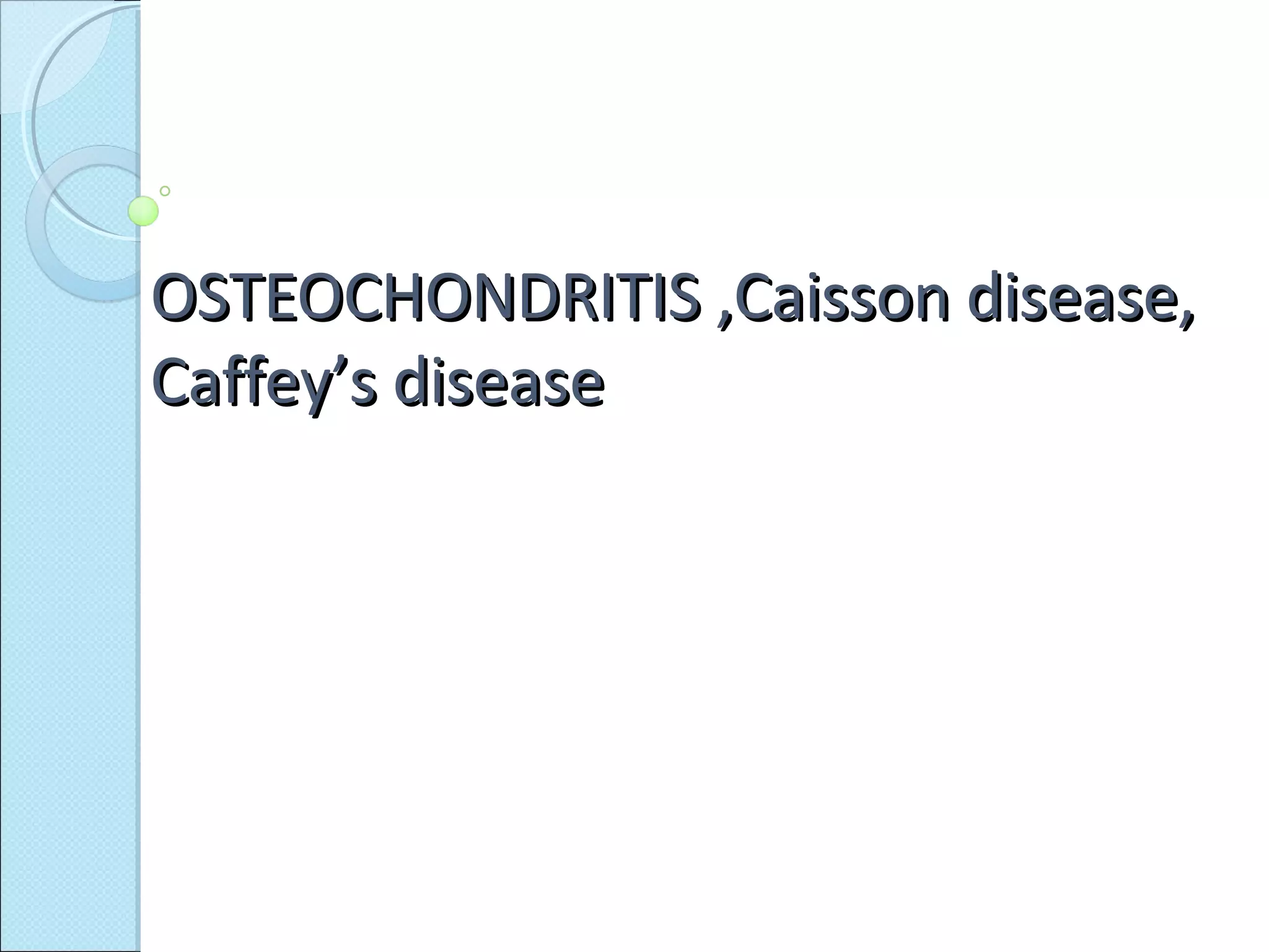
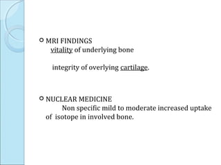
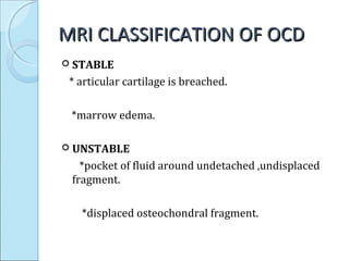
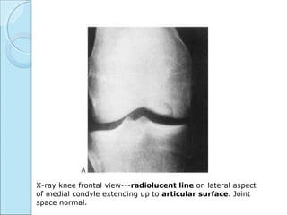
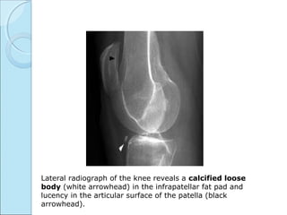
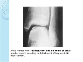
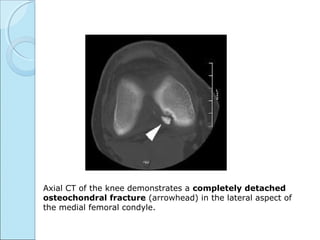
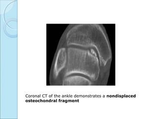
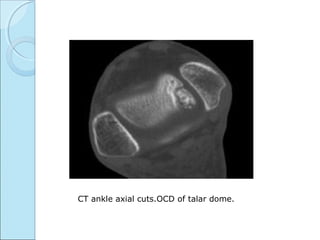
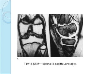
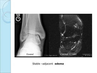
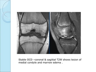
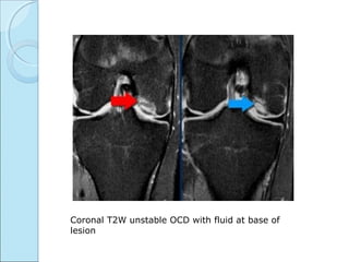
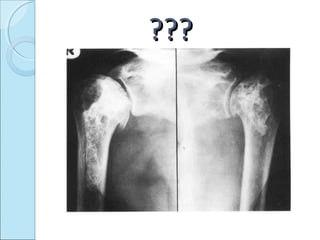
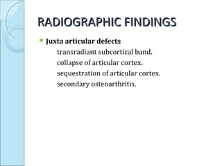
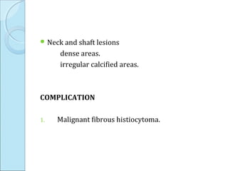
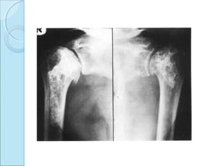
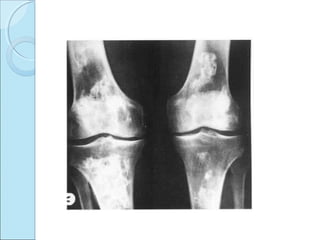
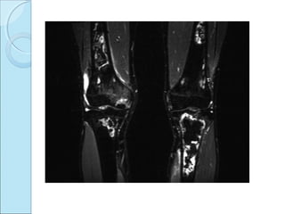
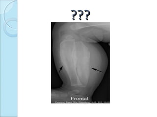
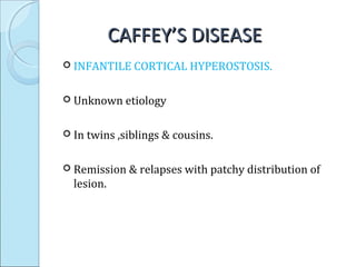
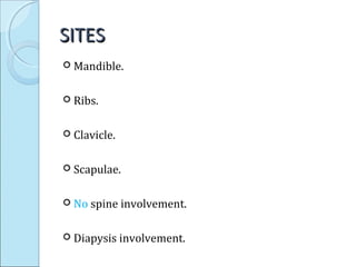
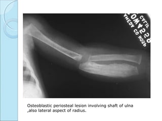
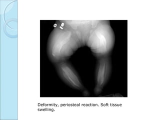
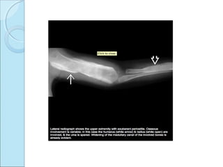
OSTEOCHONDRITIS ,Caisson disease,OSTEOCHONDRITIS ,Caisson disease,
Caffey’s diseaseCaffey’s disease
??????
OSTEOCHONDRITIS DESSICANSOSTEOCHONDRITIS DESSICANS
 Misnomer.
 Refers to post-traumatic osteochondral/chondral
fractures.
 Occuring at articular surface.
 After injury---detached portion
* remain in situ.
*Mildly displaced.
* Become loose body.
(most common cause of loose body in young
adults is OCD)
 Fragmented part—avascular
 Bed of defect ---vital
SITES INVOLEDSITES INVOLED
 CONVEX ARTICULAR SUFACES:
 Medial femoral condyles.
 Capitellum of humerus.
 Trochlear surface of talus.
 RARE SITES OF INVOLVMENT:
 Shoulder—humeral head ,glenoid.
 Scaphoid.
 Navicular.
INCIDENCEINCIDENCE
 M:F =2:1.
 One third cases are bilateral.
 Adolescence.
RADIOGRAPHIC FEATURES.RADIOGRAPHIC FEATURES.
PLAIN RADIOGRAPHS
 May be normal.
 When visible
=radiolucent ring surrounding bony fragment.
=in profile-loose body opposite to pit in bone.
Loss of sharp cortical line of articular surface.
 CT FINDINGS
Usually axial cuts.
For cortical defects & loose bodies.
Island of bone demarcated by rare field zone.
 MRI FINDINGS
vitality of underlying bone
integrity of overlying cartilage.
 NUCLEAR MEDICINE
Non specific mild to moderate increased uptake
of isotope in involved bone.
MRI CLASSIFICATION OF OCDMRI CLASSIFICATION OF OCD
 STABLE
* articular cartilage is breached.
*marrow edema.
 UNSTABLE
*pocket of fluid around undetached ,undisplaced
fragment.
*displaced osteochondral fragment.
X-ray knee frontal view---radiolucent line on lateral aspect
of medial condyle extending up to articular surface. Joint
space normal.
Lateral radiograph of the knee reveals a calcified loose
body (white arrowhead) in the infrapatellar fat pad and
lucency in the articular surface of the patella (black
arrowhead).
Ankle frontal view---radiolucent line on dome of talus
,medial aspect. resulting in detachment of fragment. No
displacement.
Axial CT of the knee demonstrates a completely detached
osteochondral fracture (arrowhead) in the lateral aspect of
the medial femoral condyle.
Coronal CT of the ankle demonstrates a nondisplaced
osteochondral fragment
CT ankle axial cuts.OCD of talar dome.
T1W & STIR---coronal & sagittal.unstable.
Stable –adjacent edema
Stable OCD –coronal & sagittal T2W shows lesion of
medial condyle and marrow edema .
Coronal T2W unstable OCD with fluid at base of
lesion
??????
CAISSON DISEASECAISSON DISEASE
 DYSBARIC OSTEONECROSIS.
 exposure to hyperbaric atmosphere.
 Undersea diving ,space craft.
 Result in decompression sickness.
MECHANISMMECHANISM
 Nitrogen bubbles liberated from bone marrow –
bone infarct by small blood vessel occlusion
 Resultant osteonecrosis..
 Shoulder most commonly involved.
PresentationPresentation
 Joint pain
 Motion aggravates pain.
RADIOGRAPHIC FINDINGSRADIOGRAPHIC FINDINGS
 Juxta articular defects
transradiant subcortical band.
collapse of articular cortex.
sequestration of articular cortex.
secondary osteoarthritis.
 Neck and shaft lesions
dense areas.
irregular calcified areas.
COMPLICATION
1. Malignant fibrous histiocytoma.
??????
CAFFEY’S DISEASECAFFEY’S DISEASE
 INFANTILE CORTICAL HYPEROSTOSIS.
 Unknown etiology
 In twins ,siblings & cousins.
 Remission & relapses with patchy distribution of
lesion.
SITESSITES
 Mandible.
 Ribs.
 Clavicle.
 Scapulae.
 No spine involvement.
 Diapysis involvement.
PRESENTATIONPRESENTATION
 Child upto 1 year.
 High grade fever & hyperirritability.
 Painful deep soft tissue swelling. occur before bone
lesion appear.
RADIOGRAPHIC FEATURESRADIOGRAPHIC FEATURES
 Marked periosteal proliferation.
 Cortical thickening.
 Soft tissue swelling.
OUTCOMEOUTCOME
 Bone returns to normal by 12 weeks.
Osteoblastic periosteal lesion involving shaft of ulna
,also lateral aspect of radius.
Deformity, periosteal reaction. Soft tissue
swelling.
Osteochondritis dessicans ,caisson disease, caffey’s disease

Osteochondritis dessicans ,caisson disease, caffey’s disease

Editor's Notes

  • #10 CLANTON & DELEE Classification of osteochondral lesions. Grade I-small area of compressed sub chondral bone.; the overlying cartilage is intact. Grade II-there is a small localized cartilage defect. Grade III-the defect extends through the cartilage and around the bone, but is only partial. Grade IV-there is a total defect around cartilage and bone, but no displacement of fragments. Grade V-a loose detached fragment is seen.
  • #19 .