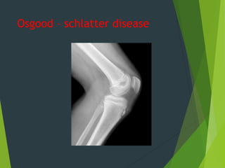
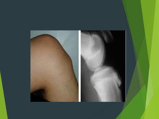
osteochondrosis
Саллам Мосаад Махмуд
502Б
Definition
It is a term used to describe the bone damage and necrosis
follow trauma or repetitive stress .
The affected segment show many features of ischemic
necrosis include loss of bone cells , increase vascularity
and sclerosis of the surrounding bone .
This conditions usually affecting the children and
adolescents.
Types of osteochondritis :
3 types
1- crushing type .
2- pulling type .
3- splitting type .
Crushing osteochondritis :
It is seen in adolescent and characterize by spontaneous
necrosis of ossific nucleus in a long bone or one of
the bones of the wrist or foot .
Clinically : pain and limitation of the joint movement
exam. Tenderness is localized to the affected bone .
X-ray : increase density in early stage but later there
will be distortion and collapse of the necrotic
segment .
The common examples of crushing osteochondritis
1- Koehler's of the navicular bone .
2- keinbock`s disease of the lunate bone .
3- Schermann`s disease of the thoracic spine .
Perthe`s disease now considered as a type of
Osteonecrosis rather than as osteochondritis .
Koehler’s disease : osteochond. Of the navicular
bone affecting children 3-5
years old ; presented as pain and limping ; local
tenderness +ve .
Treatment : by putting the foot in rest , some time use
full p.o.p. for the foot .
Koehler`s disease
Kienbock`s disease :
It is osteochond. Of the lunate bone , presented in young adult
with painful wrist , stiffness and local tenderness .
X-ray : dense fragmented lunate bone and in advanced case
osteoarthritic changes seen in the wrist .
Treatment : in early stage need splintage only , but later on
when the pain become sever arthrodesis of the
wrist is indicated .
Schermann`s disease :
Osteochond. Of the thoracic spine ; vertebral bodies become
wedge in shape due to fragmentation of the upper and
lower epiphysis of the vertebral body .
Kinnock's disease
The site of pain in
kienbock`s disease
Patient presented with smooth , rigid
kyphosis of the dorsal spine with back
pain ; the disease affecting the
adolescent .
X-ray : wedging and fragmentation of the
upper
and lower vertebral end plate .
Treatment :
Exercise and some time we use brace or
splint .
Pulling osteochondritis
( traction ) :
Excessive pull by large tendon may damage the unfused
apophysis to which it attached and this typically occur
in two place :
1-tibial tuberosity (osgood-schlatter`s
disease)
2- calcaneal apophysis (sever`s disease ).
Osgood schlutter`s disease :
It is osteochondritis of upper tibial tubercle ,affecting children
8-15 years old .
The tibial tuberosity become swallown , tender and extension
of the knee joint against resistant is painful .
X-ray : show irregular fragmentation of apophysis in which
infra patellar tendon is inserted .
Treatment : spontaneous recovery is usually occur and it is
necessary to modified child activity e.g., stop
plying foot ball and stop cycling . If pain persist then put in
plaster cylinder for about 1-2 months occasionally excision of
the fragmented piece by operation is done
Osgood – schlatter disease
Sever`s disease :
Osteochondritis of the calcanium which affect child
about 10 years old .
Patient usually presented with pain and tenderness
over the posterior aspect of the heel .
X- ray :show fragmentation of the apophysis in
which the tendoachillis is inserted into the
calcanium .
Treatment : restriction of the activity for few weeks
and heel
raising .
Saver's disease
Splitting osteochondritis (osteochondritis
dissecans )
Small fragment of articular cartilage and the
subjacent bone may separated (dissect) as an
avascular fragment .
It is occur in young adult more in male , the
commonest site is the knee joint . The cause is
repeated minor trauma lead to osteochondral
fracture , the fragment loosen its blood supply .
Clinically : the patient 15-20 years old complain from
intermittent pain , swelling , joint
effusion and in case of separation of the piece it
will act as a loose body in the joint and this will
cause locking of the joint and unexpected episode
of giving way .
Treatment
 The first step is to rest the joint.
Children must stop playing most
sports for at least two to four
months. During that time, they
also may use crutches, a brace or
a cast to reduce pressure and
protect the joint.
 Physical therapy for
osteochondritis dissecans is also
used as a non-surgical treatment.


osteochondropathy.ppt osteochondropathy.ppt

  • 1.
  • 2.
    Definition It is aterm used to describe the bone damage and necrosis follow trauma or repetitive stress . The affected segment show many features of ischemic necrosis include loss of bone cells , increase vascularity and sclerosis of the surrounding bone . This conditions usually affecting the children and adolescents.
  • 3.
    Types of osteochondritis: 3 types 1- crushing type . 2- pulling type . 3- splitting type . Crushing osteochondritis : It is seen in adolescent and characterize by spontaneous necrosis of ossific nucleus in a long bone or one of the bones of the wrist or foot . Clinically : pain and limitation of the joint movement exam. Tenderness is localized to the affected bone . X-ray : increase density in early stage but later there will be distortion and collapse of the necrotic segment .
  • 4.
    The common examplesof crushing osteochondritis 1- Koehler's of the navicular bone . 2- keinbock`s disease of the lunate bone . 3- Schermann`s disease of the thoracic spine . Perthe`s disease now considered as a type of Osteonecrosis rather than as osteochondritis . Koehler’s disease : osteochond. Of the navicular bone affecting children 3-5 years old ; presented as pain and limping ; local tenderness +ve . Treatment : by putting the foot in rest , some time use full p.o.p. for the foot .
  • 5.
  • 6.
    Kienbock`s disease : Itis osteochond. Of the lunate bone , presented in young adult with painful wrist , stiffness and local tenderness . X-ray : dense fragmented lunate bone and in advanced case osteoarthritic changes seen in the wrist . Treatment : in early stage need splintage only , but later on when the pain become sever arthrodesis of the wrist is indicated . Schermann`s disease : Osteochond. Of the thoracic spine ; vertebral bodies become wedge in shape due to fragmentation of the upper and lower epiphysis of the vertebral body .
  • 7.
  • 8.
    The site ofpain in kienbock`s disease
  • 9.
    Patient presented withsmooth , rigid kyphosis of the dorsal spine with back pain ; the disease affecting the adolescent . X-ray : wedging and fragmentation of the upper and lower vertebral end plate . Treatment : Exercise and some time we use brace or splint .
  • 11.
    Pulling osteochondritis ( traction) : Excessive pull by large tendon may damage the unfused apophysis to which it attached and this typically occur in two place : 1-tibial tuberosity (osgood-schlatter`s disease) 2- calcaneal apophysis (sever`s disease ).
  • 12.
    Osgood schlutter`s disease: It is osteochondritis of upper tibial tubercle ,affecting children 8-15 years old . The tibial tuberosity become swallown , tender and extension of the knee joint against resistant is painful . X-ray : show irregular fragmentation of apophysis in which infra patellar tendon is inserted . Treatment : spontaneous recovery is usually occur and it is necessary to modified child activity e.g., stop plying foot ball and stop cycling . If pain persist then put in plaster cylinder for about 1-2 months occasionally excision of the fragmented piece by operation is done
  • 13.
  • 15.
    Sever`s disease : Osteochondritisof the calcanium which affect child about 10 years old . Patient usually presented with pain and tenderness over the posterior aspect of the heel . X- ray :show fragmentation of the apophysis in which the tendoachillis is inserted into the calcanium . Treatment : restriction of the activity for few weeks and heel raising .
  • 16.
  • 17.
    Splitting osteochondritis (osteochondritis dissecans) Small fragment of articular cartilage and the subjacent bone may separated (dissect) as an avascular fragment . It is occur in young adult more in male , the commonest site is the knee joint . The cause is repeated minor trauma lead to osteochondral fracture , the fragment loosen its blood supply . Clinically : the patient 15-20 years old complain from intermittent pain , swelling , joint effusion and in case of separation of the piece it will act as a loose body in the joint and this will cause locking of the joint and unexpected episode of giving way .
  • 18.
    Treatment  The firststep is to rest the joint. Children must stop playing most sports for at least two to four months. During that time, they also may use crutches, a brace or a cast to reduce pressure and protect the joint.  Physical therapy for osteochondritis dissecans is also used as a non-surgical treatment. 