Mgr university bsc nursing adult health previous question paper with answers
OSMF Oral Sub mucous Fibrosis -Oral Medicine
1. OSMF
Aetiology
1. Chillies
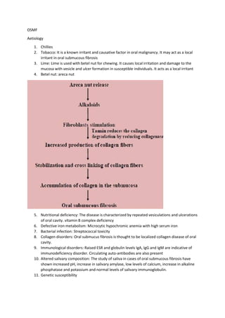
2. Tobacco: It is a known irritant and causative factor in oral malignancy. It may act as a local
irritant in oral submucous fibrosis
3. Lime: Lime is used with betel nut for chewing. It causes local irritation and damage to the
mucosa with vesicle and ulcer formation in susceptible individuals. It acts as a local irritant
4. Betel nut: areca nut
5. Nutritional deficiency: The disease is characterized by repeated vesiculations and ulcerations
of oral cavity. vitamin B complex deficiency
6. Defective iron metabolism: Microcytic hypochromic anemia with high serum iron
7. Bacterial infection: Streptococcal toxicity
8. Collagen disorders: Oral submucus fibrosis is thought to be localized collagen disease of oral
cavity.
9. Immunological disorders: Raised ESR and globulin levels IgA, IgG and IgM are indicative of
immunodeficiency disorder. Circulating auto-antibodies are also present
10. Altered salivary composition: The study of saliva in cases of oral submucous fibrosis have
shown increased pH, increase in salivary amylase, low levels of calcium, increase in alkaline
phosphatase and potassium and normal levels of salivary immunoglobulin.
11. Genetic susceptibility
2. Clinical features
1. Age and sex distribution: It affects both sexes. The age group varies, although majority of
patients are between 20 and 40 years of age
2. Site distribution: The most frequent location of oral submucous fibrosis is the buccal mucosa
and the retro-molar areas. It also commonly involves soft palate, palatal fauces, uvula,
tongue and labial mucosa. Sometimes, it involves the floor of mouth and gingiva
3. Prodromal symptoms: The onset of the condition is insidious and is often of 2 to 5 years of
duration. The most common initial symptom is burning sensation of oral mucosa, aggravated
by spicy food, followed by either hypersalivation or dryness of mouth. Vesiculation,
ulceration, pigmentation, recurrent stomatitis and defective gustatory sensation have also
been indicated as early symptoms
4. Late symptoms:
Trismus: Gradual stiffening of the oral mucosa occurs in few years after the initial symptoms
appear. This leads to inability to open the mouth completely (Fig. 9.49).
Difficulty in tongue protrusion: Later, patients have trouble in protruding the tongue
Difficulty in swallowing: When the fibrosis extends to pharynx and esophagus, the patient
may have trouble in swallowing the food
Referred pain: Referred pain in the ears and deafness, due to occlusion of Eustachian tube
and a typical nasal voice has been reported
5. Blanching of mucosa: The most common and earliest sign is blanching of mucosa, caused by
impairment of local vascularity. The blanched mucosa becomes slightly opaque and white.
The whitening often takes place in spots so that the mucosa acquires a marble like
appearance. Blanching may be localized or diffuse, involving greater part of the oral mucosa
or reticular, in which blanching consists of blanched area with intervening clinically normal
mucosa, giving it a lace-like appearance
6. Betel chewer mucosa: It is brownish red discoloration of mucosa with irregular surface that
tend to desquamate
7. Fibrous band: As disease progresses the mucosa becomes stiff and vertical bands appear.
These bands can be palpated easily and feel rough on palpation
8. Lip features: Mucosa is blanched, becomes rubbery and is characterized by the presence of
circular bands around the rima oris like a thin band. In severe labial involvement, the
opening of mouth is altered to an elliptical shape (elliptical rima oris), lips become leathery
and it become difficult to evert them
9. Buccal mucosa: The affected mucosa becomes coarse, blanched and inelastic. In advanced
cases, the mucosa becomes tough and leathery with numerous vertical fibrous bands
10. Soft palate (49%) and uvula: Involvement of soft palate is marked by fibrotic changes and a
clear delineation of the soft palate from hard palate. The mobility of soft palate is restricted.
Uvula, when involved, is shrunken and in extreme cases it becomes bud-like or hockey stick
appearance
11. Palatal fauces: in the soft palate the bands radiate from pterygomandibular raphe to the
anterior faucial pillars. The faucial pillars become thick and short and tonsils may get pressed
in between fibrosed pillars
12. Tongue The initial change is depapillation, usually in the lateral margins. Tongue becomes
smooth; its mobility, especially in protrusion, becomes impaired. Patient cannot protrude
the tongue beyond the incisal edges
13. Floor of mouth: When floor of mouth is affected, it becomes inelastic
14. Gingiva: When affected, it becomes fibrotic, blanched and inelastic
3. 15. Associated features:
Pigmentation: Hyperpigmentation or occasional loss of pigmentation is very common in
association with oral submucous fibrosis. Many times, pigmentation changes in vermilion
border are so striking that this disease can be suspected even before examining the patient
Vesicle: It is usually found in areas of redness in the soft palate, the anterior faucial pillar,
buccal mucosa or the mucosal surface of lip, particularly the lower lip.
The vesicles are painful and they soon rupture leaving behind superficial ulceration. Often,
there is history of vesiculation following the intake of spicy food, suggesting an allergic
reaction to spicy food
Ulceration: Ulceration often develops in the course of disease, particularly in advanced
cases. In advanced cases, epithelium becomes atrophie, fragile and vulnerable to ulceration
(Fig. 9.56)
Petechiae: These are small raised reddish blue spots which sometimes occur in oral
submucous fibrosis. It may be few or many. They occur mast commonly on tongue and the
labial and buccal mucosa. The petechiae are transient in nature and do not require any
specific treatment.
Management