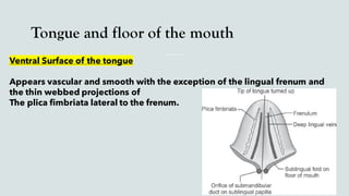
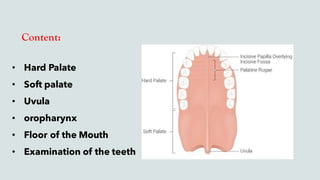
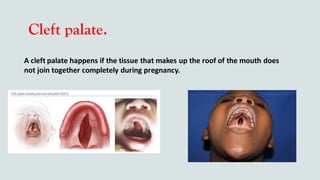
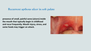
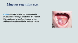
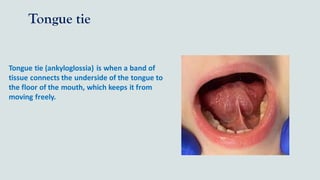
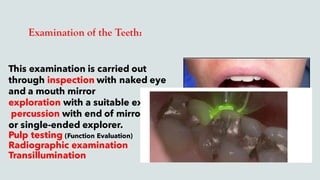
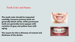
SlideShare a Scribd company logo
INTRA-ORAL
Examination
Content
Introduction
Examination Of lips and Labial Mucosa
Large lips
Lesions on the lips
Labial frenum
Buccal Mucosa
Buccal Vestibule
Common lesions
Tongue ( Dorsal – Ventral surfaces )
Tounge coating
Nerve supply
Introduction
Introduction
Topic one
Criteria of examination:
Upper & Lower lip :
- First check Vermilion border for surface lesions
- Should be Grasped gently between thumbs and index of both hands.
- Palpate the lip for submucosal masses/Nodules ( Bidigital palpation )
that are less than 1cm in diameter - Minor Salivary Glands -
Then everting the lip and examine the labial mucosa, vestibule, anterior gingiva.
Border of the mouth ( Commissure )
Large lips
May be an indication of variation of normal Like :
- Angioedema ( Allergy )
sudden diffuse swelling, firm and non pitting usually only on one lip is affected
but occasionally the whole face is involved
- Acromegaly
- included in Melkerson – Rosenthal Syndrome
“ Neurological Disorder “
Other Lesions On the lip :
1- Angular Cheilitis :
Candidal infection & low vertical dimension
2- Angular Cheilosis :
Vitamin deficiency & Protein Deficiency
3- Rhagades
in the form of Scar lines around the vermilion border in Congenital Syphilis
4- Herpes labialis
Other Lesions On the lip :
5- Smokers patch
6-Bloody Crusted Lip ( Erythema Multiform )
7- Contact Allergy due to mouth washes, Cosmetics , food
Lichen planus ( white striation in popular form, Ulcer in bullous erosive form )
8-Actinic Keratosis (Solar Keratosis )
Labial Frenum
Normally : appears as slender, midline band, it’s web like attachment at the height of
muco-labial vestibule
Level of attachment should be beyond the attached gingiva to avoid conveying the pull
action of labial muscles to marginal gingiva would result into gingival recession
A Fibrotic thick frenum helps to form diastema between central incisors
Buccal Mucosa
Bi-digital palpation of the Buccal Mucosa may reveal nodularity due to
presence of minor salivary glands
Bi-digital and Bi-manual palpation are carried out to determine the
consistency, flexibility of the cheek or buccal mucosa
Technique:
The buccal mucosa must be supported from out side by the four fingers of one
hand and the index finger of the other hand running inside the buccal mucosa
in different directions to palpate any deeply seated lesions
Similarly, Inspect the buccal vestibular mucosa and the buccal gingiva
Buccal vestibule
Inspection:
Can be inspected by retracting the cheeks whilethe mouth is opened and
then ask the patient to occludethe teeth
the buccal and labial vestibules are visualized to demonstrate their extensions,
Contour,Depth
Palpation:
the facial surfaces of maxilla,mandibleare palpatedto identify typical
elevationsor depressionsin the contour of the bone.
The palpationof the buccal vestibule can be done by slowly sliding the tip of the finger along the
alveolar surfaces at the periapicallevel
to Identify the tenderness or enlargementof periapical inflammatory lesions.
Also Signs as egg shell Crackling or Fluctuation should be noticed
On mucosal surface:
Look For Signs of inflammation ( Redness & Swelling ), Ulceration, Pigmentation and lesions
If any lesion is found, Describe them in site, size, shape and contour
Normal Variations appear as
- whitish ridge of tissues opposite the occlusal plane of teeth “ Linea Alba Buccalis “
- Fleshy swelling opposite the 2nd upper molar
“ opening of Stenson’s duct of parotid gland “
Common Lesions
1- White Lesion
Frictional Keratosis, Leukoplakia, Candidiasis, Aspirin burns, Smoker Keratosis,
papular Lichen planus
2- ulcerative lesion
Traumatic ulcer, aphthous ulcer, intra-oral herpes simplex
3-pigmented lesion
(petechia, ecchymosis) which give dark red to bluish coloration, melanoma,
amalgam tattoo
4- Warty lesions :
viral warts – papilloma
5- Neoplastic Lesion Seen as swelling or ulcer
Tongue and floor of the mouth
Dorsal Surface of the tongue
( normally appear pale pink rough surface )
1- Filiform papillae
( small whitish, hair like projections)
they may become elongated ( Hairy tongue )
they may become very short ( atrophic tongue )
2- Fungiform papillae
( the larger and more prominent
at the lateral border and tip )
3- Circumvallate papillae
nodular, irregular contours in the posterior region.
They are round and have groove around them.
this groove consists of the opening of Von Ebner Glands and also Contains
Taste buds
** the median groove or fissures on the dorsal surface of the tongue is referredto
as Fissured tongue and is common anatomic variation.
Tongue and floor of the mouth
Ventral Surface of the tongue
Appears vascular and smooth with the exception of the lingual frenum and
the thin webbed projections of
The plica fimbriata lateral to the frenum.
Normal tongue Coating
Normally tongue coating increase in the morning as salivary flow decreases
during sleep & then decreases while chewing food, speech with normal flow
of saliva.
Causes of increase tongue coating :
Drugs, mouth breathing, febrile illness,
excessive vomiting,dehydration Smoking,
Stomach upset
Decreasing tongue Coating
( Atrophy of tongue Coating )
1- Nutritional deficiency
Anemia, malnutrition – Vitamin B12 deficiency – alcoholism –
malabsorption – iron deficiency – plummer vinson syndrome)
2- Drugs as chemotherapy, antibiotics
3- Diseases as atrophic lichen planus
4- peripheralVascular changes as with
- Obliteratoin of small vessels secondary to diseases
( submucous Fibrosis ) – scleroderma
- Micro angiopathy “Diabetes Mellitus”
- syphilitic bald tongue
- S.L.E
Nerve Supply of the tongue
1- Posterior 1/3 of the Tongue:
Glossopharyngeal
( general sensation + Taste sensation )
2- Anterior 2/3 of the tongue:
General Sensation :
Lingual Branch of mandibular nerve
Taste Sensation : Chorda tympani
- Motor nerve :
All of the tongue Supplied by –
Hypoglossal Nerve
Except:
1- Palatoglossus Muscle
2- Glossopharyngeus
Supplied By :
Pharyngeal plexus “ Vagus Nerve “
Tongue lesions and
examination.
Common tongue lesions:




















Tongue infections:
Candidal infection herpes syphilis


Enlargement of the tongue:



















Examination of the tongue:







Content:
• Hard Palate
• Soft palate
• Uvula
• oropharynx
• Floor of the Mouth
• Examination of the teeth
Hard palate:
Hard palate examination done by:
A) Inspection: by two ways
a. Indirect inspection: done be the mirror.
b. Direct vision: (for better vision) the patient
mouth is opened widely.
Hard palate:
B) Palpation:
❖ The palatal alveolus is palpated at
periapical level for tender foci or hard
bone enlargement at the midline called
torus palatinus.
❖ Any abnormal sensation must be
detected as egg shell crackling or
fluctuation.
Hard palate:
❖ The normal palatal mucosa
appears pale pink and
homogenous in color.
❖ The normal anatomical
consideration must be considered
(palatal rugae, incisive papilla
and median palatine raphe)
Hard palate:
Common lesions of the hard
palate:
❖ Pizza burn
❖ Cleft palate
❖ Minor salivary gland tumor
❖ Nicotinic stomatitis
❖ Torus palatinus
❖ Inflammatory papillary
hyperplasia
❖Different types of cysts
Pizza burn.
Cleft palate.
A cleft palate happens if the tissue that makes up the roof of the mouth does
not join together completely during pregnancy.
Minor salivary gland tumor.
Usually present as a non-ulcerated,
painless submucosal mass of the oral
cavity, typically in the hard or soft palate.
Symptoms of minor salivary gland
tumors depend on tumor location,
extent, tumor type, and whether the
tumor is causing a mass effect or is
invading local structures.
Nicotinic stomatitis
❖ lesion of the roof of the mouth.
❖ The concentrated heat stream of smoke
from tobacco products causes Nicotinic
Stomatitis.
❖ These changes are observed most often in
pipe.
Torus palatinus
Torus palatinus refers to a bony growth on the roof
of your mouth.
These growths are harmless, but can be
uncomfortable and inconvenient.
Inflammatory papillary hyperplasia
is a benign lesion of the palatal mucosa. It is
usually found in denture-wearers but also has
been reported in patients without a history of
use of a maxillary prosthesis use.
Different types of cysts
Non Odontogenic
Odontogenic
Soft palate
Diagnosis of Soft palate and uvula done by:
1. Inspection: The soft palate is easily
inspected during the direct visualization of the
hard palate.
Depression of the tongue with mirror is usually
necessary to fully demonstrate the soft palate.
2. Palpation: Palpation of the soft palate
cause gagging and is not routinely
performed unless an abnormality is
observed visually.
Soft palate
The mucosa of the soft palate typically
appears reddish pink with prominence of
the underlying vascularity.
The soft palate or older persons may
appear somewhat yellow due to increased
submucosal fat.
The soft palate appears loose and mobile.
Soft palate
❖ Common lesions of the soft
palate:
❖ Herpangina
❖ Herpes zoster
❖ Recurrent apthous ulcer
❖ Petechia and ecchymosis
Herpangina
a viral illness that causes a high fever and
blister-like sores in the mouth and throat.
The illness is contagious and spreads quickly
among kids in school environments where
children are close to each other.
Herpes zoster
Herpes zoster (HZ) is a viral disease
which is primarily caused by the nerve
tissue.
Varicella zoster virus (VZV) is a DNA virus
that causes both primary and recurrent
infection.
also known as shingles, is a unique
condition induced by VZV reactivation.
Neuropathic pain, headache, malaise and
sleep disruption
Recurrent apthous ulcer in soft palate
presence of small, painful sores (ulcers) inside
the mouth that typically begin in childhood
and recur frequently. Mouth injury, stress, and
some foods may trigger an attack.
Petechia and ecchymosis
Petechiae on the palate are characteristic of streptococcal pharyngitis
Oropharyngeal region:
The most prominent structures in the oropharynx are the
tonsils
They are salmon - pink with nodular
surface
The posterior wall of oropharynx also
contains accessory lymphoid tissue
(adenoid), which appears as slightly
elevated mucosal colored papules. The
accessory lymphoid tissue at the
posterolateral border of the tongue
(lingual tonsils), palatine, and
pharyngeal tonsils which comprises
waldeyer's throat ring.
Uvula
Bifid Uvula
Deviated uvula
Abnormalities in Uvula
Floor of the mouth
Examination of the Floor of
Mouth:
Inspection: It is done by asking the
patient to raise the tongue. Normal
structures include lingual frenum,
orifices of sublingual and
submandibular salivary glands, lingual
veins any change in shape, color, and
integrity in the mucosa should be
noted.
Lingual
vestibule
Wharton’s duct openings
Sublingual
Carunculae
Palpation: Is usually performed
bimanually (i.e. by fixing the tissues
extra orally by the fingers of one hand
while the fingers of the other hand
manipulates the tissues).
The reason for palpation for this area is
to detect enlarged submandibular or
submental lymph nodes, salivary stones
along the course of Wharton's ducts
and any nodular enlargement of the
salivary glands.
Floor of the mouth
Mandibular tori: are bony, hard,
bilateral prominences of the lingual
alveolar process, commonly seen in
adults in the mandibular canine region.
Floor of the mouth
Common lesion of the floor of the mouth:
❖ Mucous retention cyst ( Ranula)
❖ Ulcer
❖ Salivary stones
❖ Tongue tie
Mucous retention cyst
Ranula is a clinical term for a mucocele or
mucous retention cyst located on the floor of
the mouth and arises from trauma to the
sublingual or submandibular salivary glands
Ulcer
Salivary stones
❖ Sialolithiasis is the medical term for salivary
gland stones (calculi).
❖ These stones can cause pain and swelling of
your salivary gland.
❖ Causes include dehydration, smoking and
certain autoimmune diseases.
Tongue tie
Tongue tie (ankyloglossia) is when a band of
tissue connects the underside of the tongue to
the floor of the mouth, which keeps it from
moving freely.
Examination of the Teeth:
This examination is carried out
through inspection with naked eye
and a mouth mirror
exploration with a suitable explorer
percussion with end of mirror handle
or single-ended explorer.
Pulp testing (Function Evaluation)
Radiographic examination
Transillumination
Pulp testing (Function Evaluation)
This test is an attempt to determine
vitality or non-vitality of the dental pulp
and it is an important diagnostic aid in
daily dental practice.
Heat testing of the pulp can be
performed by heating a small ball or
stick of gutta-percha in a flame until
tip applied to the tooth
Cold testing of the pulp can be done
through application of cold air, an ice
or cotton pledget soaked in ethyl
Electrical pulp testing:
Electrical pulp testing: offers an
advantage of more controlled, graded
stimulus compared with thermal tests,
as stimulus level can be digitally
displayed.
The physiological basis for the use of
electrical stimulation is based upon the
fact that, pain is specific sensory
experience mediated through nerve
structures that are separate from those
mediating other sensations as touch,
pressure
Transillumination
There are many commercially available
tools for transillumination. You can also
use the fiberoptic in your highspeed
handpiece. Without a bur in place
position the head of the handpiece on
the tooth surface and press to rheostat
so the light comes on. Curing lights can
also be used for crack detection.
Transillumination is one of ways to
determine if a tooth is cracked or not,
by passing a strong beam of light
through a sample and using the pattern
of light transmission for diagnosis.
Procedure: For cracked teeth we apply a strong
light source to the tooth so that the light is
traveling perpendicular to the plane of the
suspected crack. In most instances this means
we apply the light source to the labial or lingual
tooth surfaces. In a tooth that is not cracked the
light will travel uninterrupted from the buccal
surface to the lingual, which can be observed on
the occlusal table of the tooth by its uniform
illumination. When a tooth is cracked the light
hits the crack and is dispersed, therefore it does
not cross over and illuminate the side of the
tooth beyond the crack.
Transillumination
Examination of the Teeth:
The following should be
considered in examining the
teeth:
color, stains, number, form, size,
structure, erosion, abrasion,
fracture, vitality, functional
contours, carious lesions as well
as contact relationship.
Teeth Color and Stains:
The tooth color should be inspected
carefully, however primary teeth are
generally bluish white, while permanent
teeth are generally more opaque with
variation of grey and yellow hues with
aging.
The reason for this is thinness of enamel and
thickness of the dentin.
Teeth color may show alterations which
may be physiological or pathological
and intrinsic or extrinsic.
Teeth discoloration may result from
developmental disturbances as
amelogenesis imperfect,
dentinogenesis imperfect, brown
hereditary teeth and dental fluorosis or
mottling.
Examination of the Teeth:
Extrinsic and Intrinsic Teeth Staining:
dental fluorosis or mottled teeth,
tetracycline teeth staining and
erythroblastosis fetalis.
intrinsic teeth staining:
Examination of gingival and peridontal supporting structure.
Anatomy of the
Periodontium
Detection of
plaque
Examination of the
Periodontium:
Inspection
Palpation
Probing
Factors affecting periodontal
disease:
Probing (pocket) depth
Loss of attachment
Bleeding on probing
Tooth mobility
Bone Resorbtion
Furcation
Involvement
outline
Anatomy of the Periodontium
The periodontium is the supporting apparatus of the
teeth, which attaches the tooth to the bone of the
jaws and comprises the following tissues:
1- The gingiva.
2- The periodontal ligament.
3- The root cementum.
4- The alveolar bone.
Healthy Gingival Unit:

 The free gingiva
 The interdental papillae
 The attached gingiva






Healthy Gingival Unit:
Healthy vs Diseased Gingiva:
Gingival Enlargment:
Gingival margin is significantly coronal to the
CEJ
Probing depth more than 3mm(normal
sulcus is 3mm)
( Gingival enlargement = distance from
gingival margin to CEJ)
Edematous Gingiva
Fibrous Gingiva
The periodontal ligament

 0.05
to 0.25 mm
The cementum

The alveolar process:

Detection of plaque :




Disclosing Agents
Use of disclosing agents Iodine containing solutions Erythrosine
Examination of the Periodontium:






Probing And Charting:
Technique of Probing

 Mesiobuccal, midbuccal, distobuccal, distolingual, midlingual
and mesiolingual.

 10- 15 degree

Factors affecting periodontal disease:





• Probing Depth:

 Clinical attachment level (CAL)


 Deep periodontal pocket Recession or both
 Furcation involvement
Clinical significance of Probing depth
 PD > 3mm mains Pathology:


 Deep pocket associated with:



Loss Of Clinical Attatchment:
 Loss of attachment



why?
Different pocketdepthswith the same amount of attachment
loss CAL gingival level CEJ
Calculating Clinical Attachment

 1-The probing depth
 2- Gingival margin




I- Gingival margin at CEJ

II- CAL in presence of recession


III- CAL in presence of gingival enlargement

 Some clinicians prefer
Bleading on probing
 Bleading on
 Bleeding + = active disease ,inflammation
 Bleeding - = stable condition

Tooth mobility:
. Is the loosening of a tooth in its socket




Scale for Mobility Assessment
 : normal mobility
 Slight mobility
 Moderate mobility
 Severe mobility
Bone Resorption :
Assissing alveolar bone loss Radiograph
 Radiograph
 Transgingival probing (bone sounding
Disadvantages of radiograph:
 Radiograph

FURCATION INVOLVEMENT

 How do you evaluate?


Furcation Classification:



INTRA-ORAL-.pdf
INTRA-ORAL-.pdf

More Related Content

Similar to INTRA-ORAL-.pdf

TONGUE.pptx
TONGUE.pptxTONGUE.pptx
TONGUE.pptx
DrHarjeetYadav
 
OSMF Oral Sub mucous Fibrosis -Oral Medicine
OSMF Oral Sub mucous Fibrosis -Oral MedicineOSMF Oral Sub mucous Fibrosis -Oral Medicine
OSMF Oral Sub mucous Fibrosis -Oral Medicine
SarahFanny2
 
Oral cavity ppt- college seminar
Oral cavity ppt- college seminarOral cavity ppt- college seminar
Oral cavity ppt- college seminar
Hussien Ali
 
THE TONGUE IN HEALTH AND SICKNESS.pptx
THE TONGUE IN HEALTH AND SICKNESS.pptxTHE TONGUE IN HEALTH AND SICKNESS.pptx
THE TONGUE IN HEALTH AND SICKNESS.pptx
Aisha lamido
 
Oral diagnosis
Oral diagnosisOral diagnosis
Oral diagnosis
Baha'adeen Ali
 
Lab 4 developmental defect soft tissue dina patho
Lab 4 developmental defect soft tissue dina pathoLab 4 developmental defect soft tissue dina patho
Lab 4 developmental defect soft tissue dina patho
dina hameed
 
lesions of lip
lesions of liplesions of lip
lesions of lip
vinayakpathak8
 
Tongue disorders
Tongue disordersTongue disorders
Tongue disorders
Dr. Monali Prajapati
 
-Salivary glangs - totall.Description and management
-Salivary glangs - totall.Description and management-Salivary glangs - totall.Description and management
-Salivary glangs - totall.Description and management
EdouardMudekereza
 
Dr. shimla
Dr. shimlaDr. shimla
Dr. shimla
DrSHIMLAP
 
Aberrant frenum and its treatment
Aberrant frenum and its treatmentAberrant frenum and its treatment
Aberrant frenum and its treatment
Sneha Viswanathan
 
Disorders of the tongue
Disorders of the tongueDisorders of the tongue
Disorders of the tongue
Thilanka Umesh
 
Tongue
Tongue   Tongue
Tongue
Minerva Singh
 
Diseases of Tongue
Diseases of TongueDiseases of Tongue
Diseases of Tongue
sachidanand giri
 
DEVELOPMENTAL DISTURBANCES OF TONGUE
DEVELOPMENTAL DISTURBANCES OF TONGUEDEVELOPMENTAL DISTURBANCES OF TONGUE
DEVELOPMENTAL DISTURBANCES OF TONGUE
MINDSCDECOMMITTEE
 
Oral manifestations in systemic diseases
Oral manifestations in systemic diseasesOral manifestations in systemic diseases
Oral manifestations in systemic diseases
Md Roohia
 
Palatal fistula and syndromes associated with clcp part 1 by Dr. Amit Suryawa...
Palatal fistula and syndromes associated with clcp part 1 by Dr. Amit Suryawa...Palatal fistula and syndromes associated with clcp part 1 by Dr. Amit Suryawa...
Palatal fistula and syndromes associated with clcp part 1 by Dr. Amit Suryawa...
All Good Things
 
Tongue disorders
Tongue disordersTongue disorders
Tongue disorders
Praveena Veena
 
Tongue basics
Tongue  basicsTongue  basics
Tongue basics
Ahsan Kaleem
 
Examination of Nose & Throat Aditi G - Copy.pptx
Examination of Nose & Throat Aditi G - Copy.pptxExamination of Nose & Throat Aditi G - Copy.pptx
Examination of Nose & Throat Aditi G - Copy.pptx
SoumyajitJana7
 

Similar to INTRA-ORAL-.pdf (20)

TONGUE.pptx
TONGUE.pptxTONGUE.pptx
TONGUE.pptx
 
OSMF Oral Sub mucous Fibrosis -Oral Medicine
OSMF Oral Sub mucous Fibrosis -Oral MedicineOSMF Oral Sub mucous Fibrosis -Oral Medicine
OSMF Oral Sub mucous Fibrosis -Oral Medicine
 
Oral cavity ppt- college seminar
Oral cavity ppt- college seminarOral cavity ppt- college seminar
Oral cavity ppt- college seminar
 
THE TONGUE IN HEALTH AND SICKNESS.pptx
THE TONGUE IN HEALTH AND SICKNESS.pptxTHE TONGUE IN HEALTH AND SICKNESS.pptx
THE TONGUE IN HEALTH AND SICKNESS.pptx
 
Oral diagnosis
Oral diagnosisOral diagnosis
Oral diagnosis
 
Lab 4 developmental defect soft tissue dina patho
Lab 4 developmental defect soft tissue dina pathoLab 4 developmental defect soft tissue dina patho
Lab 4 developmental defect soft tissue dina patho
 
lesions of lip
lesions of liplesions of lip
lesions of lip
 
Tongue disorders
Tongue disordersTongue disorders
Tongue disorders
 
-Salivary glangs - totall.Description and management
-Salivary glangs - totall.Description and management-Salivary glangs - totall.Description and management
-Salivary glangs - totall.Description and management
 
Dr. shimla
Dr. shimlaDr. shimla
Dr. shimla
 
Aberrant frenum and its treatment
Aberrant frenum and its treatmentAberrant frenum and its treatment
Aberrant frenum and its treatment
 
Disorders of the tongue
Disorders of the tongueDisorders of the tongue
Disorders of the tongue
 
Tongue
Tongue   Tongue
Tongue
 
Diseases of Tongue
Diseases of TongueDiseases of Tongue
Diseases of Tongue
 
DEVELOPMENTAL DISTURBANCES OF TONGUE
DEVELOPMENTAL DISTURBANCES OF TONGUEDEVELOPMENTAL DISTURBANCES OF TONGUE
DEVELOPMENTAL DISTURBANCES OF TONGUE
 
Oral manifestations in systemic diseases
Oral manifestations in systemic diseasesOral manifestations in systemic diseases
Oral manifestations in systemic diseases
 
Palatal fistula and syndromes associated with clcp part 1 by Dr. Amit Suryawa...
Palatal fistula and syndromes associated with clcp part 1 by Dr. Amit Suryawa...Palatal fistula and syndromes associated with clcp part 1 by Dr. Amit Suryawa...
Palatal fistula and syndromes associated with clcp part 1 by Dr. Amit Suryawa...
 
Tongue disorders
Tongue disordersTongue disorders
Tongue disorders
 
Tongue basics
Tongue  basicsTongue  basics
Tongue basics
 
Examination of Nose & Throat Aditi G - Copy.pptx
Examination of Nose & Throat Aditi G - Copy.pptxExamination of Nose & Throat Aditi G - Copy.pptx
Examination of Nose & Throat Aditi G - Copy.pptx
 

More from MostafaElGendy37

all about the dental impression materials .
all about the dental impression materials .all about the dental impression materials .
all about the dental impression materials .
MostafaElGendy37
 
some uses of laser in dental implant .pptx
some uses of laser in dental implant .pptxsome uses of laser in dental implant .pptx
some uses of laser in dental implant .pptx
MostafaElGendy37
 
bone grafts for periodontal implants.ppt
bone grafts for periodontal implants.pptbone grafts for periodontal implants.ppt
bone grafts for periodontal implants.ppt
MostafaElGendy37
 
DENTAL IMPLANTOLOGY - OSSEOINTEGRATION.pptx
DENTAL IMPLANTOLOGY - OSSEOINTEGRATION.pptxDENTAL IMPLANTOLOGY - OSSEOINTEGRATION.pptx
DENTAL IMPLANTOLOGY - OSSEOINTEGRATION.pptx
MostafaElGendy37
 
anamolies of soft tissues of oral cavity.pptx
anamolies of soft tissues of oral cavity.pptxanamolies of soft tissues of oral cavity.pptx
anamolies of soft tissues of oral cavity.pptx
MostafaElGendy37
 
All Extra oral examination .pdf
All Extra oral examination .pdfAll Extra oral examination .pdf
All Extra oral examination .pdf
MostafaElGendy37
 

More from MostafaElGendy37 (6)

all about the dental impression materials .
all about the dental impression materials .all about the dental impression materials .
all about the dental impression materials .
 
some uses of laser in dental implant .pptx
some uses of laser in dental implant .pptxsome uses of laser in dental implant .pptx
some uses of laser in dental implant .pptx
 
bone grafts for periodontal implants.ppt
bone grafts for periodontal implants.pptbone grafts for periodontal implants.ppt
bone grafts for periodontal implants.ppt
 
DENTAL IMPLANTOLOGY - OSSEOINTEGRATION.pptx
DENTAL IMPLANTOLOGY - OSSEOINTEGRATION.pptxDENTAL IMPLANTOLOGY - OSSEOINTEGRATION.pptx
DENTAL IMPLANTOLOGY - OSSEOINTEGRATION.pptx
 
anamolies of soft tissues of oral cavity.pptx
anamolies of soft tissues of oral cavity.pptxanamolies of soft tissues of oral cavity.pptx
anamolies of soft tissues of oral cavity.pptx
 
All Extra oral examination .pdf
All Extra oral examination .pdfAll Extra oral examination .pdf
All Extra oral examination .pdf
 

Recently uploaded

Triangles of Neck and Clinical Correlation by Dr. RIG.pptx
Triangles of Neck and Clinical Correlation by Dr. RIG.pptxTriangles of Neck and Clinical Correlation by Dr. RIG.pptx
Triangles of Neck and Clinical Correlation by Dr. RIG.pptx
Dr. Rabia Inam Gandapore
 
Alcohol_Dr. Jeenal Mistry MD Pharmacology.pdf
Alcohol_Dr. Jeenal Mistry MD Pharmacology.pdfAlcohol_Dr. Jeenal Mistry MD Pharmacology.pdf
Alcohol_Dr. Jeenal Mistry MD Pharmacology.pdf
Dr Jeenal Mistry
 
Non-respiratory Functions of the Lungs.pdf
Non-respiratory Functions of the Lungs.pdfNon-respiratory Functions of the Lungs.pdf
Non-respiratory Functions of the Lungs.pdf
MedicoseAcademics
 
Pharynx and Clinical Correlations BY Dr.Rabia Inam Gandapore.pptx
Pharynx and Clinical Correlations BY Dr.Rabia Inam Gandapore.pptxPharynx and Clinical Correlations BY Dr.Rabia Inam Gandapore.pptx
Pharynx and Clinical Correlations BY Dr.Rabia Inam Gandapore.pptx
Dr. Rabia Inam Gandapore
 
For Better Surat #ℂall #Girl Service ❤85270-49040❤ Surat #ℂall #Girls
For Better Surat #ℂall #Girl Service ❤85270-49040❤ Surat #ℂall #GirlsFor Better Surat #ℂall #Girl Service ❤85270-49040❤ Surat #ℂall #Girls
For Better Surat #ℂall #Girl Service ❤85270-49040❤ Surat #ℂall #Girls
Savita Shen $i11
 
Knee anatomy and clinical tests 2024.pdf
Knee anatomy and clinical tests 2024.pdfKnee anatomy and clinical tests 2024.pdf
Knee anatomy and clinical tests 2024.pdf
vimalpl1234
 
ANATOMY AND PHYSIOLOGY OF URINARY SYSTEM.pptx
ANATOMY AND PHYSIOLOGY OF URINARY SYSTEM.pptxANATOMY AND PHYSIOLOGY OF URINARY SYSTEM.pptx
ANATOMY AND PHYSIOLOGY OF URINARY SYSTEM.pptx
Swetaba Besh
 
How to Give Better Lectures: Some Tips for Doctors
How to Give Better Lectures: Some Tips for DoctorsHow to Give Better Lectures: Some Tips for Doctors
How to Give Better Lectures: Some Tips for Doctors
LanceCatedral
 
Superficial & Deep Fascia of the NECK.pptx
Superficial & Deep Fascia of the NECK.pptxSuperficial & Deep Fascia of the NECK.pptx
Superficial & Deep Fascia of the NECK.pptx
Dr. Rabia Inam Gandapore
 
Surat @ℂall @Girls ꧁❤8527049040❤꧂@ℂall @Girls Service Vip Top Model Safe
Surat @ℂall @Girls ꧁❤8527049040❤꧂@ℂall @Girls Service Vip Top Model SafeSurat @ℂall @Girls ꧁❤8527049040❤꧂@ℂall @Girls Service Vip Top Model Safe
Surat @ℂall @Girls ꧁❤8527049040❤꧂@ℂall @Girls Service Vip Top Model Safe
Savita Shen $i11
 
Tom Selleck Health: A Comprehensive Look at the Iconic Actor’s Wellness Journey
Tom Selleck Health: A Comprehensive Look at the Iconic Actor’s Wellness JourneyTom Selleck Health: A Comprehensive Look at the Iconic Actor’s Wellness Journey
Tom Selleck Health: A Comprehensive Look at the Iconic Actor’s Wellness Journey
greendigital
 
ACUTE SCROTUM.....pdf. ACUTE SCROTAL CONDITIOND
ACUTE SCROTUM.....pdf. ACUTE SCROTAL CONDITIONDACUTE SCROTUM.....pdf. ACUTE SCROTAL CONDITIOND
ACUTE SCROTUM.....pdf. ACUTE SCROTAL CONDITIOND
DR SETH JOTHAM
 
basicmodesofventilation2022-220313203758.pdf
basicmodesofventilation2022-220313203758.pdfbasicmodesofventilation2022-220313203758.pdf
basicmodesofventilation2022-220313203758.pdf
aljamhori teaching hospital
 
Are There Any Natural Remedies To Treat Syphilis.pdf
Are There Any Natural Remedies To Treat Syphilis.pdfAre There Any Natural Remedies To Treat Syphilis.pdf
Are There Any Natural Remedies To Treat Syphilis.pdf
Little Cross Family Clinic
 
KDIGO 2024 guidelines for diabetologists
KDIGO 2024 guidelines for diabetologistsKDIGO 2024 guidelines for diabetologists
KDIGO 2024 guidelines for diabetologists
د.محمود نجيب
 
Physiology of Chemical Sensation of smell.pdf
Physiology of Chemical Sensation of smell.pdfPhysiology of Chemical Sensation of smell.pdf
Physiology of Chemical Sensation of smell.pdf
MedicoseAcademics
 
Pulmonary Thromboembolism - etilogy, types, medical- Surgical and nursing man...
Pulmonary Thromboembolism - etilogy, types, medical- Surgical and nursing man...Pulmonary Thromboembolism - etilogy, types, medical- Surgical and nursing man...
Pulmonary Thromboembolism - etilogy, types, medical- Surgical and nursing man...
VarunMahajani
 
Report Back from SGO 2024: What’s the Latest in Cervical Cancer?
Report Back from SGO 2024: What’s the Latest in Cervical Cancer?Report Back from SGO 2024: What’s the Latest in Cervical Cancer?
Report Back from SGO 2024: What’s the Latest in Cervical Cancer?
bkling
 
Ocular injury ppt Upendra pal optometrist upums saifai etawah
Ocular injury  ppt  Upendra pal  optometrist upums saifai etawahOcular injury  ppt  Upendra pal  optometrist upums saifai etawah
Ocular injury ppt Upendra pal optometrist upums saifai etawah
pal078100
 
Cervical & Brachial Plexus By Dr. RIG.pptx
Cervical & Brachial Plexus By Dr. RIG.pptxCervical & Brachial Plexus By Dr. RIG.pptx
Cervical & Brachial Plexus By Dr. RIG.pptx
Dr. Rabia Inam Gandapore
 

Recently uploaded (20)

Triangles of Neck and Clinical Correlation by Dr. RIG.pptx
Triangles of Neck and Clinical Correlation by Dr. RIG.pptxTriangles of Neck and Clinical Correlation by Dr. RIG.pptx
Triangles of Neck and Clinical Correlation by Dr. RIG.pptx
 
Alcohol_Dr. Jeenal Mistry MD Pharmacology.pdf
Alcohol_Dr. Jeenal Mistry MD Pharmacology.pdfAlcohol_Dr. Jeenal Mistry MD Pharmacology.pdf
Alcohol_Dr. Jeenal Mistry MD Pharmacology.pdf
 
Non-respiratory Functions of the Lungs.pdf
Non-respiratory Functions of the Lungs.pdfNon-respiratory Functions of the Lungs.pdf
Non-respiratory Functions of the Lungs.pdf
 
Pharynx and Clinical Correlations BY Dr.Rabia Inam Gandapore.pptx
Pharynx and Clinical Correlations BY Dr.Rabia Inam Gandapore.pptxPharynx and Clinical Correlations BY Dr.Rabia Inam Gandapore.pptx
Pharynx and Clinical Correlations BY Dr.Rabia Inam Gandapore.pptx
 
For Better Surat #ℂall #Girl Service ❤85270-49040❤ Surat #ℂall #Girls
For Better Surat #ℂall #Girl Service ❤85270-49040❤ Surat #ℂall #GirlsFor Better Surat #ℂall #Girl Service ❤85270-49040❤ Surat #ℂall #Girls
For Better Surat #ℂall #Girl Service ❤85270-49040❤ Surat #ℂall #Girls
 
Knee anatomy and clinical tests 2024.pdf
Knee anatomy and clinical tests 2024.pdfKnee anatomy and clinical tests 2024.pdf
Knee anatomy and clinical tests 2024.pdf
 
ANATOMY AND PHYSIOLOGY OF URINARY SYSTEM.pptx
ANATOMY AND PHYSIOLOGY OF URINARY SYSTEM.pptxANATOMY AND PHYSIOLOGY OF URINARY SYSTEM.pptx
ANATOMY AND PHYSIOLOGY OF URINARY SYSTEM.pptx
 
How to Give Better Lectures: Some Tips for Doctors
How to Give Better Lectures: Some Tips for DoctorsHow to Give Better Lectures: Some Tips for Doctors
How to Give Better Lectures: Some Tips for Doctors
 
Superficial & Deep Fascia of the NECK.pptx
Superficial & Deep Fascia of the NECK.pptxSuperficial & Deep Fascia of the NECK.pptx
Superficial & Deep Fascia of the NECK.pptx
 
Surat @ℂall @Girls ꧁❤8527049040❤꧂@ℂall @Girls Service Vip Top Model Safe
Surat @ℂall @Girls ꧁❤8527049040❤꧂@ℂall @Girls Service Vip Top Model SafeSurat @ℂall @Girls ꧁❤8527049040❤꧂@ℂall @Girls Service Vip Top Model Safe
Surat @ℂall @Girls ꧁❤8527049040❤꧂@ℂall @Girls Service Vip Top Model Safe
 
Tom Selleck Health: A Comprehensive Look at the Iconic Actor’s Wellness Journey
Tom Selleck Health: A Comprehensive Look at the Iconic Actor’s Wellness JourneyTom Selleck Health: A Comprehensive Look at the Iconic Actor’s Wellness Journey
Tom Selleck Health: A Comprehensive Look at the Iconic Actor’s Wellness Journey
 
ACUTE SCROTUM.....pdf. ACUTE SCROTAL CONDITIOND
ACUTE SCROTUM.....pdf. ACUTE SCROTAL CONDITIONDACUTE SCROTUM.....pdf. ACUTE SCROTAL CONDITIOND
ACUTE SCROTUM.....pdf. ACUTE SCROTAL CONDITIOND
 
basicmodesofventilation2022-220313203758.pdf
basicmodesofventilation2022-220313203758.pdfbasicmodesofventilation2022-220313203758.pdf
basicmodesofventilation2022-220313203758.pdf
 
Are There Any Natural Remedies To Treat Syphilis.pdf
Are There Any Natural Remedies To Treat Syphilis.pdfAre There Any Natural Remedies To Treat Syphilis.pdf
Are There Any Natural Remedies To Treat Syphilis.pdf
 
KDIGO 2024 guidelines for diabetologists
KDIGO 2024 guidelines for diabetologistsKDIGO 2024 guidelines for diabetologists
KDIGO 2024 guidelines for diabetologists
 
Physiology of Chemical Sensation of smell.pdf
Physiology of Chemical Sensation of smell.pdfPhysiology of Chemical Sensation of smell.pdf
Physiology of Chemical Sensation of smell.pdf
 
Pulmonary Thromboembolism - etilogy, types, medical- Surgical and nursing man...
Pulmonary Thromboembolism - etilogy, types, medical- Surgical and nursing man...Pulmonary Thromboembolism - etilogy, types, medical- Surgical and nursing man...
Pulmonary Thromboembolism - etilogy, types, medical- Surgical and nursing man...
 
Report Back from SGO 2024: What’s the Latest in Cervical Cancer?
Report Back from SGO 2024: What’s the Latest in Cervical Cancer?Report Back from SGO 2024: What’s the Latest in Cervical Cancer?
Report Back from SGO 2024: What’s the Latest in Cervical Cancer?
 
Ocular injury ppt Upendra pal optometrist upums saifai etawah
Ocular injury  ppt  Upendra pal  optometrist upums saifai etawahOcular injury  ppt  Upendra pal  optometrist upums saifai etawah
Ocular injury ppt Upendra pal optometrist upums saifai etawah
 
Cervical & Brachial Plexus By Dr. RIG.pptx
Cervical & Brachial Plexus By Dr. RIG.pptxCervical & Brachial Plexus By Dr. RIG.pptx
Cervical & Brachial Plexus By Dr. RIG.pptx
 

INTRA-ORAL-.pdf

  • 2. Content Introduction Examination Of lips and Labial Mucosa Large lips Lesions on the lips Labial frenum Buccal Mucosa Buccal Vestibule Common lesions Tongue ( Dorsal – Ventral surfaces ) Tounge coating Nerve supply
  • 6. Criteria of examination: Upper & Lower lip : - First check Vermilion border for surface lesions - Should be Grasped gently between thumbs and index of both hands. - Palpate the lip for submucosal masses/Nodules ( Bidigital palpation ) that are less than 1cm in diameter - Minor Salivary Glands - Then everting the lip and examine the labial mucosa, vestibule, anterior gingiva. Border of the mouth ( Commissure )
  • 7. Large lips May be an indication of variation of normal Like : - Angioedema ( Allergy ) sudden diffuse swelling, firm and non pitting usually only on one lip is affected but occasionally the whole face is involved - Acromegaly - included in Melkerson – Rosenthal Syndrome “ Neurological Disorder “
  • 8. Other Lesions On the lip : 1- Angular Cheilitis : Candidal infection & low vertical dimension 2- Angular Cheilosis : Vitamin deficiency & Protein Deficiency 3- Rhagades in the form of Scar lines around the vermilion border in Congenital Syphilis 4- Herpes labialis
  • 9. Other Lesions On the lip : 5- Smokers patch 6-Bloody Crusted Lip ( Erythema Multiform ) 7- Contact Allergy due to mouth washes, Cosmetics , food Lichen planus ( white striation in popular form, Ulcer in bullous erosive form ) 8-Actinic Keratosis (Solar Keratosis )
  • 10. Labial Frenum Normally : appears as slender, midline band, it’s web like attachment at the height of muco-labial vestibule Level of attachment should be beyond the attached gingiva to avoid conveying the pull action of labial muscles to marginal gingiva would result into gingival recession A Fibrotic thick frenum helps to form diastema between central incisors
  • 11. Buccal Mucosa Bi-digital palpation of the Buccal Mucosa may reveal nodularity due to presence of minor salivary glands Bi-digital and Bi-manual palpation are carried out to determine the consistency, flexibility of the cheek or buccal mucosa Technique: The buccal mucosa must be supported from out side by the four fingers of one hand and the index finger of the other hand running inside the buccal mucosa in different directions to palpate any deeply seated lesions Similarly, Inspect the buccal vestibular mucosa and the buccal gingiva
  • 12. Buccal vestibule Inspection: Can be inspected by retracting the cheeks whilethe mouth is opened and then ask the patient to occludethe teeth the buccal and labial vestibules are visualized to demonstrate their extensions, Contour,Depth Palpation: the facial surfaces of maxilla,mandibleare palpatedto identify typical elevationsor depressionsin the contour of the bone. The palpationof the buccal vestibule can be done by slowly sliding the tip of the finger along the alveolar surfaces at the periapicallevel to Identify the tenderness or enlargementof periapical inflammatory lesions. Also Signs as egg shell Crackling or Fluctuation should be noticed
  • 13. On mucosal surface: Look For Signs of inflammation ( Redness & Swelling ), Ulceration, Pigmentation and lesions If any lesion is found, Describe them in site, size, shape and contour Normal Variations appear as - whitish ridge of tissues opposite the occlusal plane of teeth “ Linea Alba Buccalis “ - Fleshy swelling opposite the 2nd upper molar “ opening of Stenson’s duct of parotid gland “
  • 14. Common Lesions 1- White Lesion Frictional Keratosis, Leukoplakia, Candidiasis, Aspirin burns, Smoker Keratosis, papular Lichen planus 2- ulcerative lesion Traumatic ulcer, aphthous ulcer, intra-oral herpes simplex 3-pigmented lesion (petechia, ecchymosis) which give dark red to bluish coloration, melanoma, amalgam tattoo 4- Warty lesions : viral warts – papilloma 5- Neoplastic Lesion Seen as swelling or ulcer
  • 15. Tongue and floor of the mouth Dorsal Surface of the tongue ( normally appear pale pink rough surface ) 1- Filiform papillae ( small whitish, hair like projections) they may become elongated ( Hairy tongue ) they may become very short ( atrophic tongue ) 2- Fungiform papillae ( the larger and more prominent at the lateral border and tip ) 3- Circumvallate papillae nodular, irregular contours in the posterior region. They are round and have groove around them. this groove consists of the opening of Von Ebner Glands and also Contains Taste buds ** the median groove or fissures on the dorsal surface of the tongue is referredto as Fissured tongue and is common anatomic variation.
  • 16. Tongue and floor of the mouth Ventral Surface of the tongue Appears vascular and smooth with the exception of the lingual frenum and the thin webbed projections of The plica fimbriata lateral to the frenum.
  • 17. Normal tongue Coating Normally tongue coating increase in the morning as salivary flow decreases during sleep & then decreases while chewing food, speech with normal flow of saliva. Causes of increase tongue coating : Drugs, mouth breathing, febrile illness, excessive vomiting,dehydration Smoking, Stomach upset
  • 18. Decreasing tongue Coating ( Atrophy of tongue Coating ) 1- Nutritional deficiency Anemia, malnutrition – Vitamin B12 deficiency – alcoholism – malabsorption – iron deficiency – plummer vinson syndrome) 2- Drugs as chemotherapy, antibiotics 3- Diseases as atrophic lichen planus 4- peripheralVascular changes as with - Obliteratoin of small vessels secondary to diseases ( submucous Fibrosis ) – scleroderma - Micro angiopathy “Diabetes Mellitus” - syphilitic bald tongue - S.L.E
  • 19. Nerve Supply of the tongue 1- Posterior 1/3 of the Tongue: Glossopharyngeal ( general sensation + Taste sensation ) 2- Anterior 2/3 of the tongue: General Sensation : Lingual Branch of mandibular nerve Taste Sensation : Chorda tympani - Motor nerve : All of the tongue Supplied by – Hypoglossal Nerve Except: 1- Palatoglossus Muscle 2- Glossopharyngeus Supplied By : Pharyngeal plexus “ Vagus Nerve “
  • 29. Enlargement of the tongue:      
  • 33. Examination of the tongue:  
  • 35. Content: • Hard Palate • Soft palate • Uvula • oropharynx • Floor of the Mouth • Examination of the teeth
  • 36. Hard palate: Hard palate examination done by: A) Inspection: by two ways a. Indirect inspection: done be the mirror. b. Direct vision: (for better vision) the patient mouth is opened widely.
  • 37. Hard palate: B) Palpation: ❖ The palatal alveolus is palpated at periapical level for tender foci or hard bone enlargement at the midline called torus palatinus. ❖ Any abnormal sensation must be detected as egg shell crackling or fluctuation.
  • 38. Hard palate: ❖ The normal palatal mucosa appears pale pink and homogenous in color. ❖ The normal anatomical consideration must be considered (palatal rugae, incisive papilla and median palatine raphe)
  • 39. Hard palate: Common lesions of the hard palate: ❖ Pizza burn ❖ Cleft palate ❖ Minor salivary gland tumor ❖ Nicotinic stomatitis ❖ Torus palatinus ❖ Inflammatory papillary hyperplasia ❖Different types of cysts
  • 41. Cleft palate. A cleft palate happens if the tissue that makes up the roof of the mouth does not join together completely during pregnancy.
  • 42. Minor salivary gland tumor. Usually present as a non-ulcerated, painless submucosal mass of the oral cavity, typically in the hard or soft palate. Symptoms of minor salivary gland tumors depend on tumor location, extent, tumor type, and whether the tumor is causing a mass effect or is invading local structures.
  • 43. Nicotinic stomatitis ❖ lesion of the roof of the mouth. ❖ The concentrated heat stream of smoke from tobacco products causes Nicotinic Stomatitis. ❖ These changes are observed most often in pipe.
  • 44. Torus palatinus Torus palatinus refers to a bony growth on the roof of your mouth. These growths are harmless, but can be uncomfortable and inconvenient.
  • 45. Inflammatory papillary hyperplasia is a benign lesion of the palatal mucosa. It is usually found in denture-wearers but also has been reported in patients without a history of use of a maxillary prosthesis use.
  • 46. Different types of cysts Non Odontogenic Odontogenic
  • 47. Soft palate Diagnosis of Soft palate and uvula done by: 1. Inspection: The soft palate is easily inspected during the direct visualization of the hard palate. Depression of the tongue with mirror is usually necessary to fully demonstrate the soft palate. 2. Palpation: Palpation of the soft palate cause gagging and is not routinely performed unless an abnormality is observed visually.
  • 48. Soft palate The mucosa of the soft palate typically appears reddish pink with prominence of the underlying vascularity. The soft palate or older persons may appear somewhat yellow due to increased submucosal fat. The soft palate appears loose and mobile.
  • 49. Soft palate ❖ Common lesions of the soft palate: ❖ Herpangina ❖ Herpes zoster ❖ Recurrent apthous ulcer ❖ Petechia and ecchymosis
  • 50. Herpangina a viral illness that causes a high fever and blister-like sores in the mouth and throat. The illness is contagious and spreads quickly among kids in school environments where children are close to each other.
  • 51. Herpes zoster Herpes zoster (HZ) is a viral disease which is primarily caused by the nerve tissue. Varicella zoster virus (VZV) is a DNA virus that causes both primary and recurrent infection. also known as shingles, is a unique condition induced by VZV reactivation. Neuropathic pain, headache, malaise and sleep disruption
  • 52. Recurrent apthous ulcer in soft palate presence of small, painful sores (ulcers) inside the mouth that typically begin in childhood and recur frequently. Mouth injury, stress, and some foods may trigger an attack.
  • 53. Petechia and ecchymosis Petechiae on the palate are characteristic of streptococcal pharyngitis
  • 54. Oropharyngeal region: The most prominent structures in the oropharynx are the tonsils They are salmon - pink with nodular surface The posterior wall of oropharynx also contains accessory lymphoid tissue (adenoid), which appears as slightly elevated mucosal colored papules. The accessory lymphoid tissue at the posterolateral border of the tongue (lingual tonsils), palatine, and pharyngeal tonsils which comprises waldeyer's throat ring.
  • 56. Floor of the mouth Examination of the Floor of Mouth: Inspection: It is done by asking the patient to raise the tongue. Normal structures include lingual frenum, orifices of sublingual and submandibular salivary glands, lingual veins any change in shape, color, and integrity in the mucosa should be noted. Lingual vestibule Wharton’s duct openings Sublingual Carunculae
  • 57. Palpation: Is usually performed bimanually (i.e. by fixing the tissues extra orally by the fingers of one hand while the fingers of the other hand manipulates the tissues). The reason for palpation for this area is to detect enlarged submandibular or submental lymph nodes, salivary stones along the course of Wharton's ducts and any nodular enlargement of the salivary glands. Floor of the mouth
  • 58. Mandibular tori: are bony, hard, bilateral prominences of the lingual alveolar process, commonly seen in adults in the mandibular canine region. Floor of the mouth
  • 59. Common lesion of the floor of the mouth: ❖ Mucous retention cyst ( Ranula) ❖ Ulcer ❖ Salivary stones ❖ Tongue tie
  • 60. Mucous retention cyst Ranula is a clinical term for a mucocele or mucous retention cyst located on the floor of the mouth and arises from trauma to the sublingual or submandibular salivary glands
  • 61. Ulcer
  • 62. Salivary stones ❖ Sialolithiasis is the medical term for salivary gland stones (calculi). ❖ These stones can cause pain and swelling of your salivary gland. ❖ Causes include dehydration, smoking and certain autoimmune diseases.
  • 63. Tongue tie Tongue tie (ankyloglossia) is when a band of tissue connects the underside of the tongue to the floor of the mouth, which keeps it from moving freely.
  • 64. Examination of the Teeth: This examination is carried out through inspection with naked eye and a mouth mirror exploration with a suitable explorer percussion with end of mirror handle or single-ended explorer. Pulp testing (Function Evaluation) Radiographic examination Transillumination
  • 65. Pulp testing (Function Evaluation) This test is an attempt to determine vitality or non-vitality of the dental pulp and it is an important diagnostic aid in daily dental practice. Heat testing of the pulp can be performed by heating a small ball or stick of gutta-percha in a flame until tip applied to the tooth Cold testing of the pulp can be done through application of cold air, an ice or cotton pledget soaked in ethyl
  • 66. Electrical pulp testing: Electrical pulp testing: offers an advantage of more controlled, graded stimulus compared with thermal tests, as stimulus level can be digitally displayed. The physiological basis for the use of electrical stimulation is based upon the fact that, pain is specific sensory experience mediated through nerve structures that are separate from those mediating other sensations as touch, pressure
  • 67. Transillumination There are many commercially available tools for transillumination. You can also use the fiberoptic in your highspeed handpiece. Without a bur in place position the head of the handpiece on the tooth surface and press to rheostat so the light comes on. Curing lights can also be used for crack detection. Transillumination is one of ways to determine if a tooth is cracked or not, by passing a strong beam of light through a sample and using the pattern of light transmission for diagnosis.
  • 68. Procedure: For cracked teeth we apply a strong light source to the tooth so that the light is traveling perpendicular to the plane of the suspected crack. In most instances this means we apply the light source to the labial or lingual tooth surfaces. In a tooth that is not cracked the light will travel uninterrupted from the buccal surface to the lingual, which can be observed on the occlusal table of the tooth by its uniform illumination. When a tooth is cracked the light hits the crack and is dispersed, therefore it does not cross over and illuminate the side of the tooth beyond the crack. Transillumination
  • 69. Examination of the Teeth: The following should be considered in examining the teeth: color, stains, number, form, size, structure, erosion, abrasion, fracture, vitality, functional contours, carious lesions as well as contact relationship.
  • 70. Teeth Color and Stains: The tooth color should be inspected carefully, however primary teeth are generally bluish white, while permanent teeth are generally more opaque with variation of grey and yellow hues with aging. The reason for this is thinness of enamel and thickness of the dentin.
  • 71. Teeth color may show alterations which may be physiological or pathological and intrinsic or extrinsic. Teeth discoloration may result from developmental disturbances as amelogenesis imperfect, dentinogenesis imperfect, brown hereditary teeth and dental fluorosis or mottling. Examination of the Teeth:
  • 72. Extrinsic and Intrinsic Teeth Staining: dental fluorosis or mottled teeth, tetracycline teeth staining and erythroblastosis fetalis. intrinsic teeth staining:
  • 73. Examination of gingival and peridontal supporting structure.
  • 74. Anatomy of the Periodontium Detection of plaque Examination of the Periodontium: Inspection Palpation Probing Factors affecting periodontal disease: Probing (pocket) depth Loss of attachment Bleeding on probing Tooth mobility Bone Resorbtion Furcation Involvement outline
  • 75. Anatomy of the Periodontium The periodontium is the supporting apparatus of the teeth, which attaches the tooth to the bone of the jaws and comprises the following tissues: 1- The gingiva. 2- The periodontal ligament. 3- The root cementum. 4- The alveolar bone.
  • 76. Healthy Gingival Unit:   The free gingiva  The interdental papillae  The attached gingiva
  • 80. Gingival Enlargment: Gingival margin is significantly coronal to the CEJ Probing depth more than 3mm(normal sulcus is 3mm) ( Gingival enlargement = distance from gingival margin to CEJ) Edematous Gingiva Fibrous Gingiva
  • 84. Detection of plaque :    
  • 85. Disclosing Agents Use of disclosing agents Iodine containing solutions Erythrosine
  • 86. Examination of the Periodontium:      
  • 88. Technique of Probing   Mesiobuccal, midbuccal, distobuccal, distolingual, midlingual and mesiolingual.   10- 15 degree 
  • 89. Factors affecting periodontal disease:     
  • 90. • Probing Depth:   Clinical attachment level (CAL)    Deep periodontal pocket Recession or both  Furcation involvement
  • 91. Clinical significance of Probing depth  PD > 3mm mains Pathology:    Deep pocket associated with:   
  • 92. Loss Of Clinical Attatchment:  Loss of attachment    why? Different pocketdepthswith the same amount of attachment loss CAL gingival level CEJ
  • 93. Calculating Clinical Attachment   1-The probing depth  2- Gingival margin    
  • 94. I- Gingival margin at CEJ 
  • 95. II- CAL in presence of recession  
  • 96. III- CAL in presence of gingival enlargement   Some clinicians prefer
  • 97. Bleading on probing  Bleading on  Bleeding + = active disease ,inflammation  Bleeding - = stable condition 
  • 98. Tooth mobility: . Is the loosening of a tooth in its socket    
  • 99. Scale for Mobility Assessment  : normal mobility  Slight mobility  Moderate mobility  Severe mobility
  • 100.
  • 102. Assissing alveolar bone loss Radiograph  Radiograph  Transgingival probing (bone sounding
  • 104. FURCATION INVOLVEMENT   How do you evaluate?  