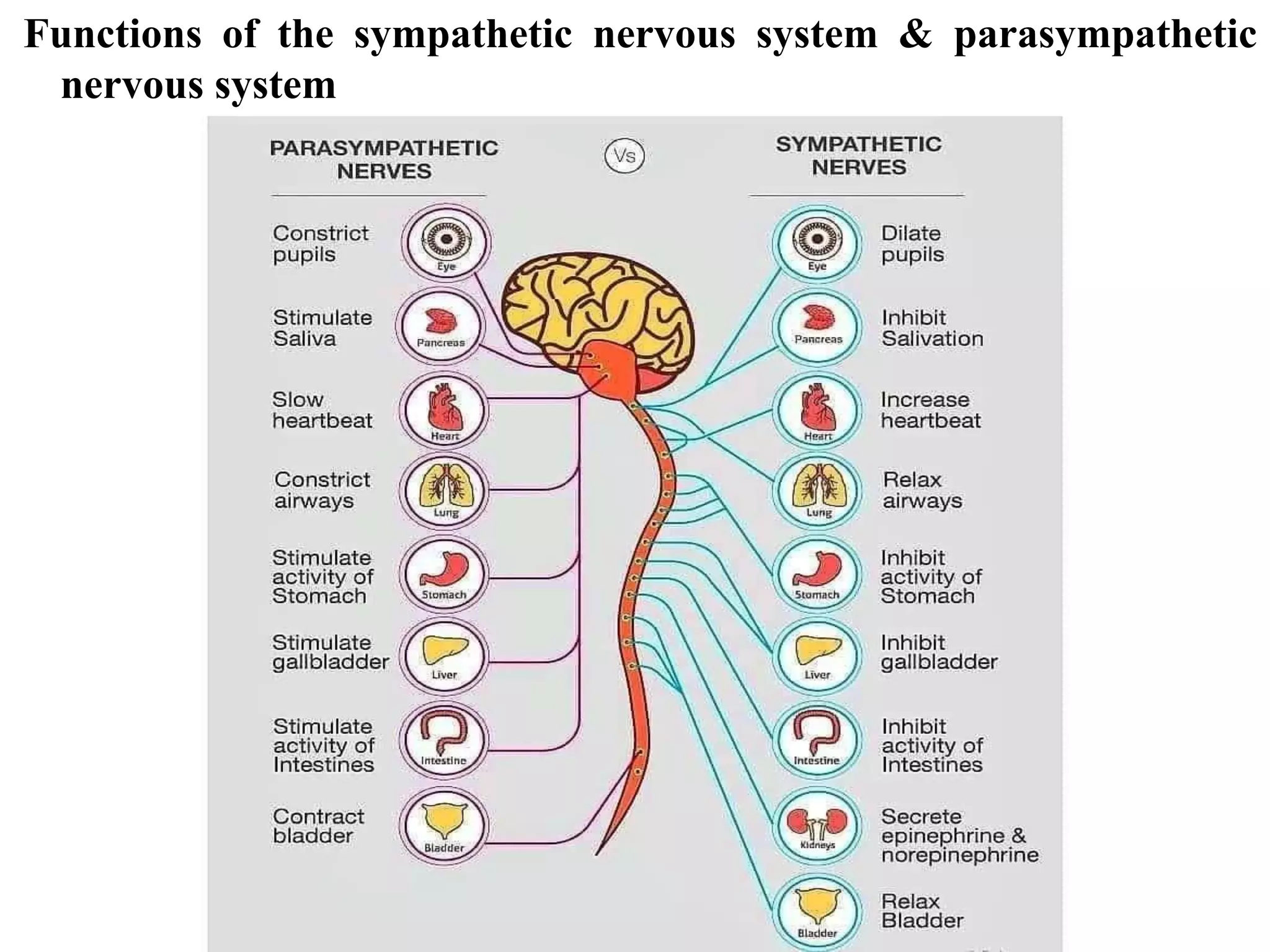
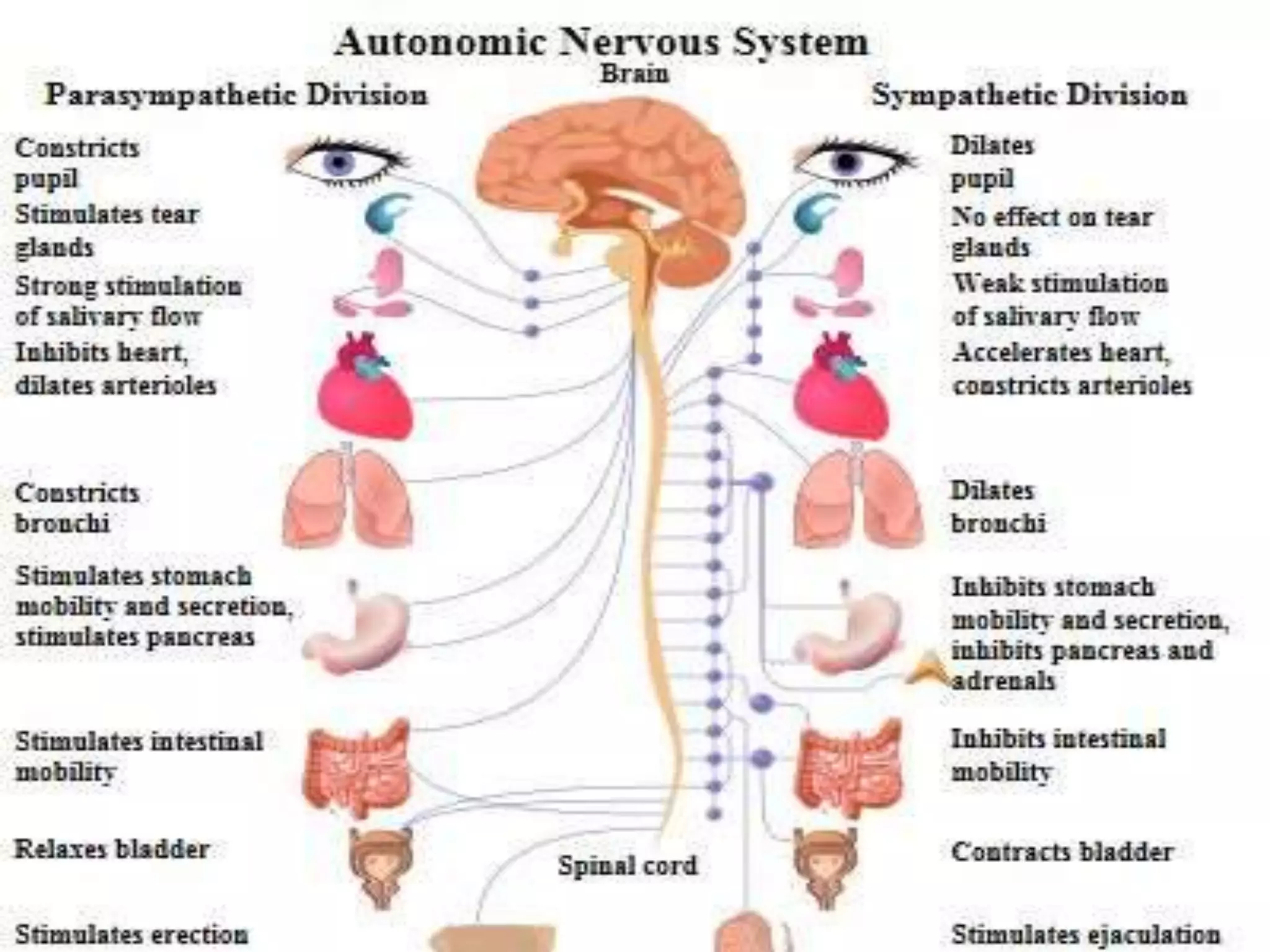
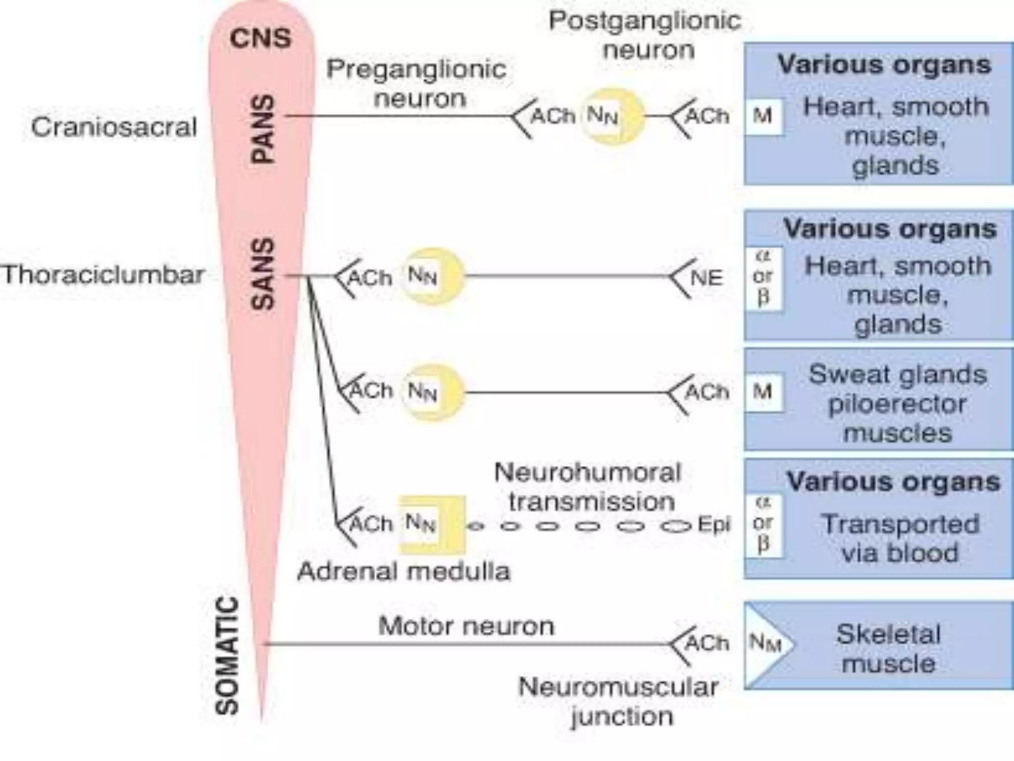
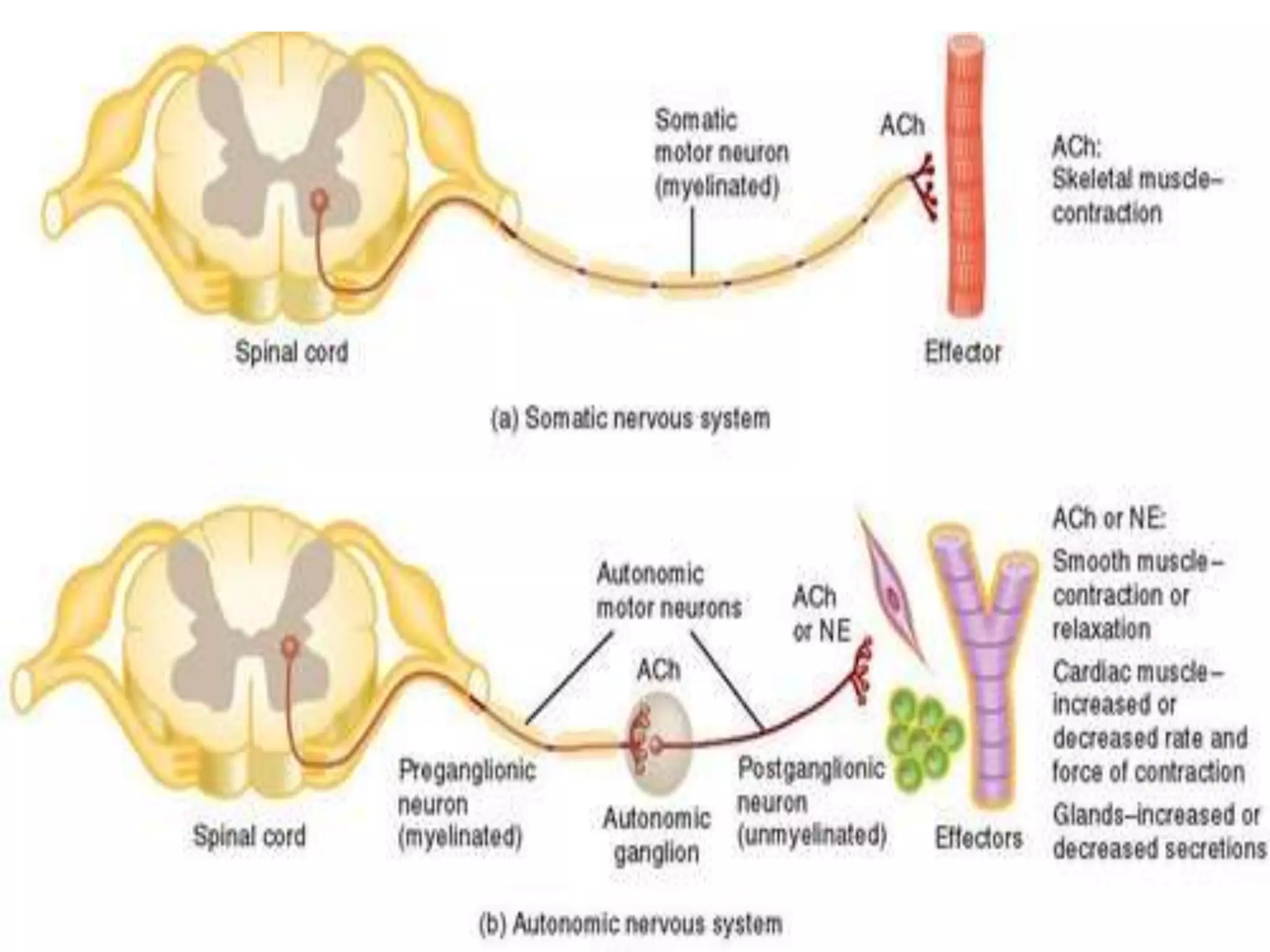
ORGANISATION & FUNCTIONS OF ANS
Autonomic nervous system (ANS) was so named by Langley (1898),
because of the fact that unlike the somatic nervous system of the
skeletal muscles, it is independent of voluntary control and thus
enjoys some degree of autonomy.
ANS innervates the heart, the smooth muscles, the glands & the viscera.
The ANS comprises
 The parasympathetic (cholinergic),
 The sympathetic (adrenergic) and
 The enteric nervous systems.
The parasympathetic system mainly participates in tissue building
reactions while
The sympathetic system enables the individual to respond to stress and
prepares the body for ‘flight or fight’.
The ANS carries nerve impulses from the CNS to the
effector organs by way of two types of efferent neurons.
The first nerve cell is called a preganglionic neuron, and its
cell body is located within the CNS.
Preganglionic neurons emerge from the brainstem or spinal
cord and make a synaptic connection in ganglia (an
aggregation of nerve cell bodies located in the peripheral
nervous system).
These ganglia function as relay stations between a
preganglionic neuron and a second nerve cell, the
postganglionic neuron.
The latter neuron has a cell body originating in the ganglion.
It is generally nonmyelinated and terminates on effector
organs, such as smooth muscles of the viscera, cardiac
muscle, and the exocrine glands.
Afferent neurons:
The afferent neurons (fibers) of the ANS
are important in the reflex regulation of
this system (for example, by sensing
pressure in the carotid sinus and aortic
arch) and in signaling the CNS to
influence the efferent branch of the
system to respond.
ANATOMY OF ANS
Efferent neurons:
ANATOMY OF SYMPATHETIC NEURONS:
The preganglionic neurons of the sympathetic system come from
thoracic and lumbar regions (T1 to L2) of the spinal cord, and they
synapse in two cordlike chains of ganglia that run close to and in
parallel on each side of the spinal cord.
The preganglionic neurons are short in comparison to the postganglionic
ones.
The sympathetic nervous system is also called the thoraco-lumbar
division because of its origins.
ANATOMY OF PARASYMPATHETIC NEURONS:
The parasympathetic preganglionic fibers arise from cranial nerves III
(oculomotor), VII (facial), IX (glossopharyngeal), and X (vagus) as
well as from the sacral region (S2 to S4) of the spinal cord and
synapse in ganglia near or on the effector organs.
Due to the origin of the parasympathetic nervous system, it is also called
the cranio-sacral division. Thus, in contrast to the sympathetic system,
the preganglionic fibers are long, and the postganglionic ones are
short, with the ganglia close to or within the organ innervated.
In most instances there is a one-to-one connection between the
preganglionic and postganglionic neurons, enabling the discrete
response of this division.
Functions of the sympathetic nervous system & parasympathetic
nervous system
Effects of stimulation of the sympathetic division:
The effect of sympathetic output is to increase heart rate and blood pressure, to mobilize
energy stores of the body, and to increase blood flow to skeletal muscles and the heart
while diverting flow from the skin and internal organs.
Sympathetic stimulation results in dilation of the pupils and the bronchioles .
It also affects GI motility and the function of the bladder and sexual organs.
Fight or flight response:
The changes experienced by the body during emergencies have been referred to as the
“fight or flight” response .
These reactions are triggered both by direct sympathetic activation of the effector
organs and by stimulation of the adrenal medulla to release epinephrine and lesser
amounts of norepinephrine.
Hormones released by the adrenal medulla directly enter the bloodstream and promote
responses in effector organs that contain adrenergic receptors .
The sympathetic nervous system tends to function as a unit and often
discharges as a complete system, for example, during severe exercise
or in reactions to fear.
This system, with its diffuse distribution of postganglionic fibers, is
involved in a wide array of physiologic activities. Although it is not
essential for survival, it is nevertheless an important system that
prepares the body to handle uncertain situations and unexpected
stimuli.
Functions of the parasympathetic nervous system
The parasympathetic division is involved with maintaining homeostasis
within the body.
To accomplish this, it maintains essential bodily functions, such as
digestive processes and elimination of wastes.
The parasympathetic division is required for life. It usually acts to
oppose or balance the actions of the sympathetic division and is
generally dominant over the sympathetic system in “rest and digest”
situations.
The parasympathetic system is not a functional entity as such and it
never discharges as a complete system. If it did, it would produce
massive, undesirable, and unpleasant symptoms, such as involuntary
urination and defecation.
Instead, discrete parasympathetic fibers are activated separately
and the system functions to affect specific organs, such as the stomach or
eye.
ANATOMY OF AUTONOMIC MOTOR PATHWAYS
1. PREGANGLIONIC NEURONS
2. GANGLIA
3. POST GANGLIONC NEURONS
PREGANGLIONIC NEURONS
Its Cell body is located within the Grey matter of CNS (Brain & Spinal
cord).
Its Myelinated axon exits within the CNS.
The pre-ganglionic neuron passes from the CNS in Spinal/Cranial Nerve.
It terminates in the ganglia.
GANGLIA:
It’s a collection of cell bodies located in a specific site within the body,
but outside the CNS.
POST-GANGLIONC NEURONS
The cell body located in an Autonomic ganglia.
The location of the ganglion is dependent upon the
division of ANS to which the neuron belongs & which organ it will
innervate.
Its axons are Unmyelinated.
The postganglionic axon passes from ganglion to effector (Cardiac
muscle, Smooth muscle, Gland) is either stimulated /inhibited.
ORGANSATION & FUNCTIONS OF ANS

ORGANSATION & FUNCTIONS OF ANS

  • 1.
    ORGANISATION & FUNCTIONSOF ANS Autonomic nervous system (ANS) was so named by Langley (1898), because of the fact that unlike the somatic nervous system of the skeletal muscles, it is independent of voluntary control and thus enjoys some degree of autonomy. ANS innervates the heart, the smooth muscles, the glands & the viscera. The ANS comprises  The parasympathetic (cholinergic),  The sympathetic (adrenergic) and  The enteric nervous systems. The parasympathetic system mainly participates in tissue building reactions while The sympathetic system enables the individual to respond to stress and prepares the body for ‘flight or fight’.
  • 3.
    The ANS carriesnerve impulses from the CNS to the effector organs by way of two types of efferent neurons. The first nerve cell is called a preganglionic neuron, and its cell body is located within the CNS. Preganglionic neurons emerge from the brainstem or spinal cord and make a synaptic connection in ganglia (an aggregation of nerve cell bodies located in the peripheral nervous system). These ganglia function as relay stations between a preganglionic neuron and a second nerve cell, the postganglionic neuron. The latter neuron has a cell body originating in the ganglion. It is generally nonmyelinated and terminates on effector organs, such as smooth muscles of the viscera, cardiac muscle, and the exocrine glands. Afferent neurons: The afferent neurons (fibers) of the ANS are important in the reflex regulation of this system (for example, by sensing pressure in the carotid sinus and aortic arch) and in signaling the CNS to influence the efferent branch of the system to respond. ANATOMY OF ANS Efferent neurons:
  • 4.
    ANATOMY OF SYMPATHETICNEURONS: The preganglionic neurons of the sympathetic system come from thoracic and lumbar regions (T1 to L2) of the spinal cord, and they synapse in two cordlike chains of ganglia that run close to and in parallel on each side of the spinal cord. The preganglionic neurons are short in comparison to the postganglionic ones. The sympathetic nervous system is also called the thoraco-lumbar division because of its origins.
  • 5.
    ANATOMY OF PARASYMPATHETICNEURONS: The parasympathetic preganglionic fibers arise from cranial nerves III (oculomotor), VII (facial), IX (glossopharyngeal), and X (vagus) as well as from the sacral region (S2 to S4) of the spinal cord and synapse in ganglia near or on the effector organs. Due to the origin of the parasympathetic nervous system, it is also called the cranio-sacral division. Thus, in contrast to the sympathetic system, the preganglionic fibers are long, and the postganglionic ones are short, with the ganglia close to or within the organ innervated. In most instances there is a one-to-one connection between the preganglionic and postganglionic neurons, enabling the discrete response of this division.
  • 6.
    Functions of thesympathetic nervous system & parasympathetic nervous system
  • 9.
    Effects of stimulationof the sympathetic division: The effect of sympathetic output is to increase heart rate and blood pressure, to mobilize energy stores of the body, and to increase blood flow to skeletal muscles and the heart while diverting flow from the skin and internal organs. Sympathetic stimulation results in dilation of the pupils and the bronchioles . It also affects GI motility and the function of the bladder and sexual organs. Fight or flight response: The changes experienced by the body during emergencies have been referred to as the “fight or flight” response . These reactions are triggered both by direct sympathetic activation of the effector organs and by stimulation of the adrenal medulla to release epinephrine and lesser amounts of norepinephrine. Hormones released by the adrenal medulla directly enter the bloodstream and promote responses in effector organs that contain adrenergic receptors .
  • 10.
    The sympathetic nervoussystem tends to function as a unit and often discharges as a complete system, for example, during severe exercise or in reactions to fear. This system, with its diffuse distribution of postganglionic fibers, is involved in a wide array of physiologic activities. Although it is not essential for survival, it is nevertheless an important system that prepares the body to handle uncertain situations and unexpected stimuli.
  • 11.
    Functions of theparasympathetic nervous system The parasympathetic division is involved with maintaining homeostasis within the body. To accomplish this, it maintains essential bodily functions, such as digestive processes and elimination of wastes. The parasympathetic division is required for life. It usually acts to oppose or balance the actions of the sympathetic division and is generally dominant over the sympathetic system in “rest and digest” situations. The parasympathetic system is not a functional entity as such and it never discharges as a complete system. If it did, it would produce massive, undesirable, and unpleasant symptoms, such as involuntary urination and defecation. Instead, discrete parasympathetic fibers are activated separately and the system functions to affect specific organs, such as the stomach or eye.
  • 12.
    ANATOMY OF AUTONOMICMOTOR PATHWAYS 1. PREGANGLIONIC NEURONS 2. GANGLIA 3. POST GANGLIONC NEURONS PREGANGLIONIC NEURONS Its Cell body is located within the Grey matter of CNS (Brain & Spinal cord). Its Myelinated axon exits within the CNS. The pre-ganglionic neuron passes from the CNS in Spinal/Cranial Nerve. It terminates in the ganglia.
  • 14.
    GANGLIA: It’s a collectionof cell bodies located in a specific site within the body, but outside the CNS. POST-GANGLIONC NEURONS The cell body located in an Autonomic ganglia. The location of the ganglion is dependent upon the division of ANS to which the neuron belongs & which organ it will innervate. Its axons are Unmyelinated. The postganglionic axon passes from ganglion to effector (Cardiac muscle, Smooth muscle, Gland) is either stimulated /inhibited.