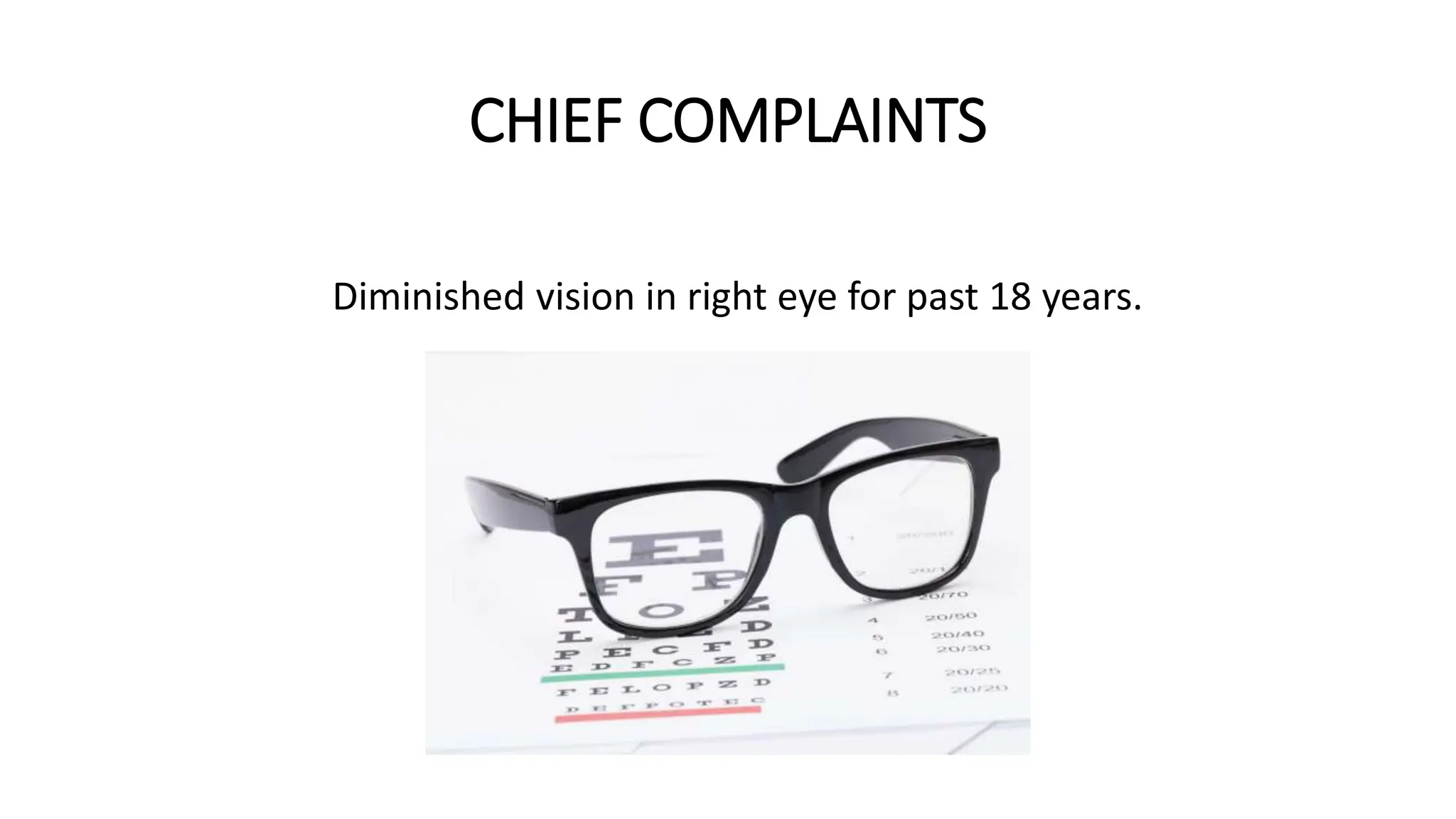
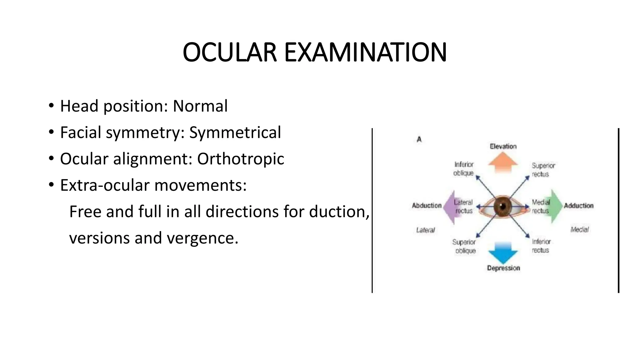
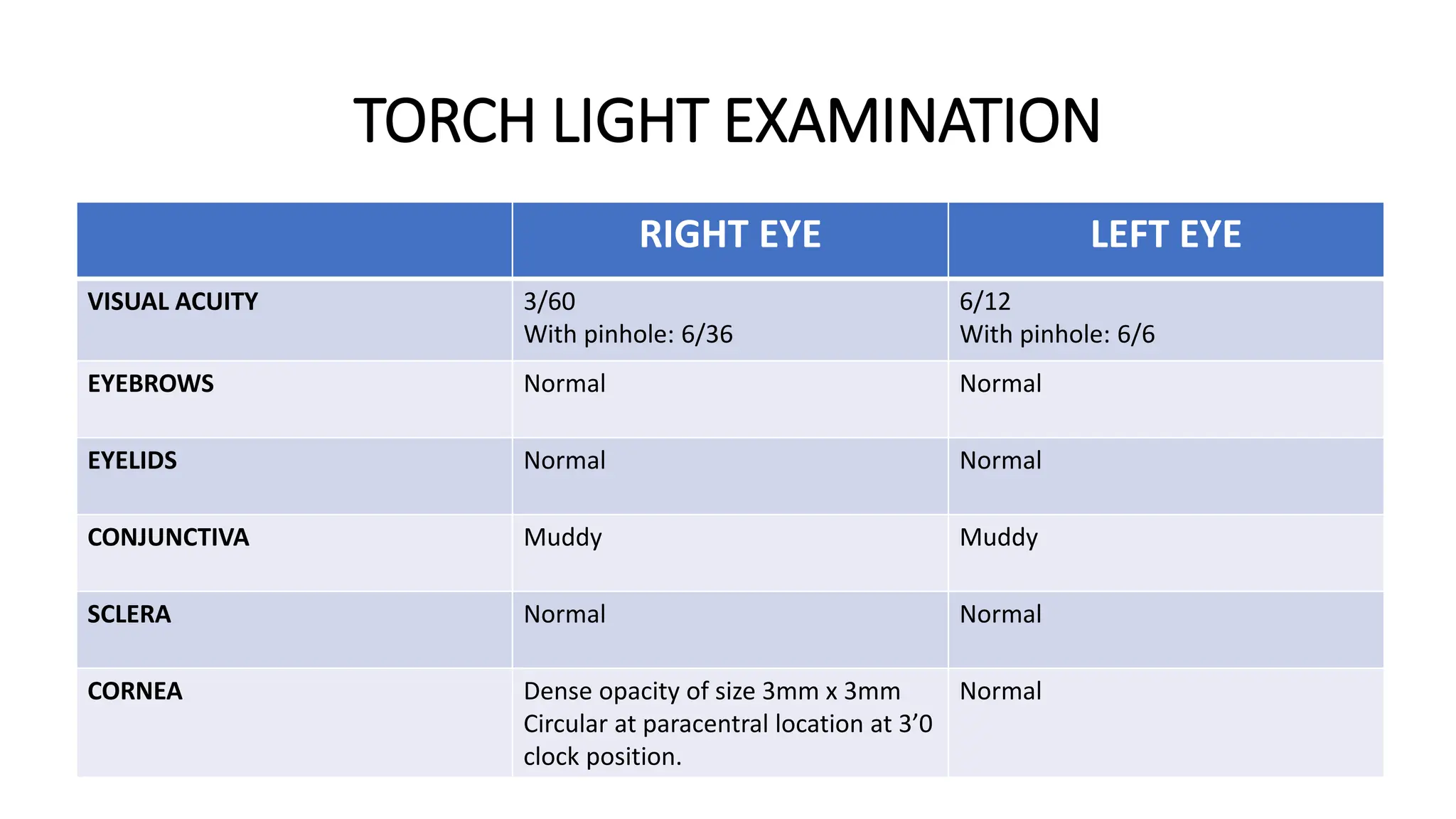
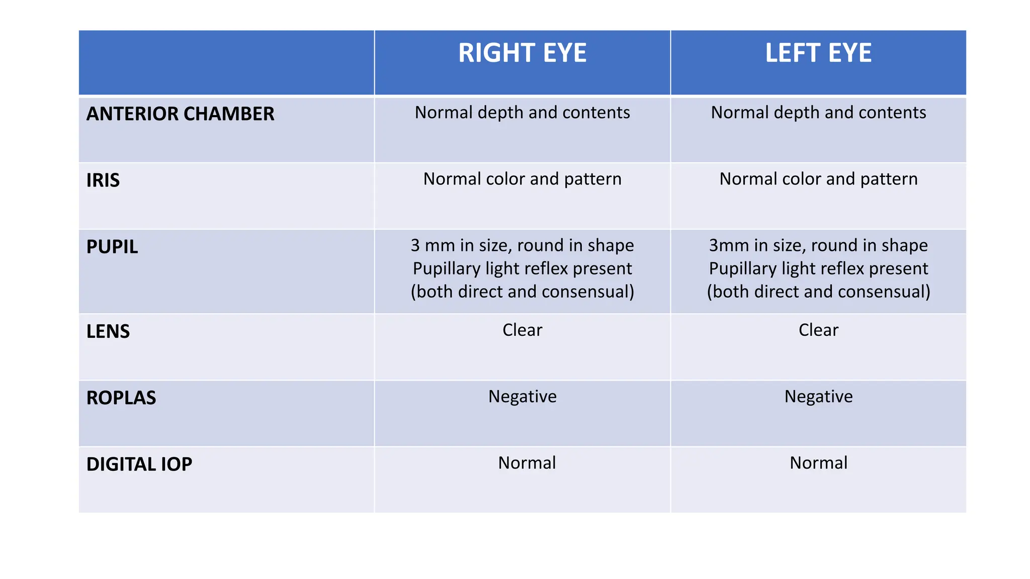
CASE PRESENTATION
MR.AROGYADAS, 55 YRS, SENGURUCHI, DATE:04/01/2023
BY G.KIRTHIK YADAV
Mr.Arogyadas a 55 year old man from senguruchi village who is a farm
worker came to OPD with chief complaints of diminished vision in right
eye for past 18 years.
AFFECTED
EYE OF
THE
PATIENT
CHIEF COMPLAINTS
Diminished vision in right eye for past 18 years.
HISTORY OF PRESENTING ILLNESS
• Mr.Arogyadas was asymptomatic 18 years back after which he developed
sudden painful diminution of vision in his right eye.
• History of trauma in right eye during the year 2005 which was 18 years
back, while working in an agricultural field.
• Defective vision was accompanied with foreign body sensation,
Photophobia, redness and excess lacrimation in right eye.
• The patient had taken eye ointment after the injury 18 years back on
prescription by a doctor after the eye trauma.
• But the patient had diminished vision which is not improving.
• No history of spectacles.
PAST HISTORY
• He is a known case of diabetes mellitus for 10 years and he is under
regular oral hypoglycemic agents.
• He is a known case of hypertension for past 7 years and he is under
regular treatment.
• No history of asthma.
• No previous surgical history.
PERSONAL HISTORY
• Mixed diet
• Normal sleep pattern
• Normal bowel and bladder habits
• No history of any addictions
FAMILY HISTORY
• No significant family history
GENERAL EXAMINATION
• Patient is conscious, coherent
cooperative and well oriented to
place and time.
• He is moderately built and nourished
• No signs of pallor, icterus, cyanosis,
clubbing, koilonychia,
lymphadenopathy or pedal oedema
• Vitals are normal
OCULAR EXAMINATION
• Head position: Normal
• Facial symmetry: Symmetrical
• Ocular alignment: Orthotropic
• Extra-ocular movements:
Free and full in all directions for duction,
versions and vergence.
TORCH LIGHT EXAMINATION
RIGHT EYE LEFT EYE
VISUAL ACUITY 3/60
With pinhole: 6/36
6/12
With pinhole: 6/6
EYEBROWS Normal Normal
EYELIDS Normal Normal
CONJUNCTIVA Muddy Muddy
SCLERA Normal Normal
CORNEA Dense opacity of size 3mm x 3mm
Circular at paracentral location at 3’0
clock position.
Normal
RIGHT EYE LEFT EYE
ANTERIOR CHAMBER Normal depth and contents Normal depth and contents
IRIS Normal color and pattern Normal color and pattern
PUPIL 3 mm in size, round in shape
Pupillary light reflex present
(both direct and consensual)
3mm in size, round in shape
Pupillary light reflex present
(both direct and consensual)
LENS Clear Clear
ROPLAS Negative Negative
DIGITAL IOP Normal Normal
DIAGNOSIS
• Paracentral Leucomatous corneal opacity in right eye involving 1/3 rd
of pupillary area at 3’o clock position.
MANAGEMENT
• Optical Iridectomy
• Keratoplasty
THANK YOU

Ophthal case presentation on senile cataract

  • 1.
    CASE PRESENTATION MR.AROGYADAS, 55YRS, SENGURUCHI, DATE:04/01/2023 BY G.KIRTHIK YADAV
  • 2.
    Mr.Arogyadas a 55year old man from senguruchi village who is a farm worker came to OPD with chief complaints of diminished vision in right eye for past 18 years.
  • 3.
  • 4.
    CHIEF COMPLAINTS Diminished visionin right eye for past 18 years.
  • 5.
    HISTORY OF PRESENTINGILLNESS • Mr.Arogyadas was asymptomatic 18 years back after which he developed sudden painful diminution of vision in his right eye. • History of trauma in right eye during the year 2005 which was 18 years back, while working in an agricultural field. • Defective vision was accompanied with foreign body sensation, Photophobia, redness and excess lacrimation in right eye. • The patient had taken eye ointment after the injury 18 years back on prescription by a doctor after the eye trauma. • But the patient had diminished vision which is not improving. • No history of spectacles.
  • 6.
    PAST HISTORY • Heis a known case of diabetes mellitus for 10 years and he is under regular oral hypoglycemic agents. • He is a known case of hypertension for past 7 years and he is under regular treatment. • No history of asthma. • No previous surgical history.
  • 7.
    PERSONAL HISTORY • Mixeddiet • Normal sleep pattern • Normal bowel and bladder habits • No history of any addictions
  • 8.
    FAMILY HISTORY • Nosignificant family history
  • 9.
    GENERAL EXAMINATION • Patientis conscious, coherent cooperative and well oriented to place and time. • He is moderately built and nourished • No signs of pallor, icterus, cyanosis, clubbing, koilonychia, lymphadenopathy or pedal oedema • Vitals are normal
  • 10.
    OCULAR EXAMINATION • Headposition: Normal • Facial symmetry: Symmetrical • Ocular alignment: Orthotropic • Extra-ocular movements: Free and full in all directions for duction, versions and vergence.
  • 11.
    TORCH LIGHT EXAMINATION RIGHTEYE LEFT EYE VISUAL ACUITY 3/60 With pinhole: 6/36 6/12 With pinhole: 6/6 EYEBROWS Normal Normal EYELIDS Normal Normal CONJUNCTIVA Muddy Muddy SCLERA Normal Normal CORNEA Dense opacity of size 3mm x 3mm Circular at paracentral location at 3’0 clock position. Normal
  • 12.
    RIGHT EYE LEFTEYE ANTERIOR CHAMBER Normal depth and contents Normal depth and contents IRIS Normal color and pattern Normal color and pattern PUPIL 3 mm in size, round in shape Pupillary light reflex present (both direct and consensual) 3mm in size, round in shape Pupillary light reflex present (both direct and consensual) LENS Clear Clear ROPLAS Negative Negative DIGITAL IOP Normal Normal
  • 13.
    DIAGNOSIS • Paracentral Leucomatouscorneal opacity in right eye involving 1/3 rd of pupillary area at 3’o clock position.
  • 14.
  • 15.