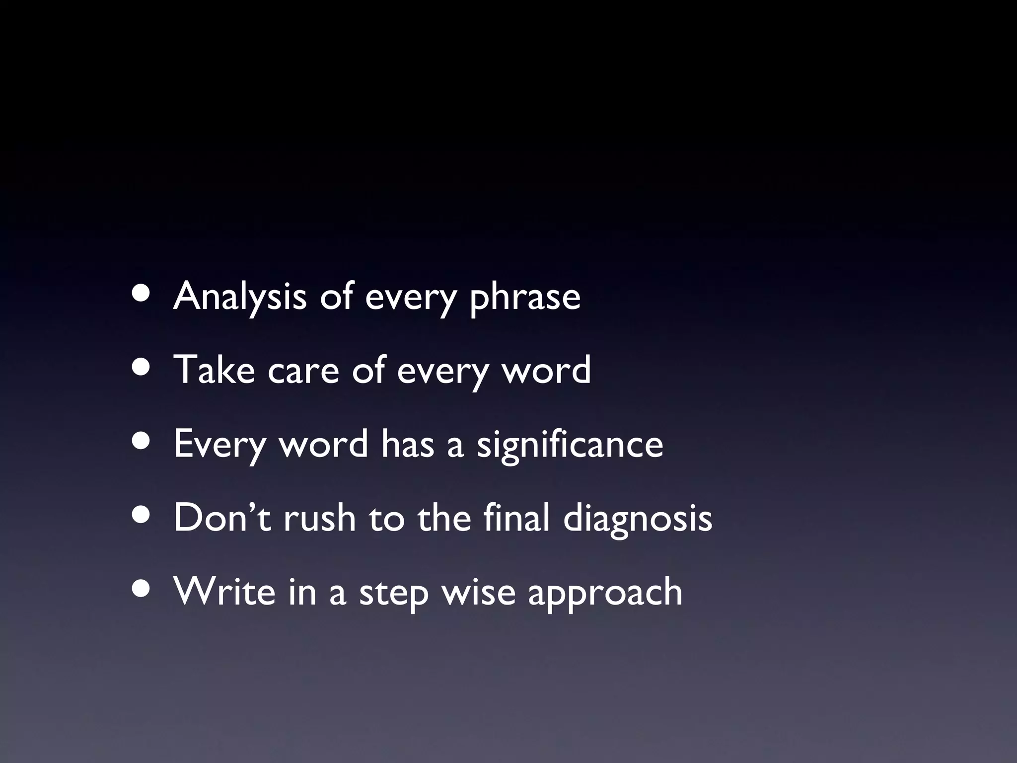
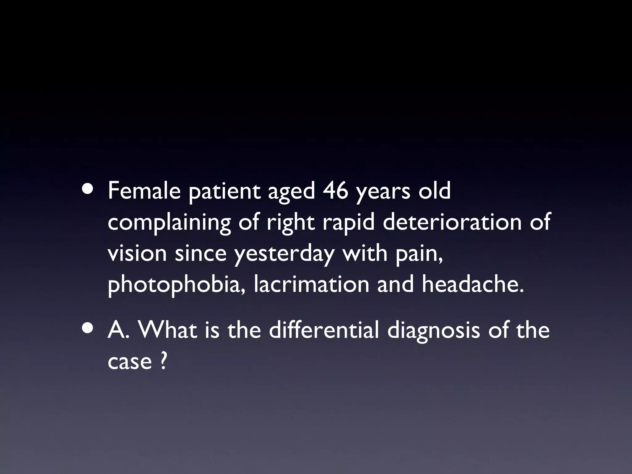
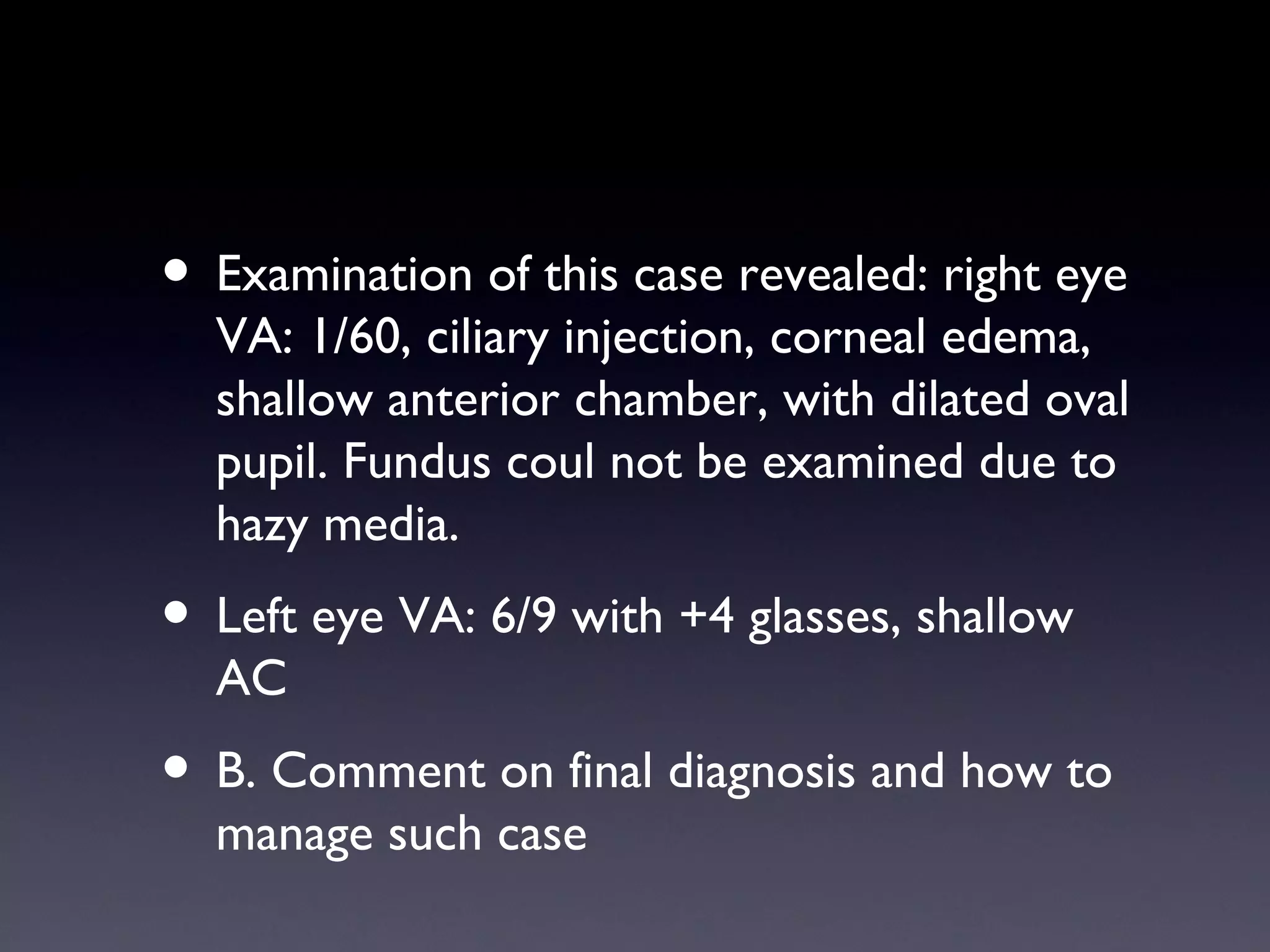
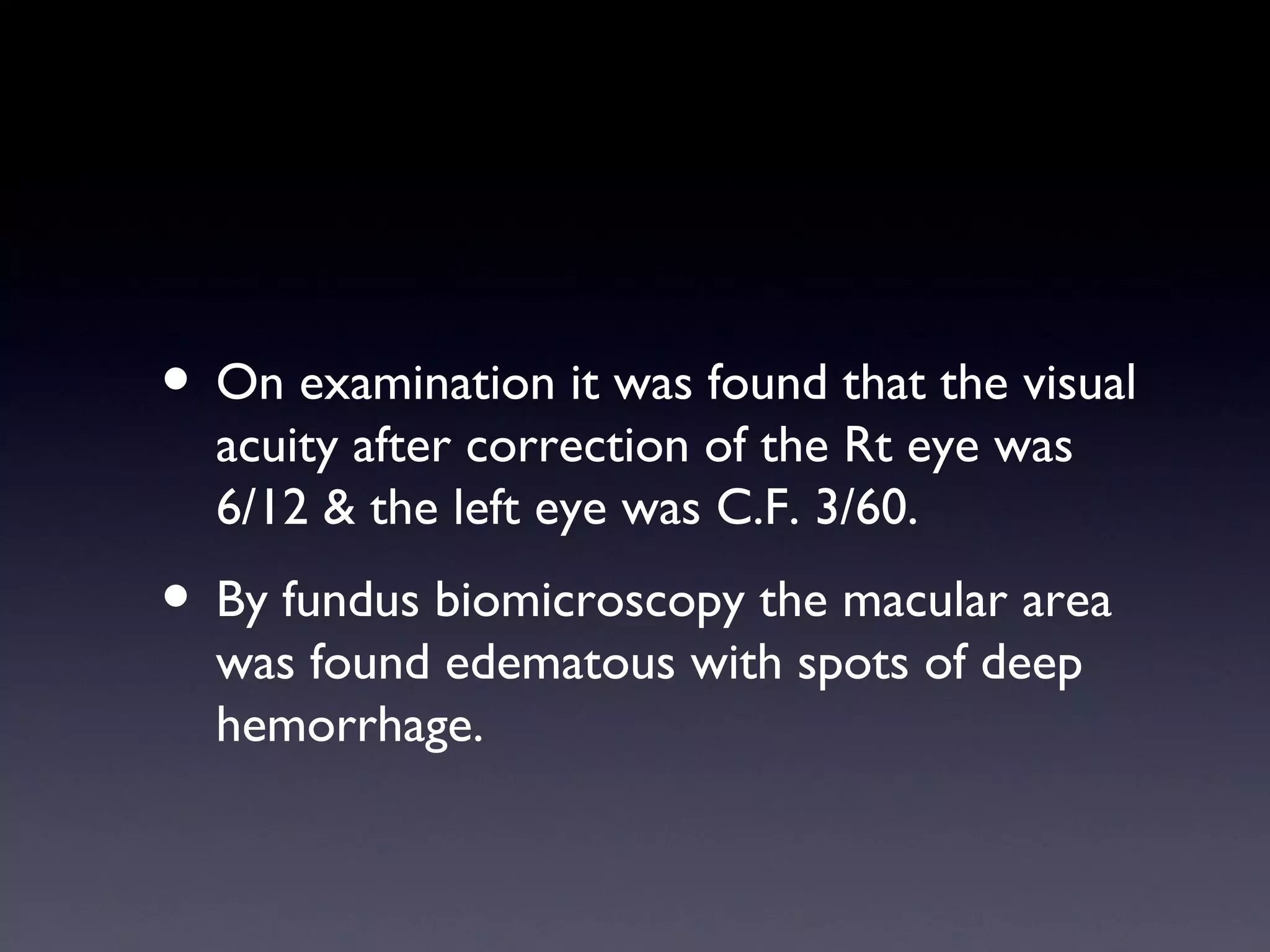
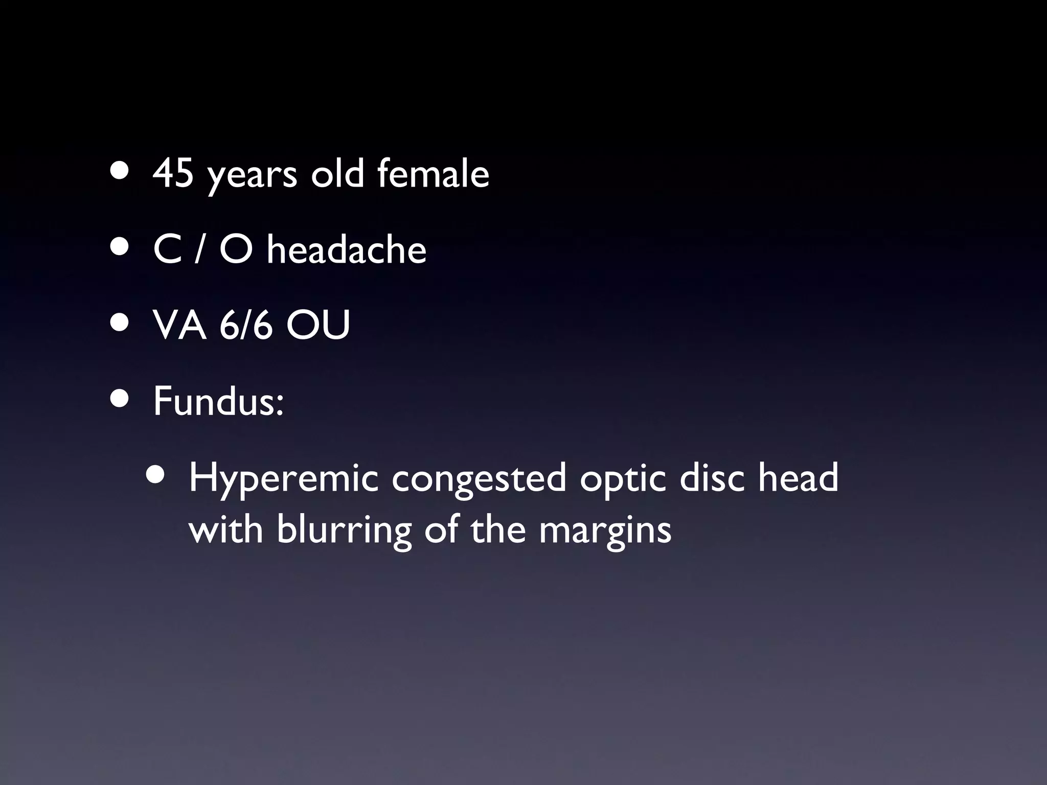
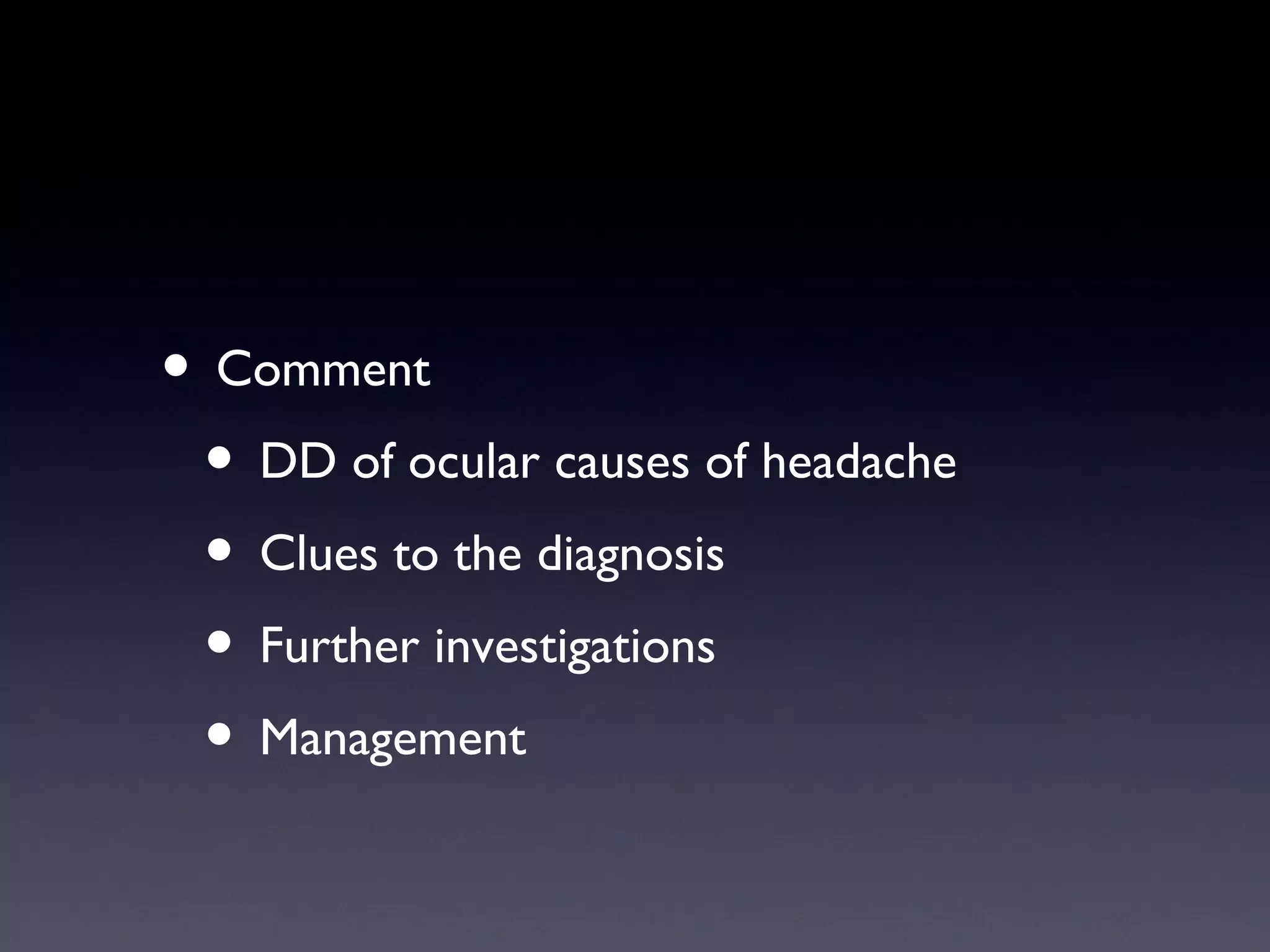
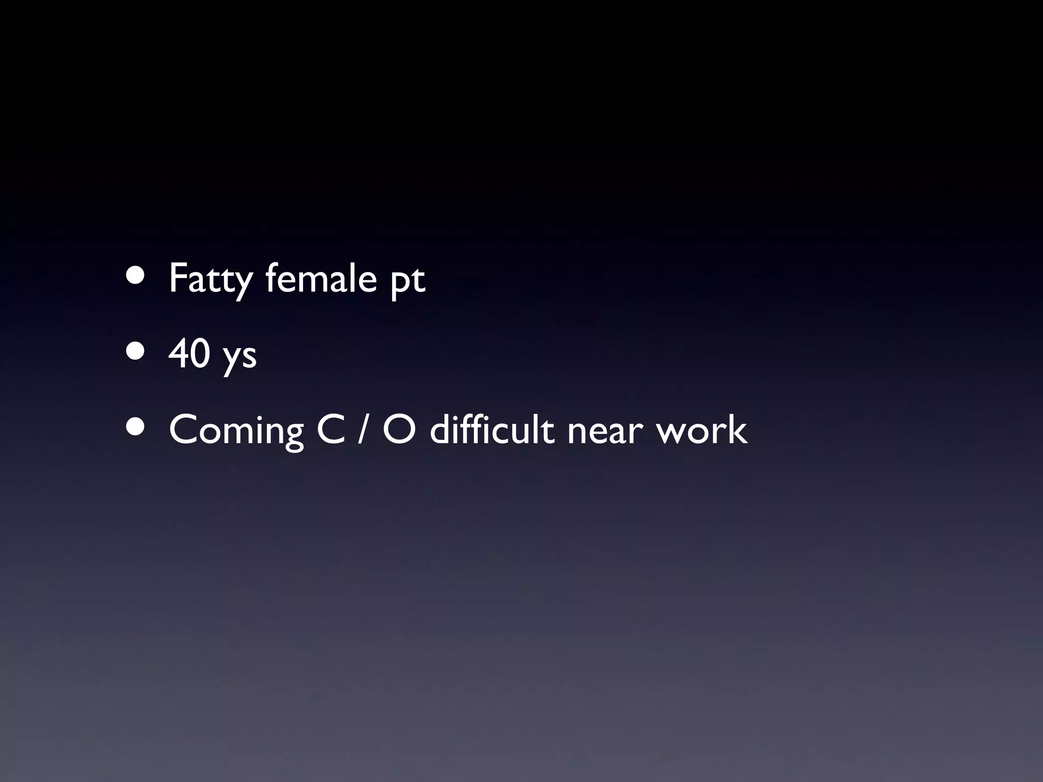
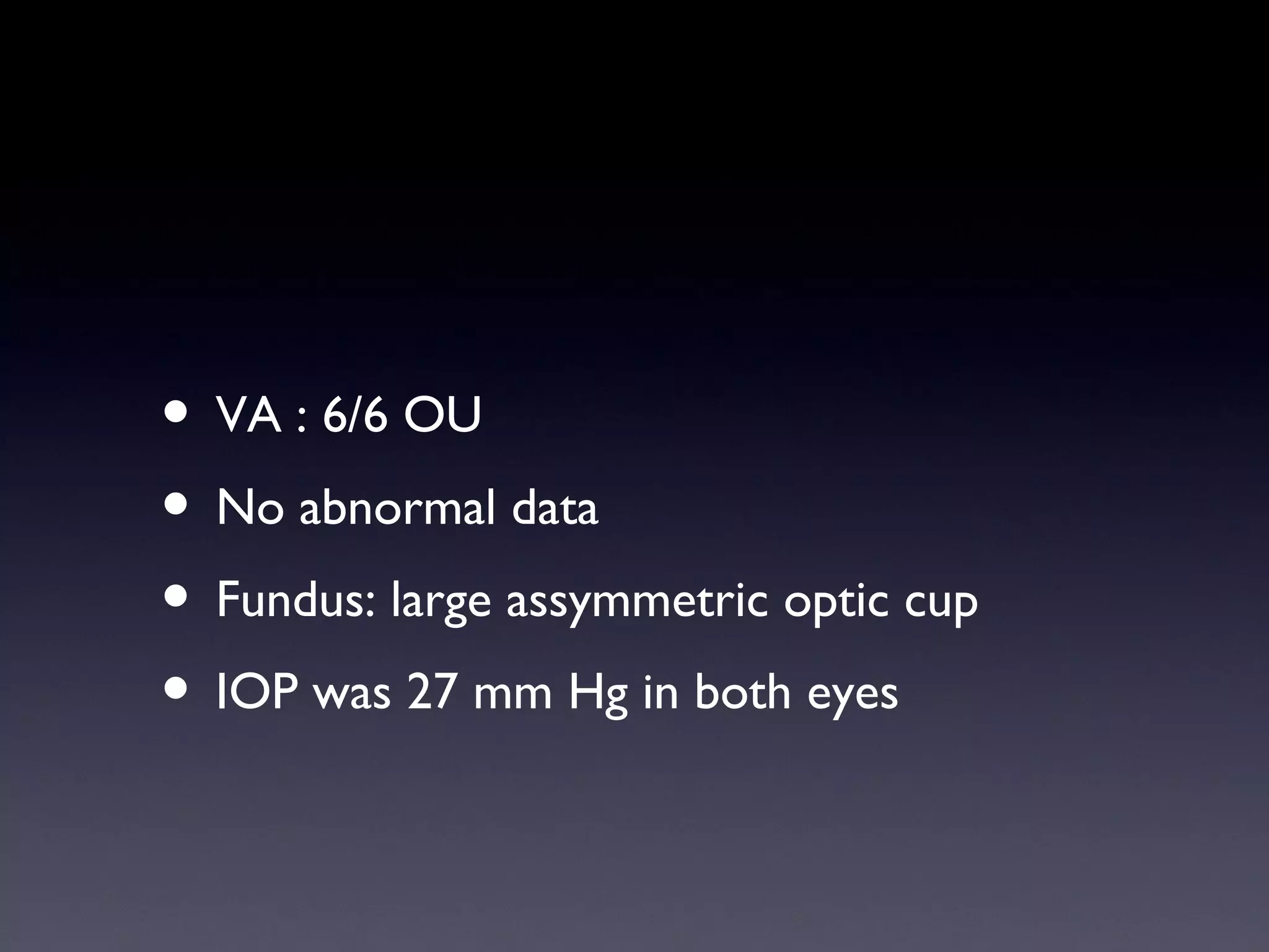
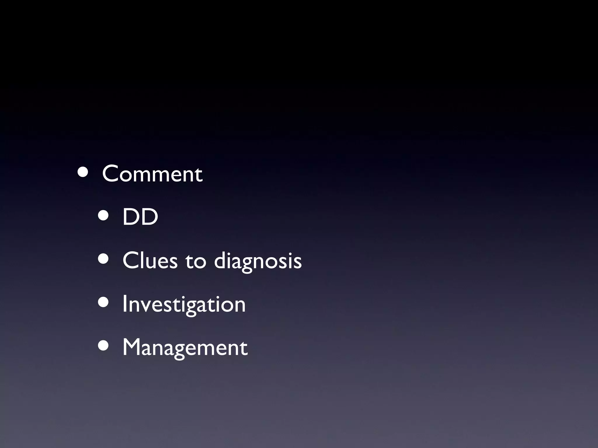
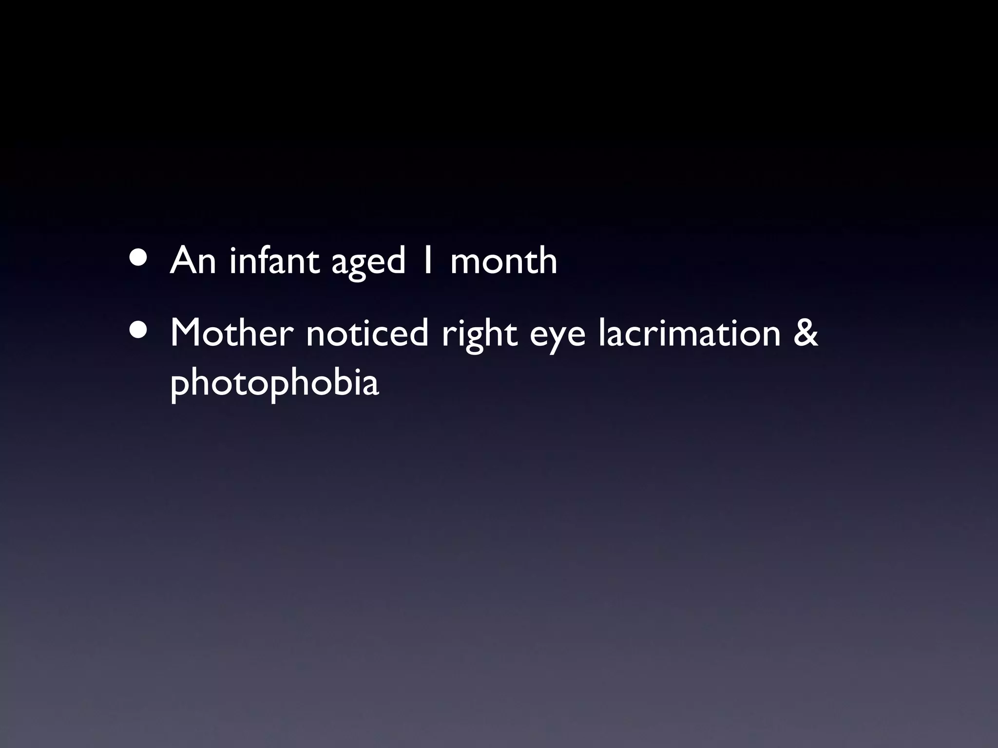
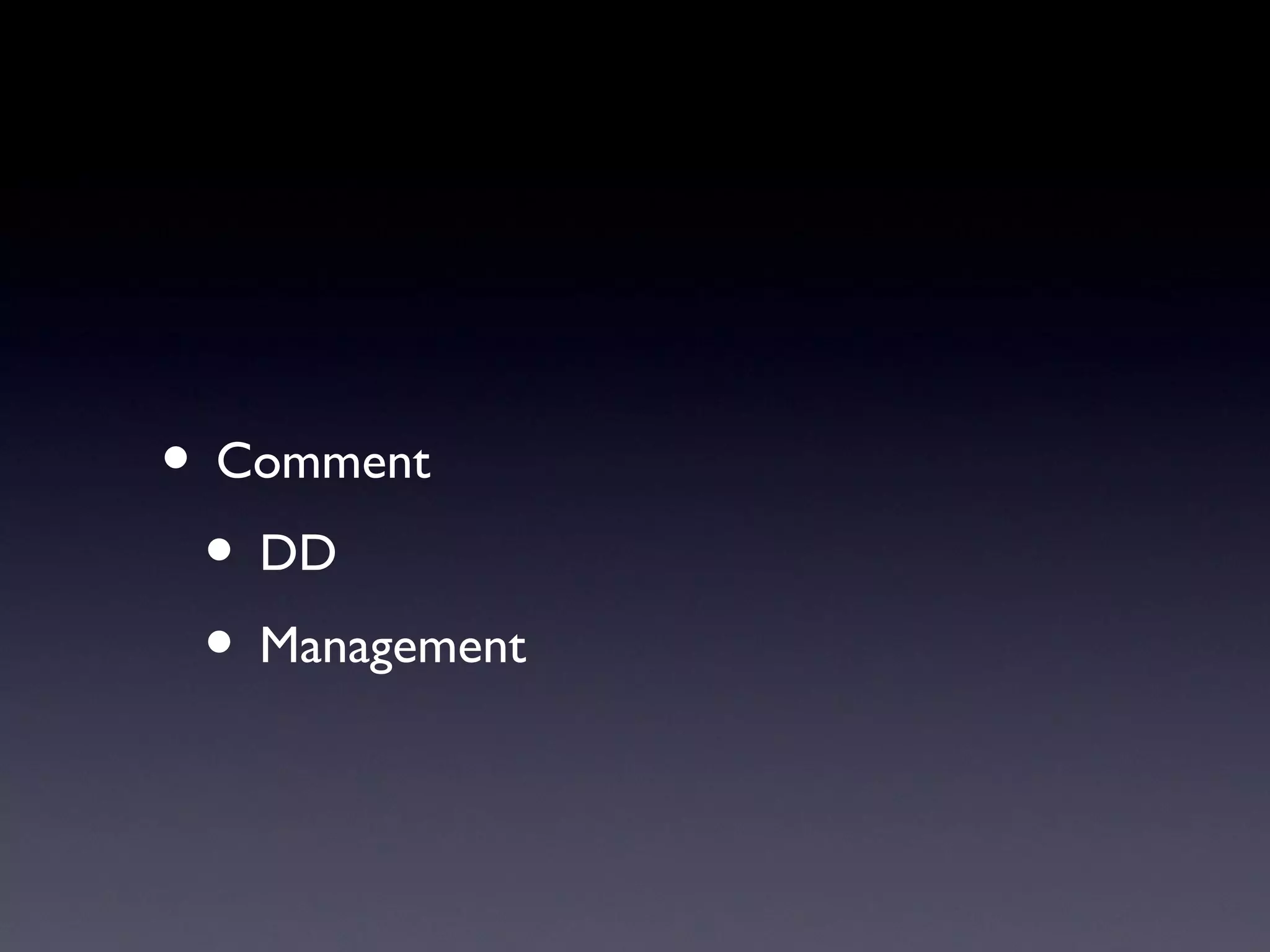
Problem Solving
Instructions
• Analysis of every phrase
• Take care of every word
• Every word has a significance
• Don’t rush to the final diagnosis
• Write in a step wise approach
Examples
Case 1
• Female patient aged 46 years old
  complaining of right rapid deterioration of
  vision since yesterday with pain,
  photophobia, lacrimation and headache.
• A. What is the differential diagnosis of the
  case ?
• Examination of this case revealed: right eye
  VA: 1/60, ciliary injection, corneal edema,
  shallow anterior chamber, with dilated oval
  pupil. Fundus coul not be examined due to
  hazy media.
• Left eye VA: 6/9 with +4 glasses, shallow
  AC
• B. Comment on final diagnosis and how to
  manage such case
Case 2
• Male pat. Farmer, aged 6oys came to
  complain of diminution of vision (fogy
  vision) of both eyes, more in the right eye.
• Gradual, progressive, painless since 6
  months in the right eye and recently in the
  left eye.
• It is more in the day light.
• No treatment received before.
• On examination:
 • VA: OD 3/60. OS 6/36 not corrected
 • Anterior segment was normal, pupil was
    regular and reactive, greyish coloration
    of the pupillary area with presence of iris
    shadow in both eyes, IOP was normal,
    fundus was normal
• Comment on the case
 • Age: senile problem
 • C / O ………...DD
 • Exclusion of other pathologies
 • Final diagnosis
 • Investigation
 • Management
Case 3
• A baby
• Aged 4 months
• Came with his parents; to complain of :
• The color of his pupil looks abnormal
• he cannot fix any object or know his
  mother
• No other data
• Comment
 • Congenital anomaly
 • DD leucocorea
 • Examination under anesthesia
 • Investigation
Case 4
• Male patient 50 years old
• Co/ Left red eye , pain photophopia ,
  lacrimation& diminution of vision .
• Past History
 • Diabetes millitus
 • Chronic inflammation of left lacrimal sac
• Comment
Case 5
• 50 years female
• Co/ sudden appearance of black curtain in
  front of left eye with marked diminution of
  vision
• Past history of wearing heavy glasses for
  long period
• Comment:
 • DD
 • Investigation
 • Management
Case 6
• A Male diabetic patient aged 50 years old.
• Came to complain of a rapid diminution of
  vision of the left eye, with photopsia and
  micropsia, the duration of diabetes was
  15ys.
• There was positive family history.
• On examination it was found that the visual
  acuity after correction of the Rt eye was
  6/12 & the left eye was C.F. 3/60.
• By fundus biomicroscopy the macular area
  was found edematous with spots of deep
  hemorrhage.
• Comment
Case 7
• 45 years old female
• C / O headache
• VA 6/6 OU
• Fundus:
 • Hyperemic congested optic disc head
    with blurring of the margins
• Comment
 • DD of ocular causes of headache
 • Clues to the diagnosis
 • Further investigations
 • Management
Case 8
• Fatty female pt
• 40 ys
• Coming C / O difficult near work
• VA : 6/6 OU
• No abnormal data
• Fundus: large assymmetric optic cup
• IOP was 27 mm Hg in both eyes
• Comment
 • DD
 • Clues to diagnosis
 • Investigation
 • Management
Case 9
• An infant aged 1 month
• Mother noticed right eye lacrimation &
  photophobia
• Comment
 • DD
 • Management
Case 10
• Male patent 60 years old
• Co/ sudden appearance of diplopia
  interferring with his daily life
• Past history of NIDDM 10ys, hypertension
• Examination:
 • Right esotropia 30°
 • Normal fundus
• Comment

Ophthalmology Problems

  • 1.
  • 2.
  • 3.
    • Analysis ofevery phrase • Take care of every word • Every word has a significance • Don’t rush to the final diagnosis • Write in a step wise approach
  • 4.
  • 5.
  • 6.
    • Female patientaged 46 years old complaining of right rapid deterioration of vision since yesterday with pain, photophobia, lacrimation and headache. • A. What is the differential diagnosis of the case ?
  • 7.
    • Examination ofthis case revealed: right eye VA: 1/60, ciliary injection, corneal edema, shallow anterior chamber, with dilated oval pupil. Fundus coul not be examined due to hazy media. • Left eye VA: 6/9 with +4 glasses, shallow AC • B. Comment on final diagnosis and how to manage such case
  • 8.
  • 9.
    • Male pat.Farmer, aged 6oys came to complain of diminution of vision (fogy vision) of both eyes, more in the right eye. • Gradual, progressive, painless since 6 months in the right eye and recently in the left eye. • It is more in the day light. • No treatment received before.
  • 10.
    • On examination: • VA: OD 3/60. OS 6/36 not corrected • Anterior segment was normal, pupil was regular and reactive, greyish coloration of the pupillary area with presence of iris shadow in both eyes, IOP was normal, fundus was normal
  • 11.
    • Comment onthe case • Age: senile problem • C / O ………...DD • Exclusion of other pathologies • Final diagnosis • Investigation • Management
  • 12.
  • 13.
    • A baby •Aged 4 months • Came with his parents; to complain of :
  • 14.
    • The colorof his pupil looks abnormal • he cannot fix any object or know his mother • No other data
  • 15.
    • Comment •Congenital anomaly • DD leucocorea • Examination under anesthesia • Investigation
  • 16.
  • 17.
    • Male patient50 years old • Co/ Left red eye , pain photophopia , lacrimation& diminution of vision . • Past History • Diabetes millitus • Chronic inflammation of left lacrimal sac
  • 18.
  • 19.
  • 20.
    • 50 yearsfemale • Co/ sudden appearance of black curtain in front of left eye with marked diminution of vision • Past history of wearing heavy glasses for long period
  • 21.
    • Comment: •DD • Investigation • Management
  • 22.
  • 23.
    • A Malediabetic patient aged 50 years old. • Came to complain of a rapid diminution of vision of the left eye, with photopsia and micropsia, the duration of diabetes was 15ys. • There was positive family history.
  • 24.
    • On examinationit was found that the visual acuity after correction of the Rt eye was 6/12 & the left eye was C.F. 3/60. • By fundus biomicroscopy the macular area was found edematous with spots of deep hemorrhage.
  • 25.
  • 26.
  • 27.
    • 45 yearsold female • C / O headache • VA 6/6 OU • Fundus: • Hyperemic congested optic disc head with blurring of the margins
  • 28.
    • Comment •DD of ocular causes of headache • Clues to the diagnosis • Further investigations • Management
  • 29.
  • 30.
    • Fatty femalept • 40 ys • Coming C / O difficult near work
  • 31.
    • VA :6/6 OU • No abnormal data • Fundus: large assymmetric optic cup • IOP was 27 mm Hg in both eyes
  • 32.
    • Comment •DD • Clues to diagnosis • Investigation • Management
  • 33.
  • 34.
    • An infantaged 1 month • Mother noticed right eye lacrimation & photophobia
  • 35.
    • Comment •DD • Management
  • 36.
  • 37.
    • Male patent60 years old • Co/ sudden appearance of diplopia interferring with his daily life • Past history of NIDDM 10ys, hypertension
  • 38.
    • Examination: •Right esotropia 30° • Normal fundus
  • 39.