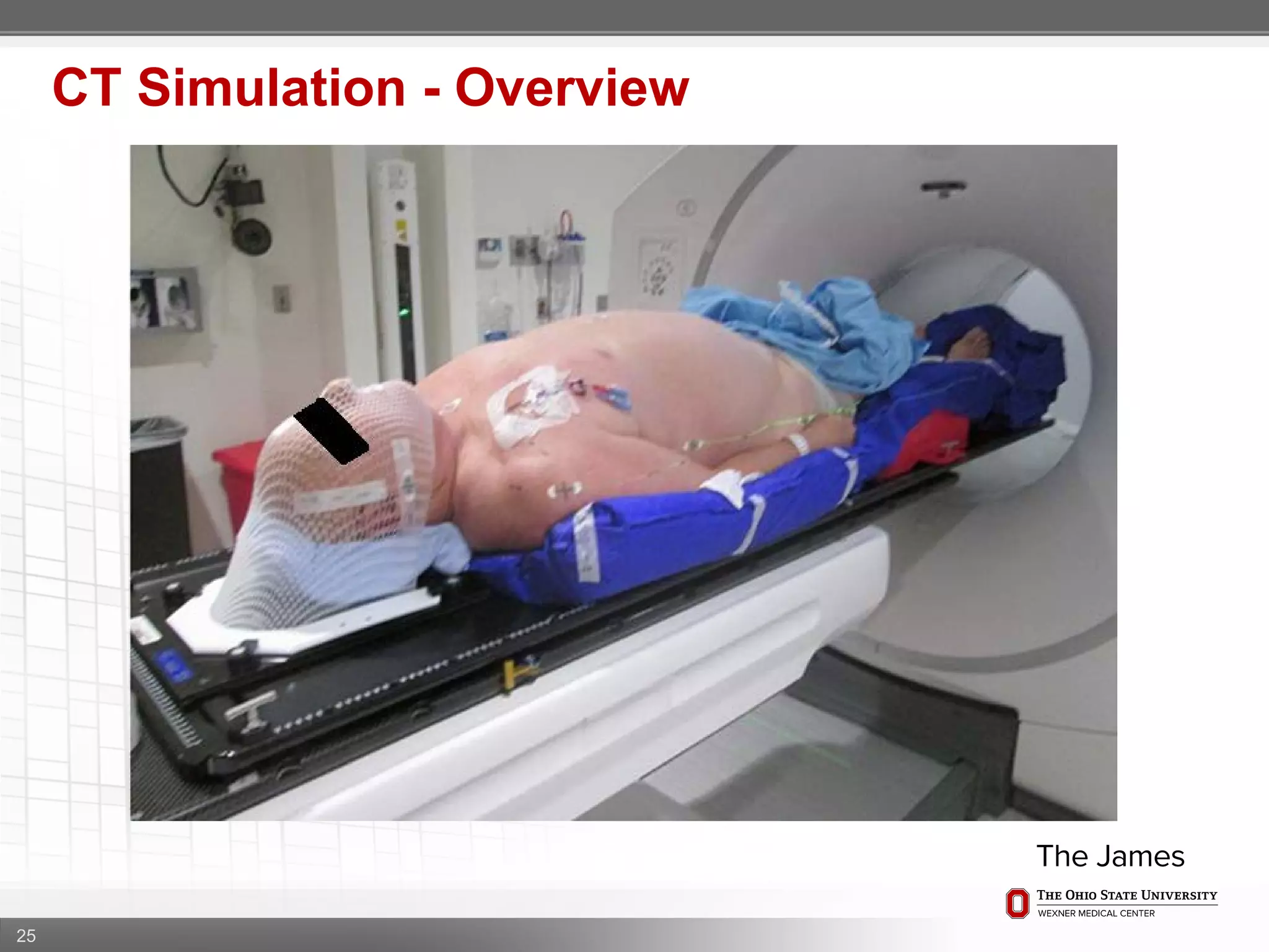
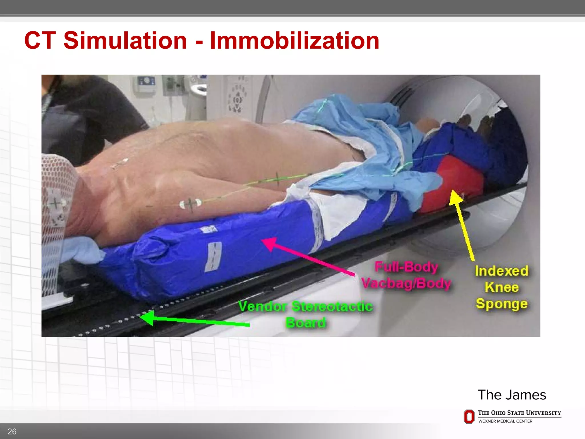
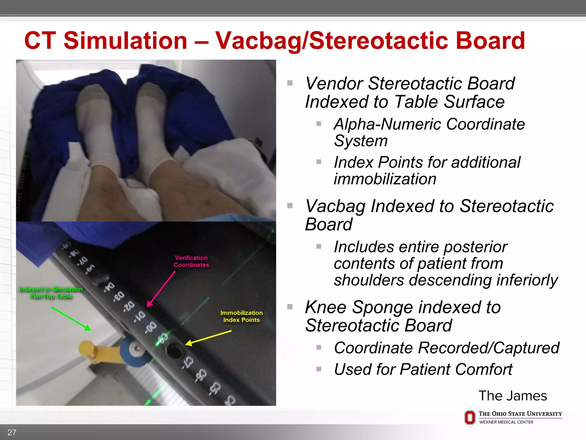
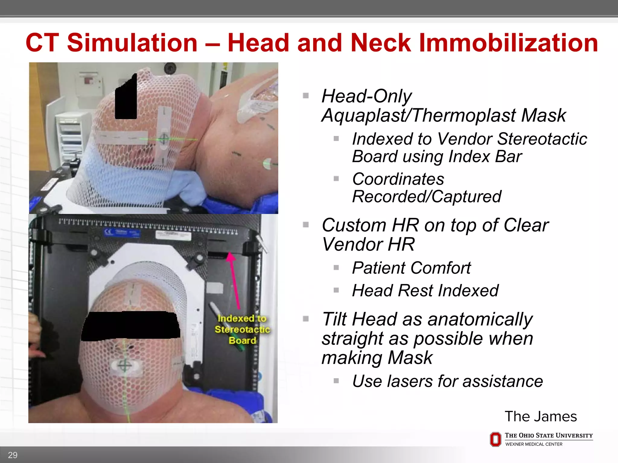
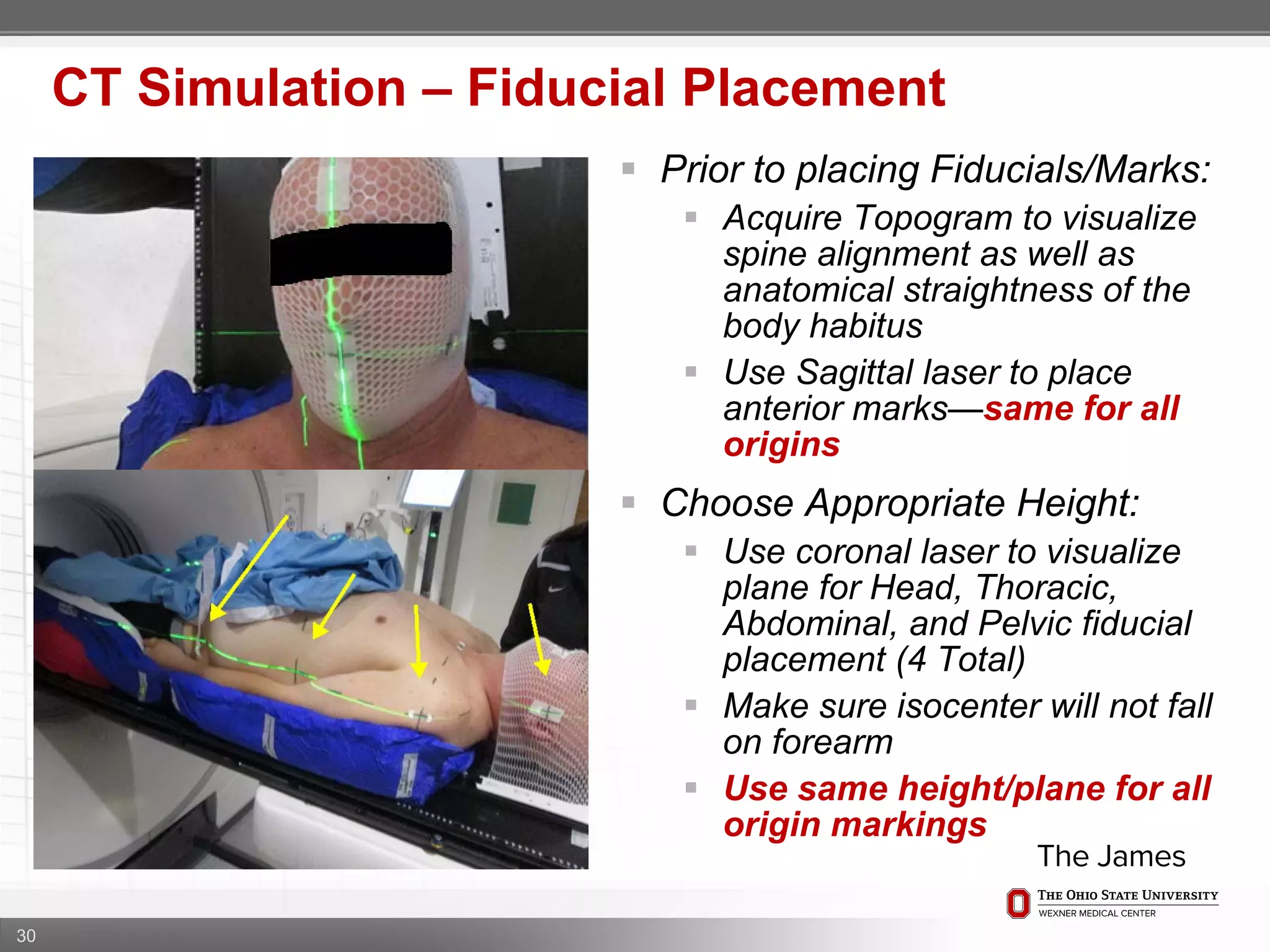
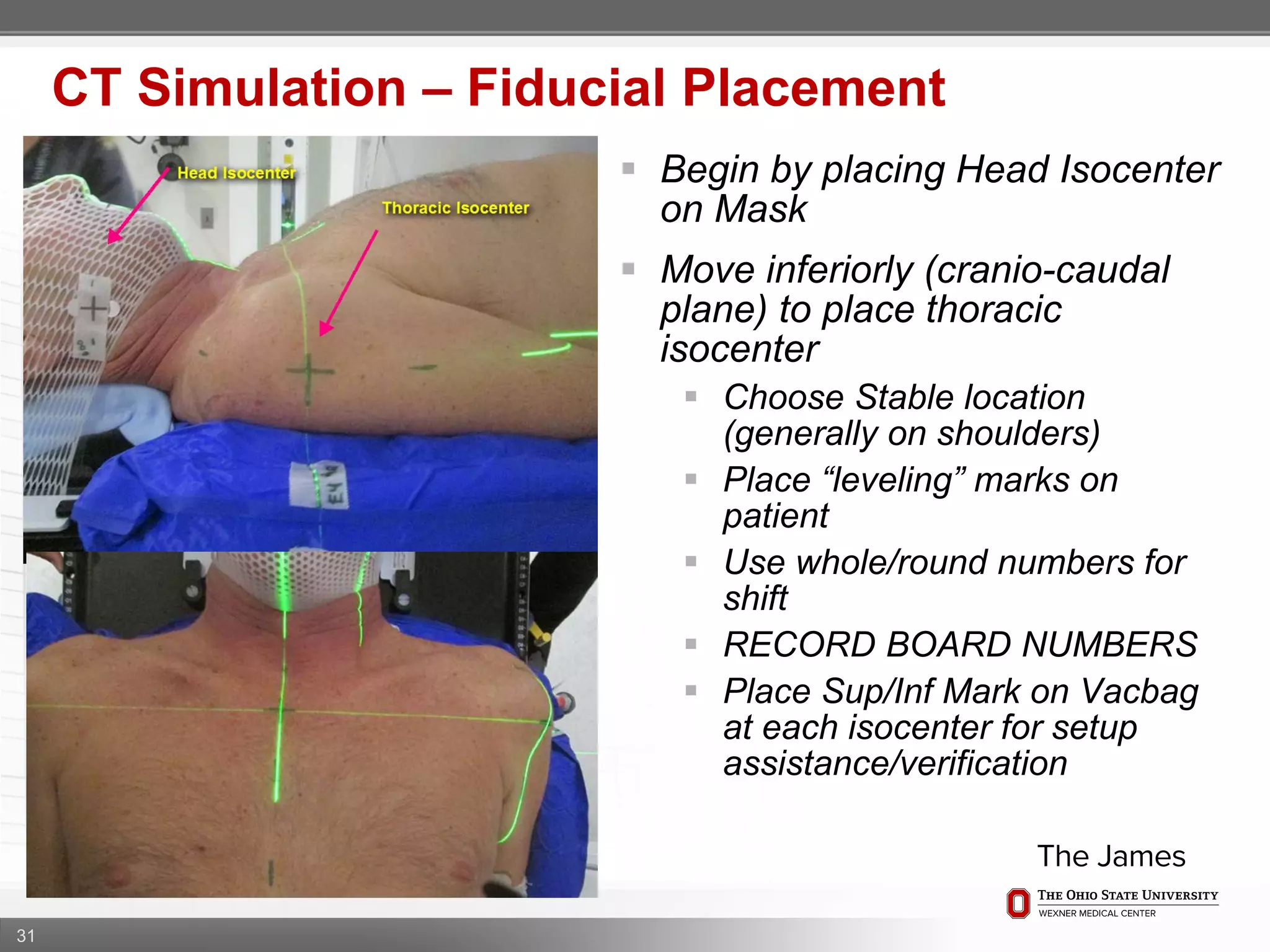
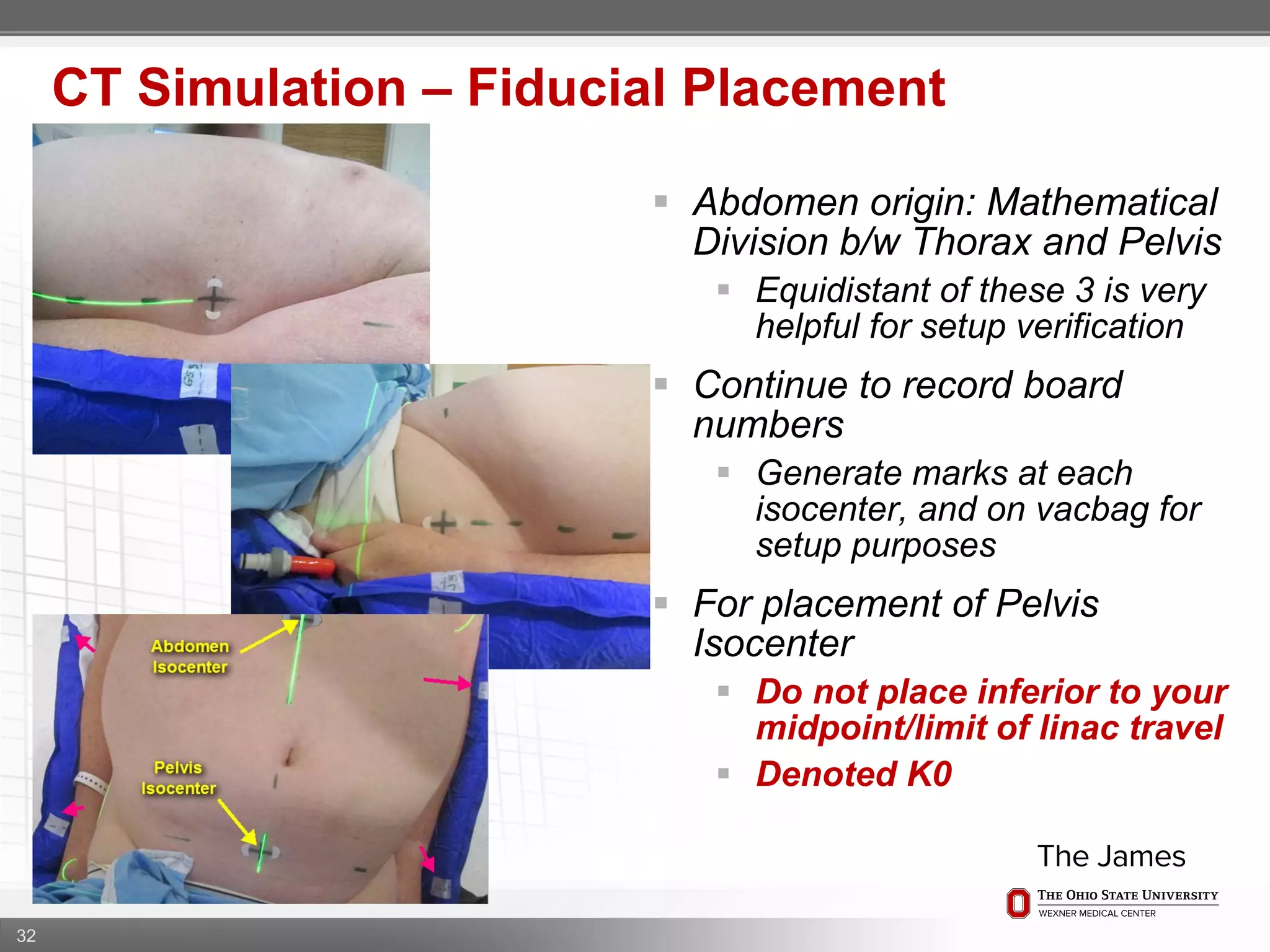
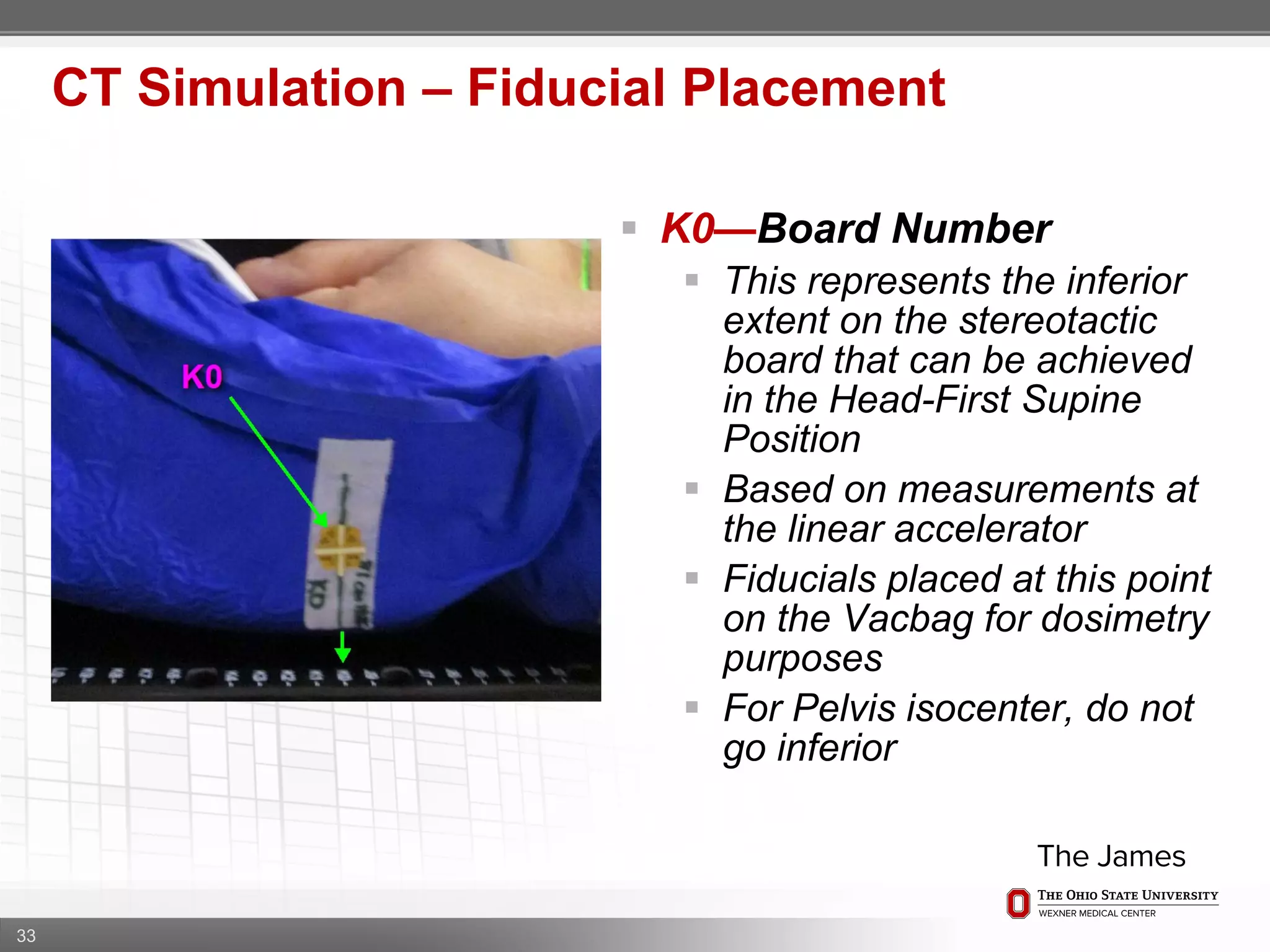
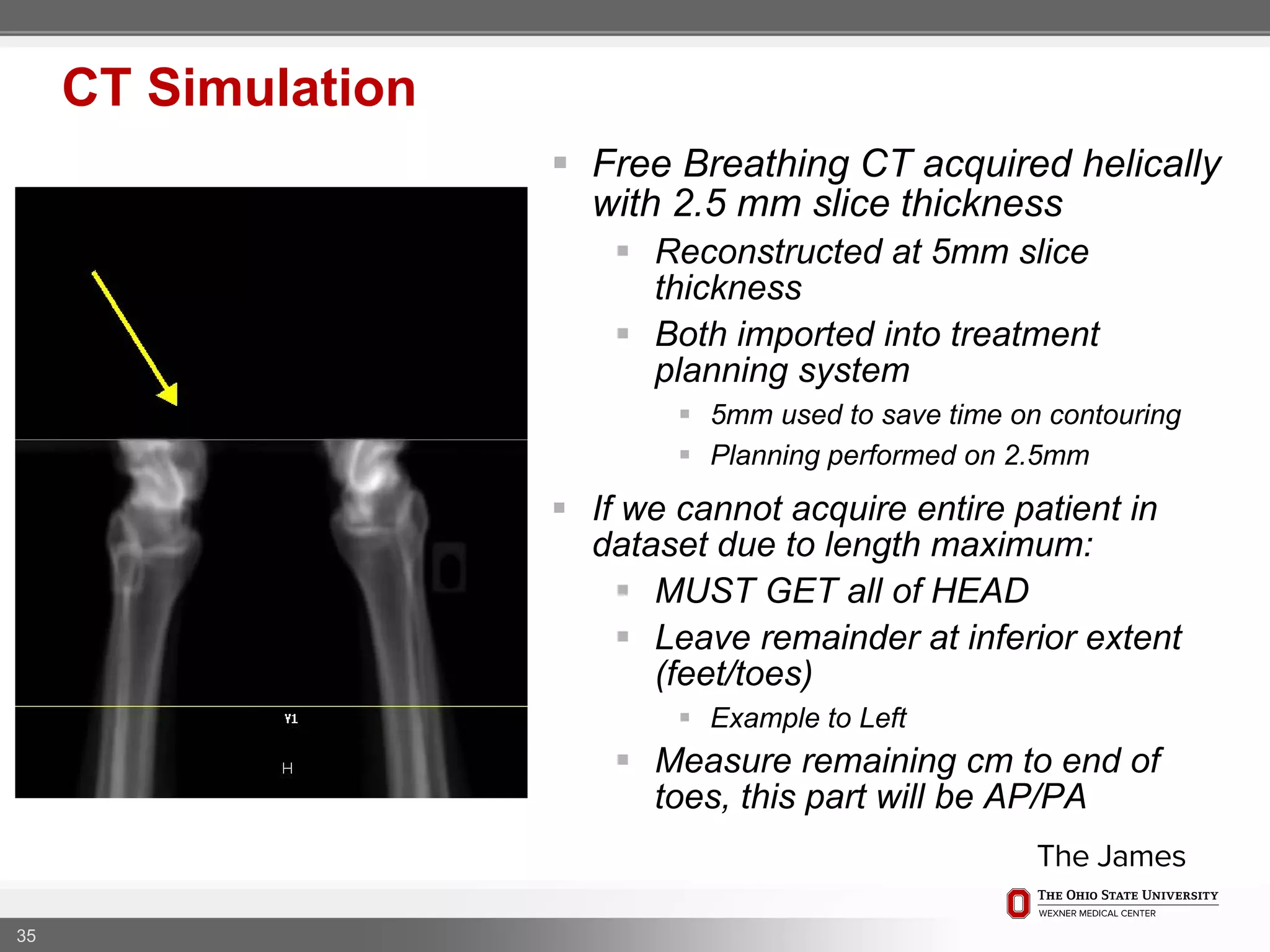
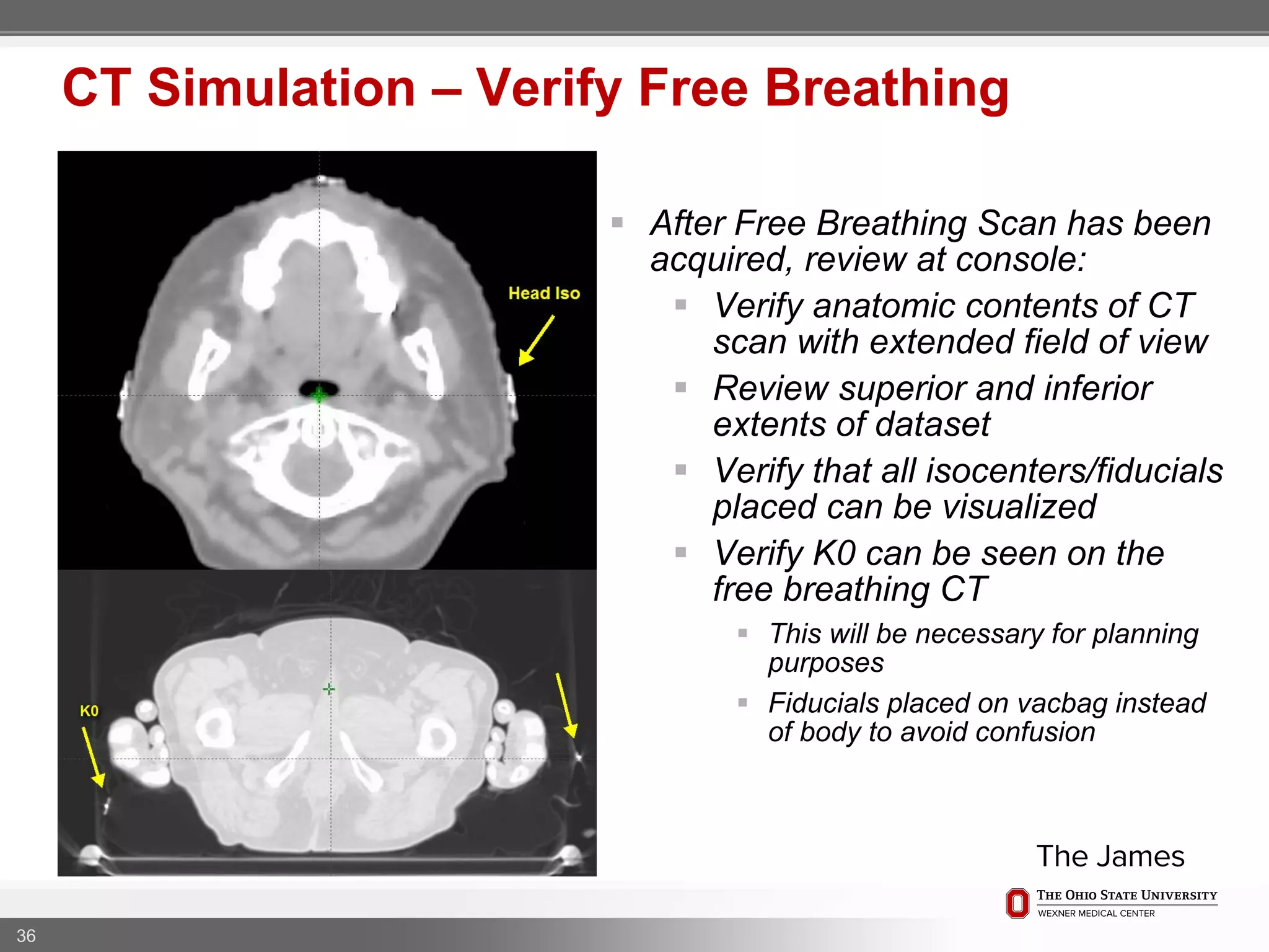
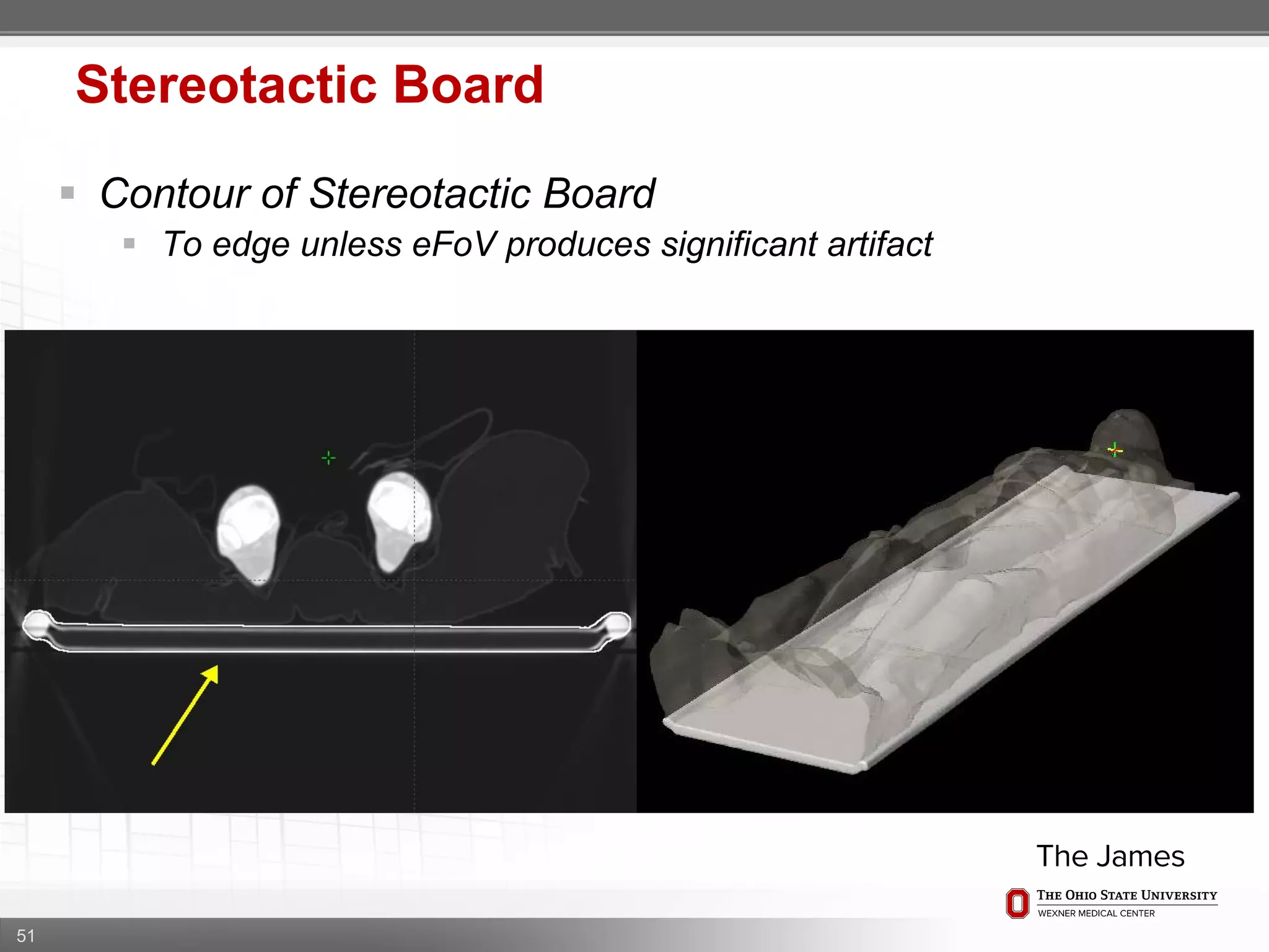
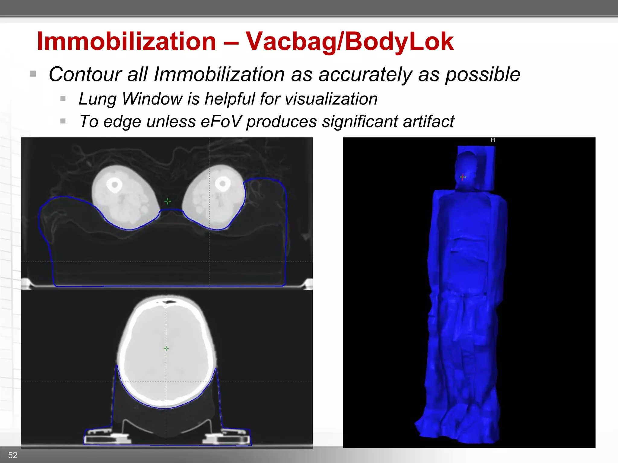
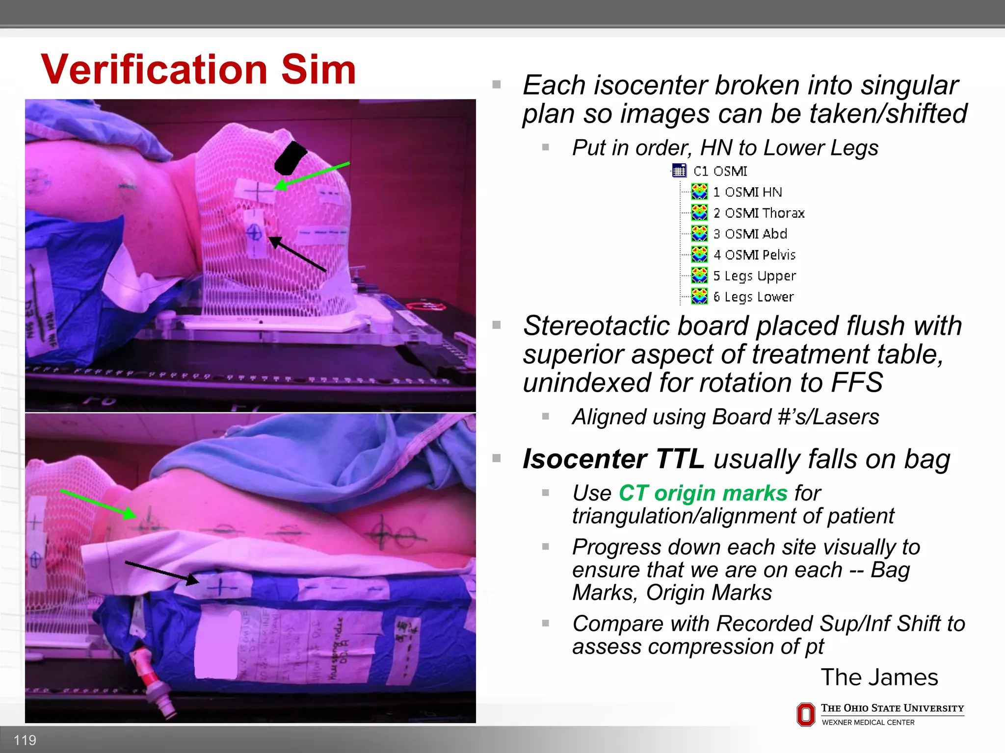
1. CT simulation involves immobilizing the patient in a vacbag on a stereotactic board with a custom headrest and mask for head and neck immobilization. Fiducial markers are placed at the head, thorax, abdomen, and pelvis to aid in daily setup verification.
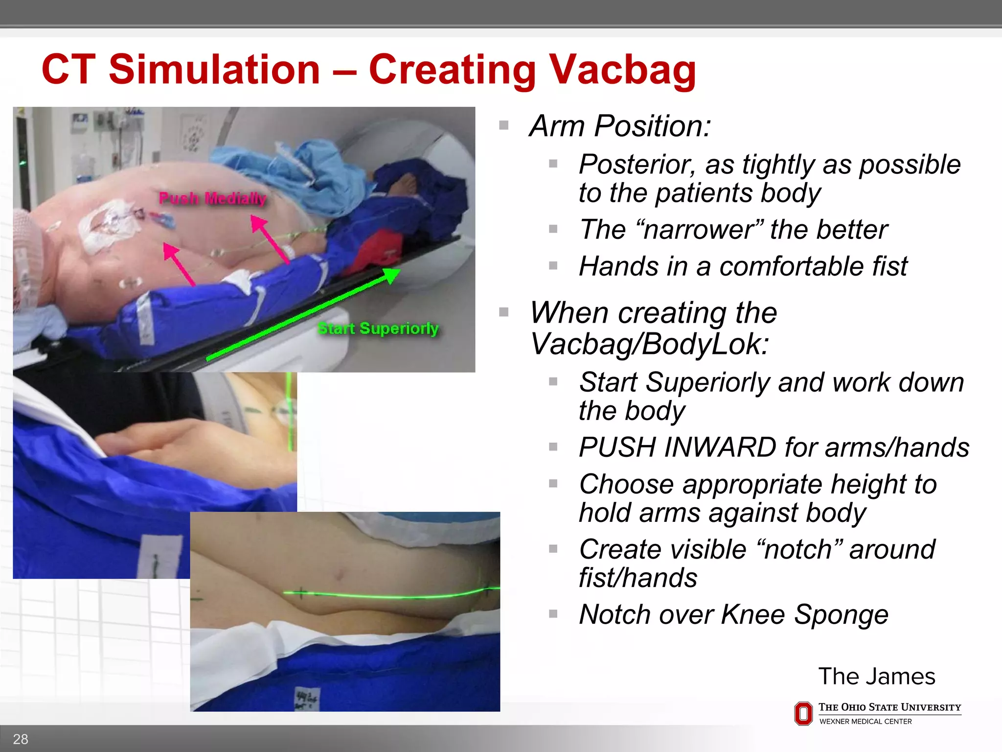
2. Care is taken to push the arms tightly against the body when creating the vacbag and to index all immobilization devices to the stereotactic board for reproducibility. Fiducials are placed at standardized coronal planes and levels are marked on the vacbag to guide setup.
3. The goal is to immobilize the patient and place reproducible fiducial markers to allow for precise targeting of the marrow and sparing
This document discusses the role of radiotherapy in treating acute leukemia in the central nervous system. It notes that intrathecal drugs do not evenly distribute throughout the brain and spinal cord, so cranial radiotherapy is used along with intrathecal chemotherapy to more fully treat the entire central nervous system. Studies from the 1960s showed that cranial radiotherapy reduced CNS relapse rates in pediatric ALL patients from 65% to 4%. More recent trials have aimed to reduce radiation doses to decrease side effects, focusing cranial radiotherapy only on high-risk patients. Doses as low as 12Gy have proven effective in some studies while maintaining low CNS relapse rates below 5%. Therapeutic radiotherapy is also discussed for treating isolated CNS
La IMRT es una modalidad avanzada de radioterapia que permite enfocar dosis más altas de radiación en el tumor minimizando la dosis en los tejidos normales adyacentes. Usa aceleradores lineales de rayos X y múltiples campos de intensidad modulada para producir una dosis individualizada con menos efectos secundarios. Se usa comúnmente para tratar cánceres de próstata, cabeza y cuello, y del sistema nervioso central.
This document discusses SSD calculations for radiation therapy treatment planning. It provides examples of calculating monitor units (MUs) needed to deliver a prescribed dose to different depths using various field sizes and source-to-surface distances (SSDs), accounting for factors like percent depth dose (PDD), inverse square, scatter, and blocking. It also gives an example of calculating the dose to the depth of maximum dose (dmax) given a prescribed dose to another depth using the ratio of PDD values.
Assessment of Intrafraction Motion during real-time tracking in the treatment...Subrata Roy
In our experience, intrafraction motion for intracranial targets treated with fiducial free, frameless cranial SRS / SRT on CyberKnife with 6-D skull tracking, is within the acceptable range, and can be reliably detected and corrected. A PTV margin of 1 mm appears adequate to account for most of the intrafraction motion in this situation. However, significant intrafraction motion occurs during treatment delivery when mask based immobilization is used, and hence the same should be accounted for, in situations where intrafraction imaging is not being practiced. Owing to the highest level of precision, excellent automation, ease of treatment planning and delivery and avoidance of anaesthesia, CyberKnife Stereotactic Radiosurgery / Radiotherapy is highly recommended as an alternative to complex cranial neurosurgical procedures.
Adaptive Radiotherapy at GenesisCare UKGenesisCareUK
Adaptive radiotherapy is a process that modifies the treatment plan based on systematic monitoring of treatment variations to improve radiation treatment. It customizes the treatment dose and field margins for each individual patient based on changes in target and organ-at-risk volumes and target position between and during fractions to safely escalate the dose. Current projects at GenesisCare UK are exploring using daily onboard imaging with plan of the day options and offline replanning to adapt for interfractional changes and intrafractional guidance with online compensation.
This document discusses reirradiation in recurrent head and neck cancer. It notes that radiation therapy plays a central role in head and neck cancer treatment but recurrence still occurs in 20-35% of patients. Reirradiation presents challenges due to prior radiation exposure and damage to normal tissues. The document discusses treatment options, appropriate patient selection, techniques like IMRT to minimize dose to organs at risk, optimal timing and dosing of reirradiation, and management of toxicities.
The document summarizes key concepts from ICRU reports 29, 50, and 62 regarding dose specification for external beam radiation therapy. It defines volumes of interest like the gross tumor volume, clinical target volume, planning target volume, and treated volume. It also describes dose reporting guidelines and distinguishes between radical and palliative treatment intents. ICRU 50 introduced standardized terminology for prescribing, recording, and reporting radiation therapy. ICRU 62 provided more detailed recommendations on treatment margins and introduced concepts like the internal target volume and conformity index.
WHY STEREOTATXY IN CRANIAL AVM / DR KANHU CHARAN PATROKanhu Charan
This document discusses stereotactic radiosurgery (SRS) for the treatment of cerebral arteriovenous malformations (AVMs). It begins by explaining what an AVM is and the risks they pose if untreated, such as bleeding in the brain. It then covers treatment options for AVMs and why SRS is often preferred for certain cases, such as when the AVM is in an eloquent or deep brain area. The document provides details on patient selection, imaging and planning for SRS, anticipated outcomes, and risks of treatment complications. It emphasizes the importance of multidisciplinary discussion and informed consent when determining if SRS is appropriate for a patient's individual AVM.
This document outlines the EMBRACE II study protocol for evaluating image-guided intensity modulated external beam radiochemotherapy and MRI-based adaptive brachytherapy in locally advanced cervical cancer. The study aims to improve local control and reduce treatment-related morbidity through dose escalation to targets and dose reduction to organs at risk using advanced radiotherapy techniques including IMRT, IGRT, adaptive brachytherapy based on weekly MRI, and chemotherapy. Primary endpoints include local control and late morbidity. Secondary endpoints include disease-free survival, overall survival, patterns of failure, and quality of life. The study involves multiple centers and aims to accrue 500 patients over 5 years to validate hypotheses regarding dose-volume relationships and identify prognostic markers
This document provides an overview of the approach to prostate SBRT planning. It discusses the evidence supporting SBRT, patient selection, immobilization techniques, imaging protocols, target delineation guidelines, dose selection, planning constraints, quality assurance procedures, and peri-treatment management. The key advantages of SBRT for prostate cancer are the short treatment time of 5 fractions, high biological effective dose achieved, and comparable oncologic outcomes to other EBRT techniques with side effects that are earlier but also resolve sooner. Careful planning and quality assurance throughout the process are emphasized.
The document discusses various considerations for magna field irradiation (TBI) including:
1) Biological factors like repair, repopulation and fractionation that are important for TBI. Fractionation increases lung tolerance up to 175%.
2) Physical factors that must be addressed like dose calibration, scatter corrections, and ensuring dose uniformity throughout the body.
3) Methods for calculating and prescribing dose including using average or single point doses. Most protocols prescribe to the umbilicus midpoint.
4) Techniques used to achieve homogeneity like bolus, compensators and beam energies. Homogeneity of ±10% is typically achieved.
5) Determining lung dose accurately through methods like CT-based calculations or
The document discusses total body irradiation (TBI), which involves delivering radiation to the entire body to condition patients for stem cell transplantation. It provides an overview of the history, concept, indications, doses, prerequisites, and treatment planning for TBI. Complications of TBI are also reviewed, including both immediate toxicities like nausea and vomiting as well as late effects such as salivary gland dysfunction and pneumonitis.
This document outlines protocols for stereotactic radiosurgery and stereotactic body radiation therapy for various anatomical sites including brain, skull base, head and neck, spine, lung, liver, pancreas, and prostate. It lists clinical trials, publications, and organizations related to stereotactic procedures for conditions such as metastases, benign and malignant tumors, and functional disorders. Key protocols mentioned include those from RTOG, Alliance, and studies on stereotactic radiosurgery versus whole brain radiation for brain metastases.
Socialización informe No. 218 de la Asociación Americana de Físicos en Medicina (AAPM): "Límites de tolerancia y metodologías para la verificación basada en la medición de Control de calidad en IMRT"
Evaluation of radiotherapy treatment planningAmin Amin
This document discusses the evaluation of radiotherapy treatment planning through the use of various tools and indices. The goals of treatment planning are to ensure the prescription dose adequately covers and conforms to the target volume while minimizing doses to surrounding healthy tissues. Key evaluation tools discussed include isodose distributions, orthogonal planes, dose volume histograms, dose statistics, homogeneity indices, and conformity/coverage indices. These tools provide both qualitative and quantitative assessments of the dose distribution and how well it meets the goals of treatment planning.
1) The document discusses perspectives on image-guided radiation therapy (IGRT) from physicians and physicists at the University of California San Diego (UCSD).
2) UCSD has implemented IGRT using both planar kV imaging and volumetric cone-beam CT (CBCT) primarily for prostate and gynecological cancers.
3) IGRT provides benefits of improved target localization and the potential for treatment adaptation if tumor size changes over the course of treatment are detected. However, proper quality assurance is needed when implementing an IGRT program.
El documento resume los informes de la Comisión Internacional de Unidades y Medidas de Radiación (ICRU) sobre la prescripción, registro y elaboración de informes en la terapia con haces de fotones. Define los volúmenes blanco como el volumen tumoral macroscópico, volumen blanco clínico, volumen blanco de planificación y volúmenes de órganos de riesgo. Describe los puntos de referencia y dosis para la terapia intracavitaria en ginecología según el informe ICRU 38.
1) SBRT for spinal metastases requires careful patient selection and treatment planning to balance pain/local control and toxicity risks. Fractionation and dose constraints must consider organs at risk like the spinal cord.
2) Common toxicities include vertebral compression fractures, pain flares, radiation myelopathy, and myositis. Mitigation strategies include pre-treatment stabilization, fractionation, steroids, and immobilization.
3) Dose constraints from trials not reporting toxicity may be too high. Partial spinal cord volumes, cauda equina differences, and serial organ considerations like the esophagus require attention.
ROLE OF RADIATION IN BRAIN TUMORS FOR NEUROSURGEONSKanhu Charan
The document discusses neuro-oncology and the role of radiotherapy. It provides statistics on cancer incidence and mortality worldwide. It then covers topics such as the neuro-oncology team, brain tumor types and characteristics, risk factors, clinical presentation, diagnostic techniques including imaging and histopathology, tumor grading, and the roles of surgery, chemotherapy, and radiotherapy in treatment.
This document describes the case of a 52-year-old male who presented with vomiting and was found to have a pituitary adenoma. An MRI showed a 2.3x1.6 cm dumbbell shaped lesion in the sella turcica extending suprasellarly and compressing the optic chiasm. The patient underwent endoscopic transphenoidal resection, with near total excision. Post-op MRI showed residual tissue in the right and left sides of the sella. The patient was planned for stereotactic radiotherapy with 25Gy in 5 fractions to treat the residual tumor. Target and organ at risk volumes were delineated on planning MRI and CT scans. Treatment planning was performed to optimize dose distribution and minimize
This document discusses Image Guided Radiation Therapy (IGRT). It begins by explaining that radiotherapy has traditionally used imaging for treatment planning and execution when the target is not on the surface. It then describes various IGRT technologies, dividing them into non-radiation based systems like ultrasound, cameras, electromagnetic tracking and MRI; and radiation based systems like EPID, CBCT, fan beam KVCT and MVCT. These systems provide improved target localization and allow for corrections. IGRT aims to reduce errors and improve precision of radiotherapy.
This document discusses recursive partitioning analysis (RPA), a statistical tool used to identify significant prognostic factors and classify patients into groups with similar outcomes. RPA has been used to analyze data from clinical trials in high-grade glioma (HGG) and brain metastasis patients. Key factors like age, performance status, extent of surgery, and molecular alterations help divide patients into prognostic classes. More recent studies have refined RPA models by incorporating additional molecular data to better stratify patients and guide treatment decisions.
This document discusses the use of stereotactic body radiation therapy (SBRT) for liver tumors. It provides details on common liver tumors including hepatocellular carcinoma and metastases. It describes SBRT as a treatment option for inoperable early stage tumors, as a bridge to transplant, and for intermediate or locally advanced stages. Key factors for patient selection and treatment planning such as tumor size, number and location, as well as liver function are summarized. The document also briefly discusses proton beam therapy and current clinical trials investigating SBRT for liver cancer.
Accelerated partial breast irradiation (APBI) delivers radiation to only the portion of the breast at highest risk of recurrence rather than the whole breast. This allows radiation to be delivered in a significantly shortened period. Several techniques for APBI exist including brachytherapy using catheters implanted in the breast, balloon brachytherapy, and external beam radiotherapy. Ongoing clinical trials are evaluating outcomes and toxicities of APBI compared to whole breast irradiation in appropriately selected patients with early-stage breast cancer.
Total body irradiation (TBI) is a form of radiotherapy used prior to bone marrow transplants to reduce the risk of transplant rejection and destroy any remaining cancer cells. TBI techniques use large photon fields, usually from cobalt-60 machines or LINACs, to irradiate the entire body. Common techniques include opposing anterior-posterior beams or lateral beams. Precise dosimetry is required due to the large fields and total body exposure, with dose uniformity targets of within ±10% across the body. In vivo dosimetry using TLD or diodes is also employed to verify accurate dose delivery. Early side effects from TBI include fatigue, nausea, hair loss and skin irritation due to the whole body irradiation
EBCTCG METAANALYSIS
INDICATION OF POST OP RADIOTHERAPY
Immobilization devices
Conventional planning
Alignment of the Tangential Beam with the Chest Wall Contour
Doses To Heart & Lung By Tangential Fields
CT simulators are essential for precise radiotherapy treatment planning. They use CT scanning to create a virtual 3D representation of the patient's anatomy. This allows clinicians to accurately localize tumors and delineate organs at risk. The CT images provide excellent soft tissue contrast and electron density data needed for treatment planning. CT simulators also facilitate patient positioning and immobilization during both simulation and treatment through reference marking and immobilization devices. Overall, CT simulation enables more accurate target localization and definition compared to conventional simulators, resulting in better optimized treatment plans.
Proper patient immobilization is crucial for accurate radiation therapy. Immobilization devices reduce setup errors and patient movement, allowing higher controlled doses to be delivered precisely to the target volume while sparing surrounding healthy tissues. Common immobilization methods include masks, molds, frames and boards for different body sites like head/neck, brain, thorax, breast and pelvis. Newer techniques like intensity-modulated radiation therapy allow improved immobilization for smaller treatment volumes and higher cure rates.
The vmat vs other recent radiotherapy techniquesM'dee Phechudi
VMAT is a new type of intensity-modulated radiation therapy (IMRT) treatment technique that uses the same hardware (i.e. a digital linear accelerator) as used for IMRT or conformal treatment, but delivers the radiotherapy treatment using a rotational or arc geometry rather than several static beams.
This technique uses continuous modulation (i.e. moving the collimator leaves) of the multileaf collimator (MLC) fields, continuous change of the fluence rate (the intensity of the X rays) and gantry rotation speed across a single or multiple 360 degree rotations
VMAT (Volumetric Modulated Arc Therapy) was first proposed in 1995 as a form of rotational IMRT. It involves delivering radiation therapy with the linear accelerator gantry rotating continuously around the patient. Intensity modulation is achieved through continuous movement of the MLC leaves as they shape the beam during rotation. VMAT can produce dose distributions similar to IMRT but with fewer monitor units and faster treatment times. Recent advances in treatment planning and linear accelerator control systems now allow for effective clinical implementation of VMAT with either single or multiple full arcs.
A summary of recent innovations in radiation oncology focussing on the priniciples of different techniques and their application. An overview of clinical results has also been given
The document discusses using a Tomotherapy unit to perform total body irradiation (TTBI) treatments. It finds TTBI with Tomotherapy is a feasible technique that offers advantages over conventional TBI, but also has some limitations. Specifically, TTBI treatments can cover the target volume well while sparing organs at risk, but planning and delivery times are long and further dosimetric validation is needed before clinical use.
This document discusses adaptive radiation therapy and the need to account for patient anatomical and biological changes over the course of treatment. It proposes using an anthropomorphic phantom simulated with LEGO Mindstorms to model organ motion from breathing and correlate internal motion with external surrogates. Preliminary results show the prototype target motion simulation can reproduce 4D motion from 4DCT scans. The LEGO approach allows experimental investigation of quality assurance and biomechanical analysis for adaptive radiation therapy in lung cancer patients.
The document discusses the capabilities of tomotherapy for radiation therapy treatment planning and delivery. It provides examples of various clinical cases that were planned and treated using tomotherapy, including mesothelioma, pancreatic cancer, head and neck cancer, and re-irradiation cases. It also discusses potential advantages of tomotherapy such as reduced margins and improved organ-at-risk sparing compared to other radiation therapy techniques.
Preoperative 3D imaging resources and intraoperative fusion are essential for complex endovascular aneurysm repair (EVAR) procedures. An integrated workflow was developed that utilizes preoperative CT angiography for sizing and planning, allows the export of planning details to the C-arm, and provides intraoperative fusion to 2D fluoroscopy for guidance. This approach simplifies the procedure, improves accuracy, and significantly reduces radiation dose and contrast use compared to traditional mobile C-arm techniques without fusion. Postoperative assessment can be performed with completion 3D angiography and contrast-enhanced ultrasound, further reducing contrast exposure and costs.
Total body irradiation (TBI) is used prior to bone marrow transplantation to eradicate diseased marrow, reduce tumor burden, and induce immunosuppression. Early regimens used single, large fractions but this caused high rates of pneumonitis. More recent fractionated regimens at lower dose rates have improved outcomes with reduced toxicity. Current myeloablative regimens use 12 Gy over 3 days while reduced-intensity regimens use 2 Gy. TBI is associated with acute toxicities and late effects including cataracts, infertility and increased risk of secondary cancers. New techniques aim to better shield organs to allow dose escalation.
This study compared outcomes of 61 gastric cancer patients treated with postoperative chemoradiotherapy using either intensity-modulated radiotherapy (IMRT) or 3-dimensional conformal radiotherapy (3D CRT). The 2-year overall survival rates were 51% for 3D CRT and 65% for IMRT, with no significant difference. Locoregional failure occurred in 15% of 3D CRT patients and 13% of IMRT patients. Both the 2-year disease-free survival and local control rates were similar between the two groups. Overall, the study found no significant differences in outcomes between IMRT and 3D CRT for adjuvant therapy of gastric cancer.
The document discusses the three years of experience with tomotherapy treatment at the Greater Poland Cancer Centre. It describes how tomotherapy allows for a wide spectrum of clinical cases to be treated and provides two specific examples, one involving treatment of medulloblastoma with craniospinal irradiation and the other involving treatment of vulva cancer. The high level of dose conformity allowed by tomotherapy helped in both cases to deliver sufficient dose to target areas while reducing dose to nearby critical organs.
This document summarizes key points about radiotherapy options for early stage prostate cancer treatment. It discusses dose escalation studies showing improved outcomes with higher radiation doses. New techniques like IMRT and IGRT have improved precision and reduced side effects. Ongoing studies are further exploring dose escalation and hypofractionated regimens, as well as combining radiotherapy with hormone therapy or novel techniques to reduce rectal toxicity.
Secondary Cancers, Health Behaviour and Cancer Screening Adherence in survivo...Cancer Institute NSW
Over 50% of patients undergoing allogeneic BMT can now be expected to become long-term survivors. Unfortunately many survivors experience an increased risk of secondary cancers, infections and chronic diseases.
Tomotherapy combines intensity modulated radiation therapy with computed tomography scanning to treat cancer. It can treat multiple tumors simultaneously with daily 3D imaging to improve accuracy and minimize side effects. Tomotherapy uses a sophisticated multi-leaf collimator and 360 degree delivery of many narrow beamlets to provide conformal, even radiation doses to tumors while avoiding healthy tissue for shorter treatments and fewer side effects than other radiation therapies. It can treat many cancer types, including mesothelioma.
Dokumen tersebut merupakan buku panduan tentang tata cara pendirian Baitul Maal wa Tamwil (BMT) yang ditulis oleh Prof. Dr. Ir. M. Amin Aziz dan diterbitkan oleh Pusat Komunikasi Ekonomi Syariah. Buku ini berisi pengantar dari direktur eksekutif PKES, sambutan dari ketua ABSINDO dan MES, serta contoh-contoh kisah sukses beberapa BMT di Indonesia.
Presentation by Scott Oliver, MD. Presented at the 2018 Eyes on a Cure: Patient & Caregiver Symposium, hosted by the Melanoma Research Foundation's CURE OM initiative.
This study prospectively evaluated 54 patients with ovarian sex cord-stromal tumors (OSCST) to develop diagnostic standards and risk-adapted treatment strategies. Most patients presented with stage IA tumors confined to the ovary and were followed regularly, while some with stage IC or higher received cisplatin-based chemotherapy. After a median follow-up of 59 months, event-free survival was 86% and overall survival was 89%. Prognosis correlated with stage, and chemotherapy seemed effective for advanced-stage tumors. The study provides a standardized approach to classify and treat OSCST.
A brief information regarding Acute lymphoblastic leukemia. It is very basic information about acute lymphoblastic leukemia, I strongly recommend other sources as well for further investigations.
Thanks
This document discusses updates to the WHO classification of meningiomas and management strategies based on grade.
The key points are:
1. The WHO 5th edition emphasizes applying criteria for atypical (grade 2) and anaplastic (grade 3) regardless of subtype. Chordoid and clear cell subtypes are now grade 2 due to higher recurrence.
2. Molecular biomarkers like SMARCE1, BAP1, KLF4/TRAF7 mutations are associated with classification and grading. TERT promoter mutations and CDKN2A/B deletions qualify a meningioma as grade 3.
3. Management depends on grade and extent of resection. Grade 1 tumors may
Uveal melanoma commonly spreads to the liver. This document discusses uveal melanoma (MUM) that has metastasized to the liver. It provides background on MUM, noting that half of patients develop metastases, usually first appearing in the liver. It describes genetic risk factors for metastasis and different risk classifications. The document advocates for locoregional therapies for liver metastases since there are no effective systemic therapies. It presents evidence that liver-directed therapies may prolong survival more than systemic treatments or surveillance alone.
This document provides guidance for physicians on appropriate use of medical imaging for common clinical
presentations. It discusses when CT, MRI, ultrasound or other modalities are most useful for evaluating abdominal
pain, chest pain, headache and other issues. The goal is to promote efficient and radiation-limiting use of imaging
to aid accurate diagnosis while avoiding unnecessary exposure or costs. Imaging choice depends on clinical
findings, but CT and MRI may help identify conditions like appendicitis, aneurysms, infections or tumors causing
abdominal, chest or neurological symptoms.
This study compared outcomes of steroid withdrawal versus continued steroid therapy in kidney and heart transplant recipients. Over 1500 patients were enrolled from 1994-2002 and followed for 5-6 years on average. Results showed that steroid withdrawal led to superior patient and graft survival rates compared to controls continuing steroids. Steroid withdrawal also reduced risks of complications like osteoporosis and cataracts. However, it was associated with a small increased risk of acute rejection episodes. Overall, the study suggests that steroid withdrawal after 6 months is safe and effective for transplant recipients.
This document provides an overview of plasma cell disorders including monoclonal gammopathy of undetermined significance (MGUS), smoldering myeloma (SMM), symptomatic multiple myeloma, solitary plasmacytoma, Waldenstrom's macroglobulinemia, and primary light chain amyloidosis. It discusses diagnostic criteria and risk factors for progression. Treatment options for active myeloma are outlined including novel agents like thalidomide, bortezomib, and lenalidomide used in various combinations. High-dose chemotherapy with autologous stem cell transplantation improves outcomes.
This document summarizes recent advances in understanding and treating primary brain tumors in adults. It focuses on gliomas and primary CNS lymphomas, which are the most common types. For gliomas, important progress includes improved imaging techniques for diagnosis and assessing tumor grade. Large clinical trials have established standard treatments and confirmed the prognostic value of specific molecular alterations. Genome-wide studies have improved understanding of tumor biology, which could lead to better classification and targeted therapies. For primary CNS lymphomas, high-dose methotrexate regimens increase survival but standards of care and the role of whole-brain radiation remain unclear and depend on patient age. Current focus is on new polychemotherapy regimens to reduce or delay whole-
The document outlines updates made in Version 3.2023 of the NCCN Guidelines for Multiple Myeloma from the previous version. Key updates include:
- Adding teclistamab-cqyv and noting belantamab mafodotin-blmf as useful in certain circumstances for patients with late relapses.
- Extensive revisions to MYEL-5 regarding response after primary therapy and follow up.
- Incorporating content from MYEL-6 into other pages regarding additional treatment for relapse or progressive disease.
- Adding a table of factors considered high risk for multiple myeloma.
Follow-Up Strategies in Focal Liver Lesions And Treatment Methodsdaranisaha
Today, advances in cross-sectional imaging have led to the detection and early recognition of incidental/focal liver lesions (FCL). In approximately 17,000 cases of chest CT, incidental liver lesions were found in 6% [1]. In general, FCL consists of hepatocytes, biliary epithelium, mesenchymal tissue, connective tissue, or metastasized cells from distant sites. Most incidental lesions are benign, some may require careful management and treatment.
Follow-Up Strategies in Focal Liver Lesions And Treatment MethodsJohnJulie1
Today, advances in cross-sectional imaging have led to the detection and early recognition of incidental/focal liver lesions (FCL). In approximately 17,000 cases of chest CT, incidental liver lesions were found in 6% [1]. In general, FCL consists of hepatocytes, biliary epithelium, mesenchymal tissue, connective tissue, or metastasized cells from distant sites. Most incidental lesions are benign, some may require careful management and treatment.
Follow-Up Strategies in Focal Liver Lesions And Treatment MethodsEditorSara
Today, advances in cross-sectional imaging have led to the detection and early recognition of incidental/focal liver lesions (FCL). In approximately 17,000 cases of chest CT, incidental liver lesions were found in 6% [1]. In general, FCL consists of hepatocytes, biliary epithelium, mesenchymal tissue, connective tissue, or metastasized cells from distant sites. Most incidental lesions are benign, some may require careful management and treatment.
Follow-Up Strategies in Focal Liver Lesions And Treatment Methodssemualkaira
Today, advances in cross-sectional imaging have led to the detection and early recognition of incidental/focal liver lesions (FCL). In approximately 17,000 cases of chest CT, incidental liver lesions were found in 6% [1]. In general, FCL consists of hepatocytes, biliary epithelium, mesenchymal tissue, connective tissue, or metastasized cells from distant sites. Most incidental lesions are benign, some may require careful management and treatment
Follow-Up Strategies in Focal Liver Lesions And Treatment Methodssemualkaira
Today, advances in cross-sectional imaging have led to the detection and early recognition of incidental/focal liver lesions (FCL). In approximately 17,000 cases of chest CT, incidental liver lesions were found in 6% [1]. In general, FCL consists of hepatocytes, biliary epithelium, mesenchymal tissue, connective tissue, or metastasized cells from distant sites. Most incidental lesions are benign, some may require careful management and treatment.
Follow-Up Strategies in Focal Liver Lesions And Treatment MethodsNainaAnon
Today, advances in cross-sectional imaging have led to the detection and early recognition of incidental/focal liver lesions (FCL). In approximately 17,000 cases of chest CT, incidental liver lesions were found in 6% [1]. In general, FCL consists of hepatocytes, biliary epithelium, mesenchymal tissue, connective tissue, or metastasized cells from distant sites. Most incidental lesions are benign, some may require careful management and treatment.
Follow-Up Strategies in Focal Liver Lesions And Treatment MethodsEditorSara
Today, advances in cross-sectional imaging have led to the detection and early recognition of incidental/focal liver lesions (FCL). In approximately 17,000 cases of chest CT, incidental liver lesions were found in 6% [1]. In general, FCL consists of hepatocytes, biliary epithelium, mesenchymal tissue, connective tissue, or metastasized cells from distant sites. Most incidental lesions are benign, some may require careful management and treatment.
Follow-Up Strategies in Focal Liver Lesions And Treatment Methodssemualkaira
Today, advances in cross-sectional imaging have led to the detection and early recognition of incidental/focal liver lesions (FCL).
In approximately 17,000 cases of chest CT, incidental liver lesions
were found in 6% [1]. In general, FCL consists of hepatocytes, biliary epithelium, mesenchymal tissue, connective tissue, or metastasized cells from distant sites. Most incidental lesions are benign,
some may require careful management and treatment. In evaluating the lesion, the patient's clinical history, underlying disease and
age factor should be considered. FCL can be detected at a rate of
10-30% in normal healthy and chronic liver disease patients, and
even in oncology patients with malignancy, FCLs can be highly
benign
Follow-Up Strategies in Focal Liver Lesions And Treatment Methods
OMSI
1. 1
Organ Sparing Marrow-Targeted Irradiation (OSMI) as an
Alternative to Traditional Total Body Irradiation (TBI)
Methods:
VMAT Treatment Planning and Clinical Implementation
Wesley Zoller, CMD
6/15/16
2. I have been a medical dosimetrist at the
James Cancer Hospital since August 2013
Graduate of Cleveland Clinic Medical Dosimetry
Program
The “New” James began treating patients
in December of 2014
Over 100,000 square feet of space
2nd floor of hospital (true 2nd floor shielding)
7 VarianTM TrueBeamsTM at main campus
2 more at our satellite, the Stefanie Spielman
Comprehensive Breast Center
PET/CT, CT, MRI in our department at campus
We treat 200-250 patients daily
60-70% VMAT/IMRT
Wide variety of disease sites
Pediatrics, HN, Thoracic, Blood-Based,
CNS, GYN, GU, Skin, Breast, GI,
Sarcomas, Stereotactic and Palliative Care
2
The James Cancer Hospital
3. I have no conflict of interest with any of the
vendors/software/equipment used for this
presentation
This presentation is not a marketing/sales
presentation regarding specific
products/software
Any software/trade names mentioned in the
presentation are resultants of accurate reporting
of methodology
All patient plans/setups in this study have been
performed at the James Cancer Hospital in
accordance with OSU protocol 13219
3
Disclosures
4. Meng Welliver, MD, PhD, Assistant Professor,
Radiation Oncology
Principal Investigator OSU 13219
Lead Attending Physician – Hematologic Cancer
Michael Weldon, MS, Medical Physicist,
Radiation Oncology
Mark Addington, CMD, Medical Dosimetrist,
Radiation Oncology
Our wonderful therapists in simulation and on the
linear accelerator
Our dedicated physics team involved in QA, 2nd
check, and treatment delivery
4
Special Thank You to our OSMI
Team
6. 6
To introduce Organ-Sparing Marrow-Targeted Irradiation
(OSMI) as an alternative to traditional Total Body Irradiation
(TBI) methods from a dosimetric perspective
To reveal VMAT planning tricks/considerations encountered
during the clinical implementation at the Ohio State University
To diagnose setup requirements/considerations associated
with VMAT OSMI treatment delivery
To differentiation dosimetric differences between planning
methods and reveal correlation to the patient experience
Learning Objectives
8. 8
Group of cancers of hematologic cells that
generally arise in bone marrow.
Commonly characterized by proliferation
of abnormal white blood cells.
2nd most common blood cancer.
According to National Cancer Institute:
60,140 New Cases predicted for 2016
24,400 Deaths (2016)
Broken down into 4 most common types:
Acute Myelogenous Leukemia (AML)
Acute Lymphocytic Leukemia (ALL)
Chronic Lymphocytic Leukemia (CLL)
Chronic Myelogenous Leukemia (CML)
Leukemia
1 National Cancer Institute
2 American Cancer Society
9. 9
Leukemia
Acute Myelogenous Leukemia (AML)
Most common leukemia in adults
Quick-onset; Aggressive, Immature myeloblasts
Acute Lymphocytic Leukemia (ALL)
Third most common leukemia in adults
Most common cancer in children (25%)
Quick-onset proliferation of Immature Lymphocytes
Chronic Lymphocytic Leukemia (CLL)
2nd most common adult leukemia, Middle-aged Elderly
Chronic, Slow-Progressing, More Mature Lymphocytes
Chronic Myelogenous Leukemia (CML)
Chronic, Slow-Progression, More Mature Myeloblasts
Median age at diagnosis – 67, Philadelphia
Chromosome (Translocation, 9 and 22)
1 National Cancer Institute
2 American Cancer Society
10. 10
History of Total Body Irradiation (TBI)
Myeloablative ability of radiation has been studied in animals/humans since 19023
Bone Marrow Transplant (BMT) first used as a rescue therapy for acute radiation
exposures in the 1950’s
By 1957-59, attempting to use therapeutic BMT/Stem Cell Transplant with prep chemo
and radiation regimens for treatment of leukemia4
In 1994, evidence found little difference tumor cell kill of fractionated vs single fx
Improved late toxicity and immunosuppressive effects with fractionation – Dr. Cosset5
In 2000, 12 Gy → 2Gy/fx BID (8 hrs apart)
with 6MV or Co-60 unit 6
Lungs blocked to ≤ 10Gy to reduce
pneumonitis
Able to reduce acute/late toxicities (symptom
management)
Publication, Established this regimen for use
for ablative TBI6
11. 11
Traditional Total Body Irradiation (TBI)
For many patients with AML, ALL, and MDS:
Chemo allogeneic stem cell transplant
(HSCT) remains only curative therapy7
HSCT - Hematopoietic Stem Cell
Transplantation
Allogeneic – coming from matching donor
Patients must undergo conditioning prior to
stem cell transplant in order to:
Prevent relapse
Eradicate Residual cancer cells
Ensure Engraftment
New cells grow, make healthy stem cells
Achieve Immunosuppression
12. 12
Traditional TBI Continued
Goal of TBI – Achieve Homogeneous dose
distribution throughout patient’s body7
Extended Distance/Opposed Fields
Potential Pulmonary and GI toxicities
limiting factors for regimen
TBI conditioning regimen associated with
less relapse7
TBI Toxicity profile:
Fatigue
Nausea
Emesis
Parotid tenderness
Pruritus
Pneumonitis
Early onset cataracts
Increased risk for
secondary malignancies
13. 13
Traditional TBI Method at The James
12 Gy in 6 fractions BID, 6 MV
Separation measured for:
Head, Neck, Thorax,
Umbilicus, Upper Arm, Wrist,
Thigh, Ankle
Dose Prescribed mid-plane
Umbilicus (Hand Calculation)
500 cm SSD
Both AP and PA
Patient on customized cart,
laying on side (AP/PA)
Gantry Lateral
Collimator Setting 40 x 40 cm2
Collimator turned to 45
Degrees (pt height)
14. 14
Beam Spoiler can be used
Decrease skin sparing
Generate Electron Contamination
Diodes placed by physics for in-vivo dosimetry
Monitor dose to various areas
Adjustments if necessary
Even dose distribution desirable
Lead Panels placed at 300 cm block screen to
achieve homogeneity
Limit Lung mean less than 10 Gy
Lung Blocks Used (example right)
Lung Blocks/Tray placed at 300cm block
screen
Image verification prior to treatment
Traditional TBI Method at The James
15. 15
Traditional TBI Method at The James
AP Field PA Field
Patient against backstop for stability
500 cm SSD to Umbilicus
Spoiler can be placed front of table
500 cm SSD aligned with back
17. 17
Premise of Organ Sparing Marrow-Targeted
Irradiation (OSMI)
Contributors (Tech): MLC’s/Intensity-
Modulation, Improved Imaging/IGRT,
Improved Setups.7
Contributors (Field/Study): Improved
understanding of true targets for TBI.7
Treat Skeletal Bone (Marrow), Lymphatic Chains,
Testes, CNS, Extramedullary Organs (if applicable)
Limit dose to sensitive organs such as liver, lungs,
bowel, kidneys, lenses, and oral cavity
Potential Benefits of OSMI7:
1. Myeloablative radiation-based conditioning for
older patients or patients who otherwise would not
be eligible due to other co-morbidities
2. Allows for focal dose escalation in high risk
disease areas
3. Patient who can otherwise tolerate full dose TBI,
OSMI potentially offers less acute and late
toxicities, including secondary malignancies
18. 18
History of Organ Sparing Marrow-Targeted
Irradiation (OSMI)
Theoretically, OSMI planning can be achieved
using several methods7:
IMRT, Helical Tomotherapy, LINAC-Based VMAT
Intensity Modulated Total Marrow Irradiation
(IM-TMI):
2006-2012 Hypothetical IMRT planning studies,
assess technique dosimetrically8,9
Helical Tomotherapy
Ideal for long volumes, couch motion w/ treatment
Patient safely treated using Tomo since 2010-1112
Tomotherapy only available in ≈20% of centers
Linac-Based VMAT – OSMI
2011-2012: Hypothetical VMAT planning studies,
assess technique dosimetrically, dose verification in
phantom10-11
Linac-based VMAT OSMI more readily available
OSU 13219
19. 19
OSU 13219
Slides 19-24 reference OSU Protocol 13219, Reference 7
First patient enrolled completed radiation
June 2015
20. 20
Hypothesis:
OSMI can be administered by conventional linear accelerators in a safe, feasible, well-
tolerated manner to patients with high-risk hematologic malignancies who are not eligible
for conventional myeloablative TBI preparative regimens.
Primary Endpoints:
To assess feasibility and tolerability of OSMI based HSCT -- Transplant-Related Mortality
(TRM) at day 30
Assess rate of grade II/III organ toxicity attributable to conditioning occurring within D30.
>10% Day 30 TRM or any grade III organ toxicity related to OSMI or conditioning at
D30 or D100 will be considered unacceptable
Trial will be suspended to perform a careful review and to decide next steps. Day 30 TRM will
be assessed in each cohort separately.
TRM at 10% or less at D30, and 15-20% at D100 consider it feasible and move
forward to phase 2.
21. 21
Hematopoietic Cell Transplantation-Specific
Comorbidity Index (HCT-CI)
Stratification of pts into low, intermediate, and high
risk group for non-relapse mortality
OSU 13219
22. 22
Preparative Regimen Overview:
OSMI 200 cGy BID Day -6 through Day -4
Cyclophosphamide 60 mg/kg q24h Day -3 through Day -2
If unrelated donor, give ATG 2.25 mg/kg q24h Day -3 through Day -2.
If deemed necessary, sequence of OSMI and cyclophosphamide administration
could be reversed
Patient to be consulted in Radiation Oncology a minimum of 3 weeks
prior to transplant
Simulation:
Supine in Treatment Position, Whole-Body Vacbag
Custom Head Rest
Head and Shoulder Mask to minimize motion
3-mm or less slice thickness from vertex to mid-thigh
Four sets of appropriately-spaced triangulation skin marks will be placed on the
patient to aid in alignment
– Head (mask), Thorax, Abdomen, Pelvis
OSU 13219
23. 23
OSU 13219
Radiation Administration:
Linacs with energy ≥ 6 MV photons
IMRT or VMAT radiation
Phantom study will be carried out to ensure adequate dose to the target areas
Quality assurance will be performed per department standards
Radiation Treatment Planning: 12 Gy, 6 fractions, BID ( ≥ 6 hours apart)
Upper Body: Modulated Target all marrow-containing bones and perspective
sanctuary sites (if applicable) from vertex through ischial tuberosity/hands
This target area will be defined as CTV
PTV = CTV + 5-10 mm margin (area dependent)
90% PTV should receive ≥ 90% of 12 Gy prescription dose
99% CTV should receive > 10 Gy
Dose to lung, liver, kidneys, lenses, esophagus, oral cavity, GI will be limited to as
low as possible
Total mean Lung Dose should not exceed 10 Gy
Lower Body: AP/PA Bilateral lower extremities from Isch Tub/Hands Inferiorly
Uses intra-fractional feathering technique at junctions with borders
No specific radio-sensitive organs to spare
27. 27
CT Simulation – Vacbag/Stereotactic Board
Vendor Stereotactic Board
Indexed to Table Surface
Alpha-Numeric Coordinate
System
Index Points for additional
immobilization
Vacbag Indexed to Stereotactic
Board
Includes entire posterior
contents of patient from
shoulders descending inferiorly
Knee Sponge indexed to
Stereotactic Board
Coordinate Recorded/Captured
Used for Patient Comfort
28. 28
CT Simulation – Creating Vacbag
Arm Position:
Posterior, as tightly as possible
to the patients body
The “narrower” the better
Hands in a comfortable fist
When creating the
Vacbag/BodyLok:
Start Superiorly and work down
the body
PUSH INWARD for arms/hands
Choose appropriate height to
hold arms against body
Create visible “notch” around
fist/hands
Notch over Knee Sponge
29. 29
CT Simulation – Head and Neck Immobilization
Head-Only
Aquaplast/Thermoplast Mask
Indexed to Vendor Stereotactic
Board using Index Bar
Coordinates
Recorded/Captured
Custom HR on top of Clear
Vendor HR
Patient Comfort
Head Rest Indexed
Tilt Head as anatomically
straight as possible when
making Mask
Use lasers for assistance
30. 30
CT Simulation – Fiducial Placement
Prior to placing Fiducials/Marks:
Acquire Topogram to visualize
spine alignment as well as
anatomical straightness of the
body habitus
Use Sagittal laser to place
anterior marks—same for all
origins
Choose Appropriate Height:
Use coronal laser to visualize
plane for Head, Thoracic,
Abdominal, and Pelvic fiducial
placement (4 Total)
Make sure isocenter will not fall
on forearm
Use same height/plane for all
origin markings
31. 31
CT Simulation – Fiducial Placement
Begin by placing Head Isocenter
on Mask
Move inferiorly (cranio-caudal
plane) to place thoracic
isocenter
Choose Stable location
(generally on shoulders)
Place “leveling” marks on
patient
Use whole/round numbers for
shift
RECORD BOARD NUMBERS
Place Sup/Inf Mark on Vacbag
at each isocenter for setup
assistance/verification
32. 32
CT Simulation – Fiducial Placement
Abdomen origin: Mathematical
Division b/w Thorax and Pelvis
Equidistant of these 3 is very
helpful for setup verification
Continue to record board
numbers
Generate marks at each
isocenter, and on vacbag for
setup purposes
For placement of Pelvis
Isocenter
Do not place inferior to your
midpoint/limit of linac travel
Denoted K0
33. 33
CT Simulation – Fiducial Placement
K0—Board Number
This represents the inferior
extent on the stereotactic
board that can be achieved
in the Head-First Supine
Position
Based on measurements at
the linear accelerator
Fiducials placed at this point
on the Vacbag for dosimetry
purposes
For Pelvis isocenter, do not
go inferior
34. Simulated on PET/CT Scanner
182 cm Scan Length
Approximately 6 feet of dataset
Extended Field of View
eFoV 70 cm
To acquire immobilization and
extremities
As mentioned, start with Topogram
to validate alignment of spinal
column
34
CT Simulation
35. Free Breathing CT acquired helically
with 2.5 mm slice thickness
Reconstructed at 5mm slice
thickness
Both imported into treatment
planning system
5mm used to save time on contouring
Planning performed on 2.5mm
If we cannot acquire entire patient in
dataset due to length maximum:
MUST GET all of HEAD
Leave remainder at inferior extent
(feet/toes)
Example to Left
Measure remaining cm to end of
toes, this part will be AP/PA
35
CT Simulation
36. 36
CT Simulation – Verify Free Breathing
After Free Breathing Scan has been
acquired, review at console:
Verify anatomic contents of CT
scan with extended field of view
Review superior and inferior
extents of dataset
Verify that all isocenters/fiducials
placed can be visualized
Verify K0 can be seen on the
free breathing CT
This will be necessary for planning
purposes
Fiducials placed on vacbag instead
of body to avoid confusion
37. 37
CT Simulation – Analysis of Breathing
After the free breathing CT has been
determined as acceptable:
Acquisition of motion analysis
CT(s) are now necessary using
some kind of motion management
system
Acceptable Options:
4D CT Lungs/Upper Abdomen
Inhalation/Exhalation Breath-Hold
CT Scans
Uses:
Assess the need for ITV for
anterior rib targets
Determine required margins
Assess motion of liver/spleen
39. 39
Contouring - Overview
Target Structures
CTV Ant Ribs/ITV
Ant Ribs
PTV Ant Ribs *CTV Liver *PTV Liver
CTV Post Ribs PTV Post Ribs *CTV Brain *PTV Brain
CTV HN PTV HN *CTV Testes *PTV Testes
CTV Pelvis/Abd PTV Pelvis/Abd *CTV Spleen *PTV Spleen
CTV Arm L PTV Arm L *Only to Extramedullary/Sanctuary if
Applicable
CTV Arm R PTV Arm R
CTV Total PTV Total
40. 40
CTV Head/Neck PTV Head/Neck
CTV+0.5 cm
Cropped 3mm from External (excluding
areas where crop brings PTV inside CTV)
Includes bony contents of skull,
base of skull (Excluding Facial
Bones, Mandible, Brain)
41. 41
CTV Ant Ribs PTV Ant Ribs
CTV+0.5 cm (ITV+0.5 if applicable per
physician based on analysis of Motion
Management)
Cropped out of Lung Total/3mm skin
Includes bony contents of Anterior
Thorax in addition to Ribs
(Sternum, Clavicles, etc.)
42. 42
CTV Post Ribs PTV Post Ribs
CTV+0.5 cm
Cropped out of Lung Total/3mm skin
Includes bony contents of
Posterior Thorax in addition to
Ribs (Scapula, etc.)
43. 43
CTV Pelvis/Abd PTV Pelvis/Abd
CTV+0.5 cm
Cropped 3mm from External
Includes inferior to thorax/ribs,
including Spinal Column and Pelvic
Girdle through sup femur (AP/PA)
44. 44
CTV Arms L/R PTV Arms L/R
CTV+1.0 cm
Not cropped from skin, will use Arm
Flash structure to account for setup
Humeral Head superiorly
descending through hands/fingers
45. 45
CTV/PTV to Sanctuary/Extramedullary
CTV Brain PTV Brain CTV Spleen PTV Spleen
CTV Testes PTV Testes CTV Liver PTV Liver
Normal Brain CTV +
Contents of
Bony Skull
(Simply Include
in Total PTV)
ITV of Spleen
Drawn on 4D
sets or EBH/IBH
and merged)
ITV + 0.5 cm
Cropped 3mm
from Surface
Both Testes CTV + 1.0 cm
Not Cropped,
Skin Flash
Structure added
during planning to
account for setup
ITV of Liver
Drawn on 4D
sets or EBH/IBH
and merged)
ITV + 0.5 cm
Cropped 3mm
from Surface
47. 47
CTV/PTV Liver - Example
Liver ITV (GREEN)
Contoured Liver on
Exhalation Breath Hold CT
Contoured Liver on
Inhalation Breath Hold CT
Contoured Liver on Free
Breathing CT
MERGED ALL THREE ON
Free Breathing (Planning)
CT
Note: 4D CT is ideal method
for motion delineation—
compared to EBH/IBH
48. 48
CTV Total PTV Total
The Sum of All PTV’s—including
sanctuary/risk sites if applicable per
physician NOTE: Do not re-expand
The Sum of All CTV’s—including
sanctuary/risk sites if applicable per
physician
49. 49
Contouring - Overview
Normal Structures and Immobilization
Bladder Skin Lens R Immobilization/
Vacbag/BodyLok
Bowel Rectum Lens R Larynx
Lung L Spinal Cord Kidney L Oral Cavity
Lung R Spinal Cord PRV Kidney R *Brain
Lung Total Stomach Kidney Total *Liver
Esophagus Eye R (Globes) Heart *Spleen
Esophagus-PTV Eye L (Globes) External *Genitalia
Body (Eclipse) Couch (Eclipse) Stereotactic Board **Only if
Applicable
50. 50
Applicable Normal Tissue
Structures in chart contoured
per RTOG Guidelines
Skin Contour – 5mm rind
from surface
Spinal Cord PRV – 5mm
expansion on Spinal Cord
Brain/Genitalia contoured
as normal structures when
applicable per physician
Liver/Spleen – ITV not
necessary if non-targets.
Normal Tissue contoured on
Free Breathing CT
Correct any artifact
Normal Tissue Contouring - Notes
Carina used for
Visualization/Alignment at
Linac (as well as specific
vertebral body contours)
Couch/Body Contours
exclusive to Vendor Specific
TPS
Override density for objects
that won’t be present for
treatment (indexing bar)
Examples to Follow:
Stereotactic Board
Immobilization (Vacbag)
Bowel
52. 52
Immobilization – Vacbag/BodyLok
Contour all Immobilization as accurately as possible
Lung Window is helpful for visualization
To edge unless eFoV produces significant artifact
55. 55
Treatment Planning for OSU 13219
Helpful Tips:
Linac with 6MV and higher energy setting (10MV, 15MV, or 18MV)
Assists with AP/PA
Linac with Large Field Size capabilities is preferable
40x40 cm field may be necessary
Linac with CBCT capability is useful for setup
Planning done on 2.5 mm dataset
Radiation Treatment Planning:
12 Gy, 6 fractions, BID ( ≥ 6 hours apart)
90% PTV should receive ≥ 90% of 12 Gy prescription dose
99% CTV should receive > 10 Gy
Dose to lung, liver, kidneys, lenses, esophagus, oral cavity, GI will be
limited to as low as possible
Total mean Lung Dose should not exceed 10 Gy
Upper Body VMAT; Lower Body AP/PA
56. All planning isocenters will be placed in the same
coronal and sagittal planes
Superior/Inferior Shift between fields for ease of
treatment
For sagittal placement, choose neutral sagittal
plane that mirrors the body, bifurcating vertebral
column
For coronal isocenter placement, choose neutral
coronal plane that lies mid-plane ant/post in the
thorax.
For lung sparing/rib coverage, will need centrally
located rotational axis
For TPS with single origin, place origin at head
fiducials
When placing isocenter, it is often necessary to
shift.
Do not feel the need to stick with origin as iso
The details matter, make it perfect
56
Treatment Planning
57. 57
Treatment Planning
(Lower Extremities)
Begin planning with the Lower Extremities
Bilateral Legs in single open fields, AP/PA
No radiosensitive avoidances
Reminder: ALL isocenters will have same
coronal and sagittal planes, even if in
space or immobilization for legs
6MV, 10MV, 15MV, or 18MV all acceptable
based on necessity/thickness
Treated/Planned FEET-FIRST-SUPINE
1) Start with Inferior Portion of Legs
2) Maximize the inferior independent Y jaw so you
have 3-5 cm of flash over the toes
The upper jaw will be feathered with the
superior legs
3) Set width to have margin over lateral aspect of
long bones, usually 38-40 cm X collimator
58. 58
Treatment Planning
(Lower Extremities)
4) Move to placement of Upper Leg Fields
5) Superior field border will be placed inferior to
hands or ischial tuberosities (whichever is most
inferior)
AP/PA not wide enough to treat hands, even
stretched at 40cm
Bony pelvis will benefit from VMAT due to OAR
6) Set width to have margin over lateral aspect of
long bones, usually 38-40 cm X collimator
7) Find good match location between Upper and
Lower Leg Fields
Usually equidistant between fields is best
Do not maximize adjacent independent jaws to
20cm as we will need to feather the junction
8) If patient height does not allow for 2 isocenters
for lower body, add 3rd intermediate isocenter
equidistant between Upper and Lower
Will have additional junction
59. 59
Lower Extremities (Feathering the Junctions)
Superior field border of Upper Legs
will need to be feathered
Create more gradual gradient for
VMAT matching
This junction entails switching from
HFS to FFS
May be helpful to utilize intra-fractional
feathering technique
Subfields/control points within a plan
stepped back at 0.5-1.0 cm intervals
Does not require “shifting the junction”
after a set number of fractions
Reduces margin for error
If treatment planning system allows,
this upper field border will be used as
a “base-dose plan” during optimization
of VMAT inferior field
60. 60
Lower Extremities (Feathering the Junctions)
Field junctions between Upper/Lower Leg fields will need to be feathered
Desirable to match across long bone
May be helpful to utilize intra-fractional feathering technique
Subfields/control points within a plan stepped back at 0.5-1.0 cm intervals
When Upper Field’s bottom border shifted superiorly, Lower Field’s top
border must be shifted superiorly to match
Creates diamond shape centered in spongy bone
61. 61
X= collimator setting for
independent jaw
H= height anterior from
TTL to bone match point
D= distance sup/inf to
match location from iso
TTL= height of isocenter
for entire OSMI
Lower Extremities (Feathering the Junctions)
Easy if bone junction in same coronal plane as TTL
If bone lies anterior or posterior…
62. 62
Lower Extremities (Feathering the Junctions)
Prescribe such that 11-12Gy covers the contents of long bones within the
fields
YOU’RE HALF DONE—KIND OF!!
63. VMAT Considerations (RapidArcTM/TrueBeamTM):
For our linacs at the James, an individual MLC leaf can only travel 15 cm
beyond the jaw (X)
Treating with a large VMAT field sizes (40cm x 40 cm) is not
applicable for OSMI
If X jaws are set to 30cm, each leaf bank can only theoretically travel
to CAX, no further
63
VMAT Considerations
Unless Jaw is reduced
This leaves other half of field blocked
behind jaw
May not run into as many
complications with different disease
sites (smaller treatment volumes)
OSMI requires each arc to be very
productive in terms of modulation
Large treatment area/volume
64. 64
VMAT Considerations
Quick Example: MLC Bank on R side is leaving open to treat chest wall
MLC Bank on L Side would need to block lung or set vertebral column
65. 65
VMAT Considerations
The MLC cannot travel to block R Lung, or even vertebral column due to large field size
66. 66
This leads to open sectors/gaps, causing dose to flare out away from target
Take that same 30 cm field, and break it up into an upper field (15 cm) and lower field (15
cm)
Now MLC can travel completely across the field, allowing blocking where necessary
VMAT Considerations
67. Planning Arcs: Collimator settings of 90
degrees and 0 degrees
Due to length of target, and placement
of multiple isocenters, fields will be
overlapping/sharing treatment areas
0/90 degrees produce more predictable
dose sharing in overlapping regions
based on our experiences thus far
Corners create a “shearing effect” dose
gradient between fields, especially at
superior and inferior border
Gradient in an oblique plane
Irregular matching in vertebral bodies
Larger Margin for Error at delivery
MLC Leakage associated with 0 degree
Collimator Setting is of minor concern
Often contained in dose calculation in
modern TPS; Treating entire body
67
VMAT Considerations
68. OSMI VMAT Upper Body—HEAD FIRST SUPINE.
1) Select location of Pelvis Isocenter
Lower field can appropriately cover the area being
matched by the upper legs with the Y1 jaw
Reminder: Do not place pelvis isocenter inferior to K0
It may be helpful to contour a single slice for K0 for
easy viewing in a BEV
2) Derive 3 other Isocenters locations
(4 total) such that the shifts are Equidistant
Usually 20-25 cm spaces between each isocenter
depending on patient’s height
Keep in mind, Head isocenter must have enough range
in upper jaw to cover vertex
68
Treatment Planning (Upper Body)
69. Helpful tips to help place the
pelvis isocenter:
Convert the 11Gy range from the
upper legs into a structure for
visualization
Ensure that the inferior border
covers the gap/junction
Contour the K0 slice of external
to make sure isocenter
placement does not extend
inferiorly
69
Treatment Planning (Upper Body)
70. Preliminary Upper Body Optimization
We will run an optimization with all isocenters in a
single plan/study
This is to create field sharing between overlapping
VMAT fields
The goal is to soften the gradient between fields
Our TPS limits us to 10 VMAT fields in a single
plan
This plan is INITIAL RUN (PRELIMINRY)
The final product will benefit from having a few
additional arcs
This is based on difficulties created by plan
width/organ sparing
ALL FIELDS RUN WITH SAME ENERGY
TPS Version Requirement
70
Treatment Planning (Upper Body)
71. Head and Neck Isocenter
Initial Run – 10X, 2 Full Arcs
Both at collimator 0
Arc 1: X1=0; X2=15; Arc 2: X1=15; X2=0 (Y Jaws – Machine Maximum (38-40 cm)
71
Preliminary Upper Body Optimization
72. Chest Isocenter
Checkerboard
Arrangement
Initial Run – 10X, 4 Full
Arcs
Two at collimator 90
Two at collimator 0
Arc 1: X1=0; X2=15
Arc 2: X1=15; X2=0
Arc 3: X1=0; X2=15
Arc 4: X1=15; X2=0
(Y Jaws – Machine
Maximum (38-40 cm)
72
Preliminary Upper Body Optimization
73. Abdomen Isocenter
Initial Run – 10X, 2 Full Arcs
Both at collimator 90
Arc 1: X1=0; X2=15; Arc 2: X1=15; X2=0 (Y Jaws – Machine Maximum (38-40 cm)
73
Preliminary Upper Body Optimization
74. Pelvis Isocenter
Initial Run – 10X, 2 Full Arcs
Both at collimator 90
Arc 1: X1=0; X2=15; Arc 2: X1=15; X2=0 (Y Jaws – Machine Maximum (38-40 cm)
74
Preliminary Upper Body Optimization
75. 75
Planning and Optimization Structures
Arm Flash
Override Density to Tissue
Created as a “Skin Flash” Tool for Arms/Hands
3mm expansion of PTV, Included in BODY/Calculation Region
This negates need to crop back PTV from surface, cutting into
margin
76. 76
Planning and Optimization Structures
Planning PTV’s based on “SECTORS”
One set for each isocenter
PTV’s “cut off” within region of Arcs/Shared Arcs
Allows for independent optimization/dose control
Extremity and Body Targets broken up into two parts
Drive Dose to Arms (Width creates difficulty)
77. 77
Planning and Optimization Structures
OAR “Ramp-Down” Structures
Stair Case fall-off within encased OAR by setting stiffer objectives
Primarily use for Brain and Lung Total
78. 78
Planning and Optimization Structures
Various Rings and Avoidances
If pushing 90% Isodose, start Ring/Avoidance 5mm away for HN, 1 cm
away for other isocenters
Anything closer, difficult to control
Examples:
79. 79
Optimization of Preliminary
You will use the “Upper Legs” dose in Optimization of VMAT Upper
Body
“Base Dose Plan” – Different ways to do this based on TPS
80. 80
Optimization and Completion of Preliminary
Ideal Treatment Goals for
Preliminary:
D99% CTV > 10 Gy
D90% PTV > 10.8 Gy
Lung Mean < 8 Gy (9 Gy)
Kidney Mean < 8 Gy (9 Gy)
Bowel Mean < 8 Gy (9 Gy)
Heart Mean < 8 Gy (9 Gy)
Stomach Mean < 8 Gy (9 Gy)
Brain Mean < 10 Gy
Liver Mean < 10 Gy
Lens < 7 Gy Max (8 Gy)
Oral Cavity mean < 8 Gy
Esophagus-PTV mean < 10Gy
OAR acceptable if not met on
preliminary, same goals for
final plan
81. Copy the Preliminary and break it into 3 separate plans
HN Isocenter
Thorax/Abdomen Isocenters (Same Plan)
Pelvis Isocenter
Note: Record the OAR mean doses for the preliminary
plan, you should attempt to improve these with the final
draft
1) Now re-optimize the Head and Neck Plan alone with 6 MV
No change to Fields; 2 full arcs with 0 collimator
6X will help get better dose to periphery of cranium, spare medial brain
NOTE: You will use the Preliminary Thorax Plan as a Base Dose
Plan to recreate shared field gradient
81
Treatment Planning (Final Plans)
82. 2) Now re-optimize the Pelvis plan alone with 4 Total Arcs instead of
just 2
Add 2 full arcs with 0 collimator; Keeping 2 full arcs with 90 collimator
Still use 10 MV; Checkerboard Arrangement
NOTE: You will use a Sum of Upper Legs and the
Thorax/Abdomen Isocenters Plan as a Base Dose Plan to recreate
shared field gradients
82
Treatment Planning (Final Plans)
83. 3) Now re-optimize the Thorax/Abdomen plan alone with 4 Total
Arcs each instead of just 2 for the Abdomen (4 for Thorax still)
Abdomen: Add 2 full arcs with 0 collimator; Keeping 2 full arcs with
90 collimator
Still use 10 MV; Checkerboard Arrangement
Thorax: Do not change fields
NOTE: You will use a Sum of Re-Optimized HN and Re-Optimized
Pelvis Plan as a Base Dose Plan to recreate shared field gradients
83
Treatment Planning (Final Plans)
84. 84
Final Plans Should Include:
Head and Neck: 2 arcs, 0 collimator; 6X (alternating X1
and X2 with 15 cm/0 cm)
Thorax: 4 full arcs, 2 0 collimator, 2 90 collimator, 10X
(alternating X1 and X2 with 15 cm/0 cm)
Abdomen: 4 full arcs, 2 0 collimator, 2 90 collimator,
10X (alternating X1 and X2 with 15 cm/0 cm)
Pelvis: 4 full arcs, 2 0 collimator, 2 90 collimator, 10X
(alternating X1 and X2 with 15 cm/0 cm)
Sum all plans, Upper Body and Lower Body
For imaging/shift purposes, we must break all isocenters
into individual plans
Treatment Planning (Summary)
119. 119
Verification Sim Each isocenter broken into singular
plan so images can be taken/shifted
Put in order, HN to Lower Legs
Stereotactic board placed flush with
superior aspect of treatment table,
unindexed for rotation to FFS
Aligned using Board #’s/Lasers
Isocenter TTL usually falls on bag
Use CT origin marks for
triangulation/alignment of patient
Progress down each site visually to
ensure that we are on each -- Bag
Marks, Origin Marks
Compare with Recorded Sup/Inf Shift to
assess compression of pt
120. 120
Verification Sim After getting patient visually aligned
based on CT marks, shift to HN
Isocenter
SSD’s Verified
Make Shift to Thorax Isocenter
Take AP Setup Image
Visualize alignment, make adjustments
Record table parameters
Apply measured inferior shift to
Pelvis Isocenter
Take AP Setup Image
Visualize alignment, trends and make
adjustments
Record table parameters
GOAL: Verify that patient is
aligned Cranio-Caudal prior to
CBCT/treatment
Re-setup if necessary
121. 121
Verification Sim
Use table longitudinal coordinates
from recorded Pelvis parameters to
assess patient compression or
extension in the sup/inf direction
Ideally, Pelvis longitudinal position
differs from planned shift from head
isocenter by 3mm or less
Maximum allowable – 5mm
Assess Thorax Isocenter in
reference to Head Isocenter
Thorax longitudinal position should
not differ from planned head
isocenter shift (based on TPS) by
more than 3mm
If Patient longitudinal extension or
compression exceeds tolerance,
adjust/re-setup
122. 122
Verification Simulation
Shift back to Head Isocenter
Acquire CBCT and match
MD Approval
Capture couch parameters, Mark Isocenter
Apply inferior shifts to Thorax Isocenter
Use captured couch parameter to verify
Acquire CBCT and match
MD Approval, Mark Isocenter
Shift to Abdomen Isocenter
CALCULATED: Based on
longitudinal split of Pelvis Couch
Parameter and Thorax Couch
parameter
Take Orthogonal images
CBCT only if necessary
123. 123
Verification Simulation
Abdominal Orthog Pictured left
Used to save additional CT
Verification (Pelvis and Thorax imaged)
Shift to Pelvis Isocenter
Use captured parameters
Acquire CBCT and match
MD Approval
Mark Isocenter
Verify that all isocenters are marked
Check SSD’s at all isocenters
Take plenty of photos
Getting ready to flip to Feet-First
Supine
Mark Inferior Match Field on Skin
Mark K0 on bag
124. 124
Verification Simulation (Match on skin)
Setup field created for Pelvis field called “LB Skin Match”
This field is created to match most superior field (1st field) of Upper Legs
Make 40 cm wide
Before rotating to Feet-First, project this Field HFS at Pelvis iso and mark
on the vacbag (and/or patient)
Used as a verification of positioning after rotation
K0 will also be marked on the vacbag
From Pelvis Isocenter, shifts to K0 will be calculated, with remainder from K0 to Upper
Leg Isocenter. Once patient is moved from HFS to FFS, this remainder will be applied
125. 125
Verification Sim (Lower Body, Feet-First)
Rotate un-indexed stereotactic board to
feet first supine
Use lasers to straighten board, verify alignment
Bottom of board flush with superior aspect of
table
Apply shift remainder from K0 to Upper
Legs isocenter
Verify position using SSD and SETUP SKIN
MATCH
Acquire AP/Lateral images at upper leg
isocenter
MV port field, larger (helps to get pelvis or knee)
Apply necessary shifts (mark)
Shift inferiorly to lower leg isocenter
Repeat imaging/ mark process
126. 126
Treatment Delivery
Same procedure as verification
simulation
Begin Head-First Supine (VMAT)
Triangulation based on CT marks
Use isocenter marks from v-sim
Thoracic and Pelvic AP images
Begin treatment after CBCT and
continue inferiorly
Same setup rules apply
5mm maximum difference pelvis iso to
head iso compared to planned value
Entire Verification Plan Process
2-3 hours
Treatment Delivery
1.5-2 hours each session (BID)
Based on setup, Imaging
128. 128
Setup Analysis (Treatment Plan)
By having overlapping fields (with base dose planning), dose sharing/fall-off
between fields is not as steep
Abdominal Total: Plan Dose falls from 12 Gy to 6.4 Gy in 2.5 cm
Fall-off Rate = 22.4 cGy/mm
Thoracic Total: Plan Dose falls from 11.8Gy to 6.4 Gy in 3.5 cm
Fall-off Rate = 15.4 cGy/mm
130. 130
Setup Analysis
Based on couch longitudinal
coordinates, we are able to assess
the degree of compression or
extension
Sup/Inf plane
This has the ability to derail a plan
with multiple isocenters
Coordinates acquired from AP
setup fields of Thorax and Pelvis
Compared to Superior/Inferior
distance between isocenter in the
treatment plan
If difference is >5mm, we will
adjust setup
Our patients have been very
consistent in length, as seen to left
132. Ongoing Study Results Pending
5 of desired 25 patients accrued and treated at time of this presentation
Words from Meng Xu Welliver, MD, PhD
Principal Investigator for OSU 13219
Dr. Welliver consented to interview regarding our experiences thus far
How are patients tolerating radiation thus far—specifically
compared to traditional TBI?
“Patients tend to have less GI toxicities—nausea, vomiting,
poor appetite—which are the most acute toxicities (associated
with TBI).”
Have there been any complications thus far? What could be the
cause of this?
“We’ve seen mucositis from esophagitis in the neck region. We
try to limit the dose in the esophagus but it is difficult since it is
close to the cervical spine target.”
132
Words from the Principal Investigator
133. What have you done for side effect management for these
patients?
“All patients have needed IV pain medication for
esophagitis/mucositis.”
Does this study open an option to those with none previously?
“Yes, those who are more elderly and sicker patients can now
undergo myeloablative RT (as opposed to RIC methods).”
When might Reduced Intensity Conditioning (RIC) methods be
used, such as a “mini-TBI”, and how does this compare to
myeloablative conditioning such as TBI/OSMI?
“RIC methods, including mini-TBI, have been successfully used
for lower risk patients, and may be used for the elderly or those
who have a co-morbidity. For high risk patients, recurrence of
disease can be roughly 50% with RIC, compared to about 25%
with TBI.”
Note: “Mini-TBI” traditionally 2Gy in 1 fraction, conditioning method
133
Words from the PI (continued)
134. What is the criteria to treat sanctuary/extramedullary sites in
addition to bone marrow, versus marrow only?
“We would only treat these sites if they are at high risk of relapse.
This would include: 1) ALL and 2) If there is CNS or testicular
involvement prior to chemotherapy. As for Liver and Spleen—if
they are suspicious or biopsy proven to have disease, then we will
treat them.”
How satisfied have you been with treatment setup and delivery
thus far?
“Very satisfied. As you know, we have modified set-up and
delivery quite a bit—like removing the mask after finishing
treatment of the head/neck region, and limiting the number of
CBCT, etc.”
134
Words from the PI (continued)
135. Would you say that OSMI has been more or less labor intensive for
the care team when compared to traditional TBI?
“No, not for the care team, but definitely more labor intensive from
the treatment planning team.”
What have you learned so far as a result of this study?
“4 out of 5 high risk patients are disease free thus far. The
remaining 1 out of 5 patients (has since) died from infection—not
likely from OSMI.”
Note: All 5 received OSMI RT from June-October 2015
135
Words from the PI (continued)
138. Training/Stability is vital: Get a physicist, dosimetrist and therapist
dedicated to the physician’s OSMI team—responsibility generates
ownership
Communicate to make sure the details are covered for each step of the process,
Consult -> Simulation -> Planning -> Physics Review -> Delivery
Experts in each modality throughout the process
Lean on each other through implementation
Due to size, structure set may be too large for treatment console
Delete optimization/unnecessary structures after plan approval
Reproducibility has been very good, helps to take AP images first
Leads to smaller corrections for CBCT; Verification of length/compression
Arms most difficult to reproduce (use 1 cm margin, can use 1.5 cm if necessary—no
OAR)
Trying to plan “predictable’ shared fall-off between fields is difficult
Collimator 0 or 90 degrees improved this
138
Recommendations/Tips
139. The narrower the patient, the better the plan is going to be
Arms, width beyond 50cm is very difficult to drive dose
6X creates a diamond shape for body VMAT, large hot spots >25-30%
10X helped alleviate this (< 20% hotspots)
Trying to pump dose to arms at oblique angles
Order fields superior -> inferior for treatment machine
Progress down the line at the machine, avoid confusion
Verification day is crucial
Time consuming, but table parameters can be saved and images taken
Good idea of what setup will be like daily for particular patient
Plenty of Photos at every stage!
Each step is very time-consuming and labor intensive
Give plenty of time for contouring, planning, setup on these cases
IMRT QA can be tedious due to 14 VMAT arcs
Portal Dosimetry at James
139
Recommendations/Tips
141. 1. National Cancer Institute. Leukemia - Health Professional Version. National Institutes for
Health. http://www.cancer.gov/types/leukemia. Updated April 5, 2016. Accessed June 1,
2016.
2. American Cancer Society. Leukemia. American Cancer Society, Inc.
http://www.cancer.org/cancer/leukemia/. 2016. Accessed June 1, 2016.
3. Hall EJ, Giaccia AJ. Radiobiology for the radiologist. Philadelphia: Lippincott Williams &
Wilkins. Edition 6th. 2006.
4. Hall EJ. Radiobiology for the radiologist. Hagerstown, MD: Medical Dept. Edition 1st.
1973.
5. Hall EJ. Radiobiology for the radiologist. Philadelphia: Lippincott Williams & Wilkins.
Edition 3rd. 1988.
6. Hall EJ. Radiobiology for the radiologist. Hagerstown, MD: Harper & Row. Edition 2nd.
1978.
7. Welliver M. A feasibility study of organ-sparing marrow-targeted irradiation (OSMI) to
condition patients with high-risk hematologic malignancies prior to allogeneic hematopoietic
stem cell transplantation. The Ohio State University. Version 13. January 25, 2016.
141
References
142. 8. Aydogan B, Mundt AJ, Roeske JC. Linac-based intensity modulated total marrow
irradiation (IM-TMI). Technol Cancer Res Treat 5:513-19. 2006.
9. Yeginer M, Roeske JC, Radosevich JA, et al. Linear accelerator-based intensity-
modulated total marrow irradiation technique for treatment of hematologic malignancies:
a dosimetric feasibility study. Int J Radiat Oncol Biol Phys 79:1256-65. 2011.
10. Ayodgan B, Yeginer M, Kavak GO, et al. Total marrow irradiation with RapidArc
volumetric arc therapy. Int J Radiat Oncol Biol Phys 81:592-9. 2011.
11. Surucu M, Yeginer M, Kavak GO, et al. Verification of dose distribution for volumetric
modulated arc therapy total marrow irradiation in a humanlike phantom. Med Phys
39:281-8. 2012.
12. Rosenthal J, Wong J, Stein A, et al. Phase 1/2 trial of total marrow and lymph node
irradiation to augment reduced-intensity transplantation for advanced hematologic
malignancies. Blood 117:309-15. 2011.
13. RTOG Radiation Therapy Oncology Group. Contouring Atlases. RTOG.
https://www.rtog.org/CoreLab/ContouringAtlases.aspx. 2016. Accessed June 1, 2016.
142
References (Continued)
143. Thank You!
To learn more about Ohio State’s cancer
program, please visit cancer.osu.edu or
follow us in social media:
143
Contact Information:
Wesley Zoller, CMD
Wesley.Zoller@osumc.edu
(614) 293-5747