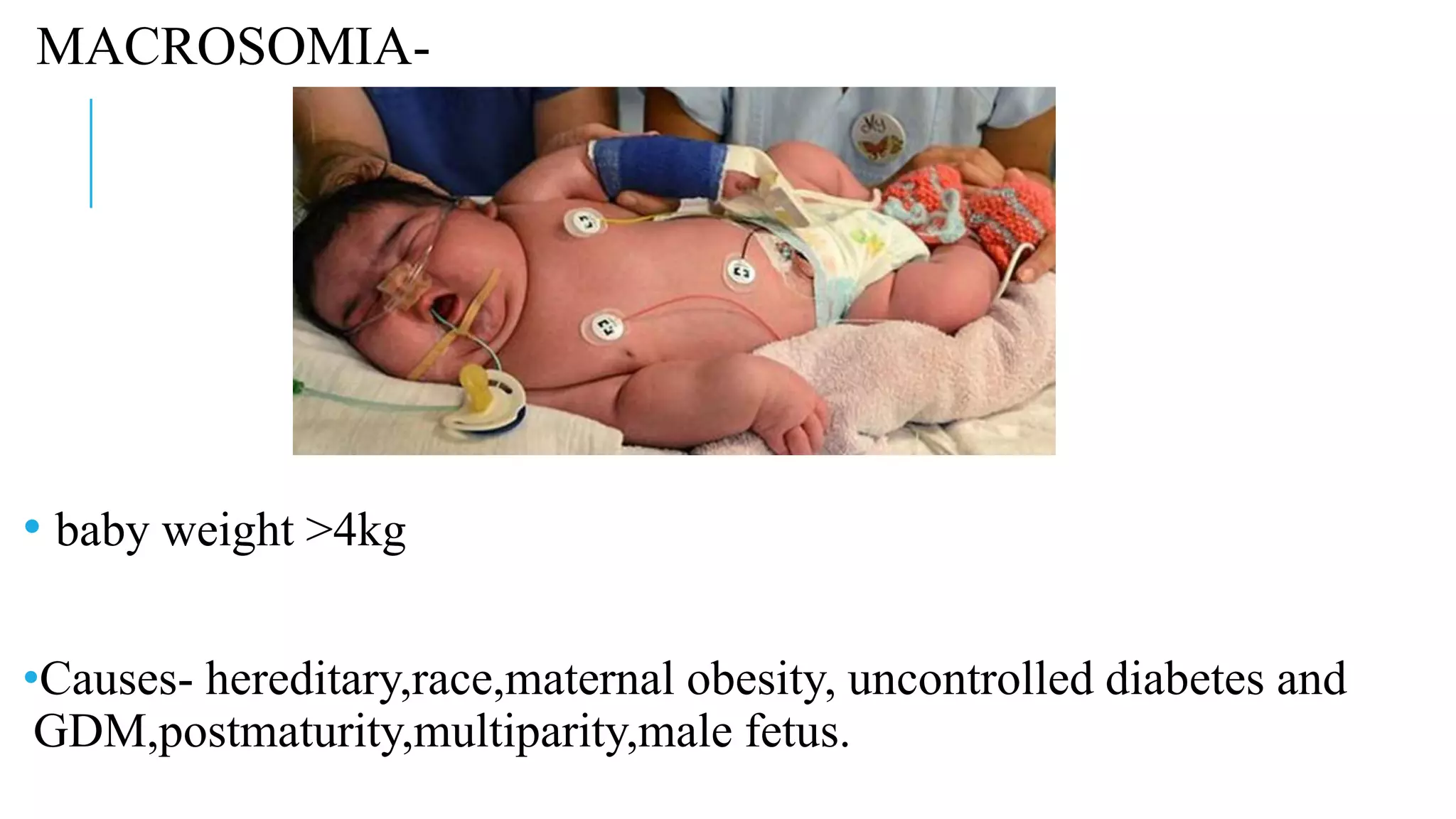
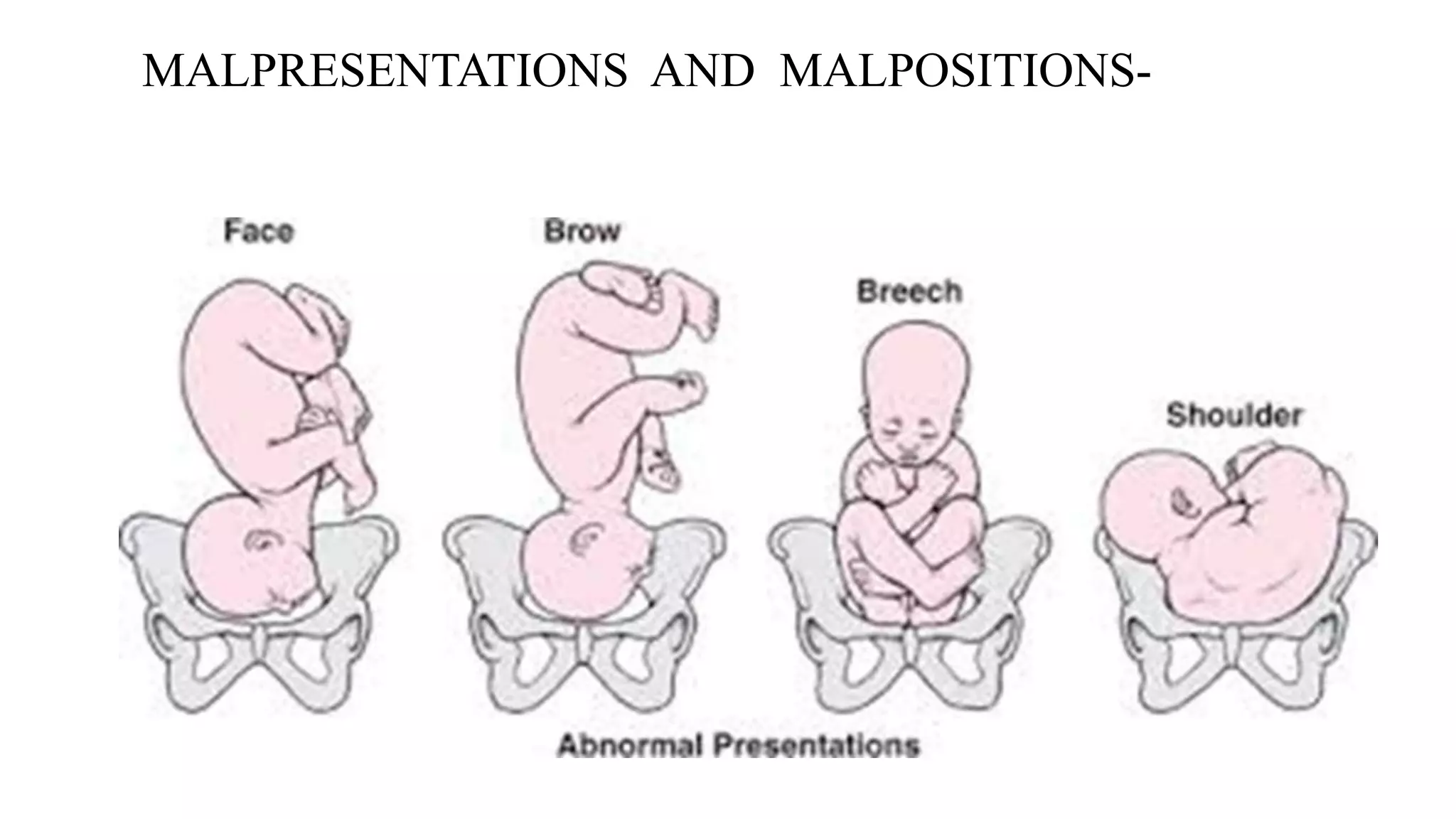
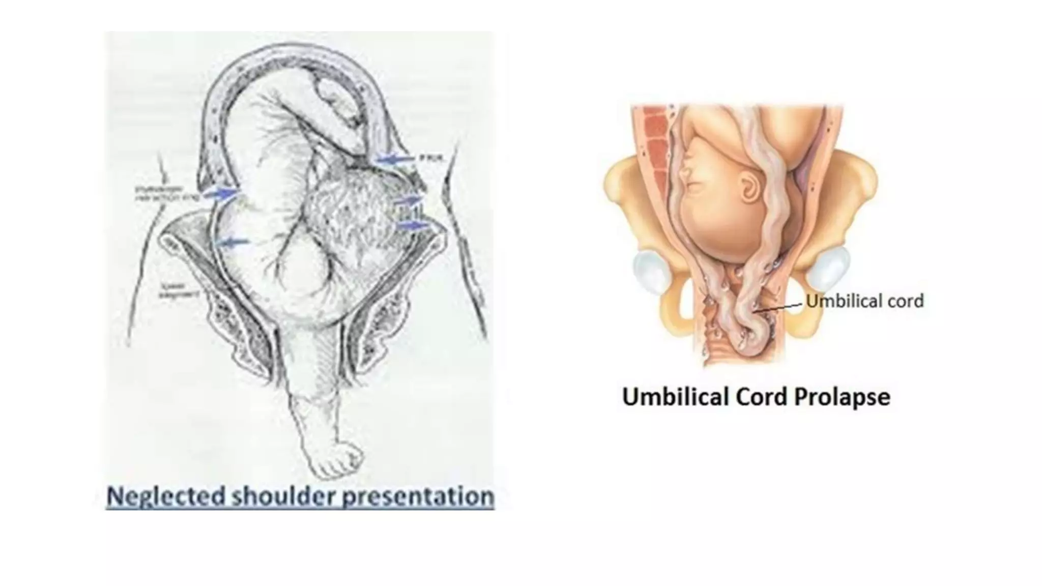
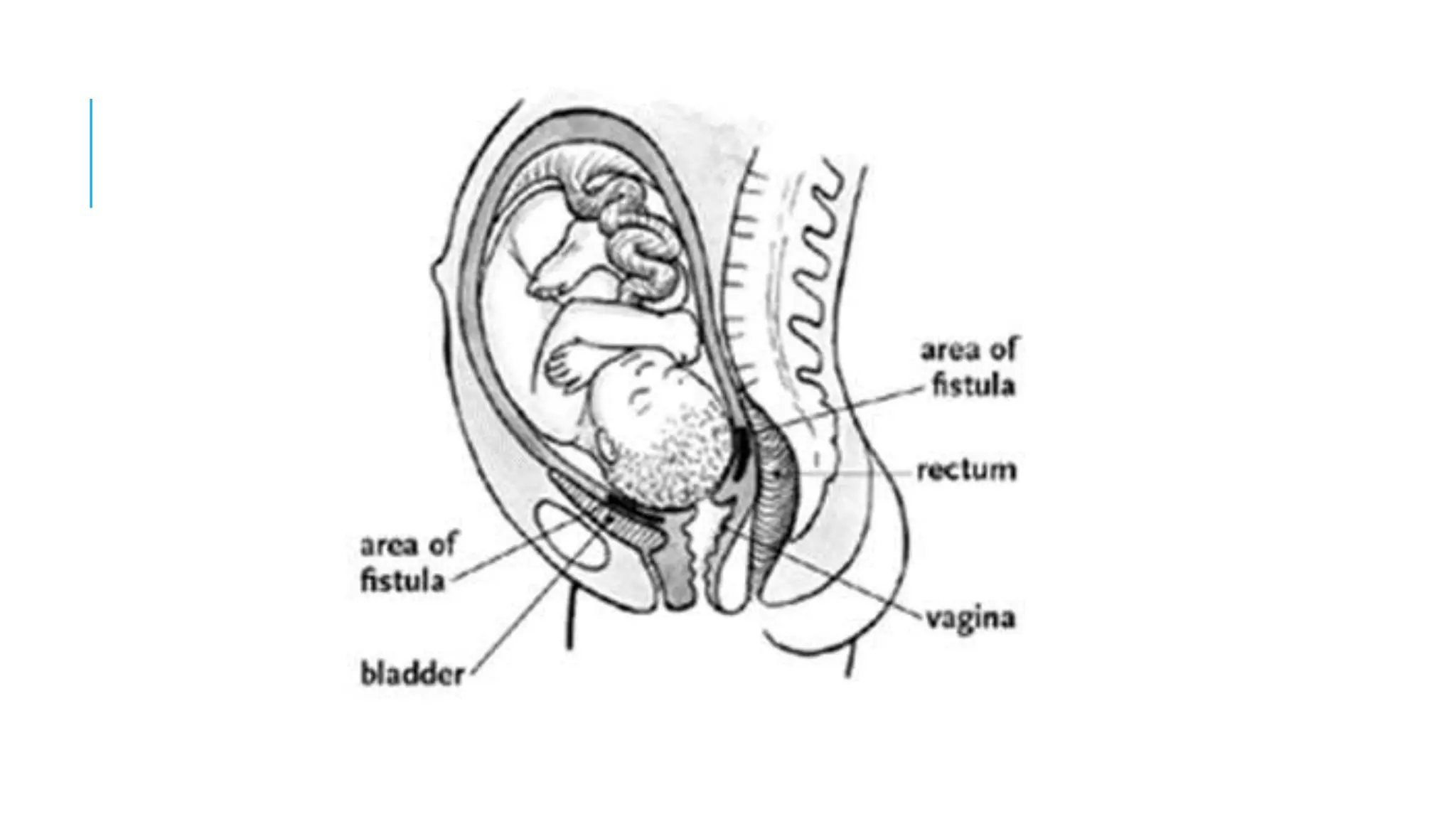
OBSTRUCTED
LABOUR
Dr. TEJAL
VAIDYA
OBSTRUCTED LABOUR
Definition- obstructed labour is one where in spite of good uterine
contractions,the progressive descent of presenting part is arrested
due to mechanical obstruction.
Either due to factors in the fetus or in the birth canal or both,so
that further progress is almost impossible without assistance.
Incidence – about 1-2 % in referral hospitals (in developing
countries). It contributes to 8% of all maternal deaths in the world.
CAUSES -
1) fault in passenger
2) fault in passage
FAULTS IN PASSENGER –
Macrosomia
Congenital anomalies of the fetus- hydrocephalus,
anencephaly,ascites,tumours, neck swellings,conjoined twins.
Malpresentations and malpostions – transvere lie, brow
presentation ,face presentation, occipito posterior position,compound
presentation,locked twins.
Shoulder dystocia
MACROSOMIA-
• baby weight >4kg
•Causes- hereditary,race,maternal obesity, uncontrolled diabetes and
GDM,postmaturity,multiparity,male fetus.
•Fetal hazards- dystocia due to CPD, shoulder dystocia,brachial
plexus injury, asphyxia ,birth trauma,meconium aspiration.
•Maternal hazards- soft tissue injury ,PPH, puerperal sepsis.
•Prophylactic early induction and elective caesarean section.
HYDROCEPHALUS
• Excessive accumulation of CSF in the ventricles resulting
in enlargement of the skull.
• 0.5-1.5 litre
• 1 in 2000 deliveries and 5% recurrence
• Head circumference >5o cm
• Head is high up cannot be pushed down in pelvis. FHS high
up.
• USG- fontanelle and suture wide,ventriculomegaly,thin
vault bones, dangling choroids sign .
•p/v- gaping sutures and fontanelle, crackling sensation on pressing
•Dangers- dystocia is inevitable ,fetal prognosis is poor
•Diagnosed mostly in later half of pregnancy
•Principle – decompress the head
•Cephalocentesis – p/v or usg guided through the abdominal approach ,
using a wide bore 17 G needle.
•Breech(30%) – perforation and decompression through sub–occipital
region
•During caesarean delivery – drain CSF by needle before incision of
uterus in diagnosed cases
•MTP cannot be done after 20 weeks
•Prognosis- use of ventriculoamniotic shunt is limited
ANENCEPHALY
•Deficient development of vault of skull and brain tissue
•Pituitary gland is hypoplastic or absent, atrophic adrenal glands
•70% females
•Diagnosis- increased alphafetoprotein in amniotic fluid, USG- absent
cranial vault, angiomatous brain tissue,associated hydramnios.(10 weeks)
p/a- unable to palpate the head
•Complications- hydramnios(70%),malpresentation – face and
breech,postmaturity, shoulder dystocia,obstructed labour head and
shoulders try to engage together because of short neck
•Indication for termination irrespective of gestation age as it is
incompatible with life
Iniencephaly- failure of formation of cervical and thoracic vertebra and
base of skull with abnormal formed brain tissue.
Enlargement of fetal abdomen-
• Ascitis, distended bladder, enlargement of
kidneys by tumor,umbilical hernia can cause
dystocia .
• USG findings of ascitis – Buddha position
• Decompression of abdomen with wide bore
needle
Neck tumor ,neck masses and cord around neck
• Congenital goiter , bronchocele,cystic hygroma,
Thoracic lymphangioma
CONJOINT TWINS-
•Incomplete twinning result in monster babies leading dystocia .
MALPRESENTATIONS AND MALPOSITIONS-
Transverse lie
•Long axis of fetus perpendicular to maternal spine
•Dorsoanterior is most common (60%)
•Dorsoposterior – chances of fetal extension and arm prolapse with cord
prolapse
•Formation of pathological retraction ring and obstructed labour
Neglected shoulder presentation-
•Series of complication that may arise out of shoulder presentation when
labour is left uncared.
•Impacted shoulder lead to obstructed labour , rupture of uterus with
clinical evidence of dehydration ,ketoacidosis , shock ,haemorrhage
,peritonitis and sepsis.
•Marked increase in the fetal loss is due to cord prolapse,tonic
contractions and rupture uterus.
•Management – aims at prevention of condition by managing the
shoulder presentation in early labour. First resuscitation of mother
followed by delivery of the baby.
•In cases of rupture- laparotomy followed by delivery through caesarean
section
FACE PRESENTATION-
•Variety of cephalic presentation with complete extension of head
•Denominator is mentum and most common is LMA
•Engaging diameter – fully extended –submentobregmatic(9.5cm)
partially extended –submentovertical(11.5cm)
•Mentoposterior (20-25%)- anterior rotation occurs in only 20-30%
cases rest, incomplete anterior rotation, nonrotation or short posterior
rotation occurs.
•No possibility of spontaneous delivery in persistent mentoposterior
position as short neck cannot clear off the total length of the sacrum .the
thorax is thrust in resulting the bregmaticosternal diameter (18cm)to
occupy pelvis and cause obstruction.
BROW PRESENTATION-
•Variety of cephalic presentation when head lies in between full
extension and full flexion.
•Engaging diameter is mentoverticle (14cm).
•Leads to obstructed labour,prolonged labour and uterine rupture.
•There is no mechanism of labour in an average size baby with normal
pelvis.
Occipito –posterior position -
SHOULDER DYSTOCIA-
•Defined to describe the wide range of obstetric maneuvers to deliver the
fetus after the head has been born and gentle traction has failed to deliver
the shoulders. (0.1-1%)
•Previous shoulder dystocia
•Macrosomia ,postmaturity
•Diabetis and obesity
•Induced labour , prolonged 1ST ans 2ND stage
•Secondary arrest of labour
•Anencephaly , fetal ascites
•Mid pelvic instrumental delivery
•Multiparity
COMPLICATIONS –
Fetal- asphyxia, brachial plexus injury due to stretch, humerus fracture
clavicle fracture or sternocleidomastoid hematoma.
Maternal complications-
•PPH (11%)
•Cervical lacerations
•Vaginal tear
•Perineal tear (3rd and 4th degree)
•Uterine rupture
•Bladder injury
•Sacroiliac joint dislocation
Diagnosis –
1. Definite recoil of the head back against the perineum (turtle neck sign)
2. Inadequate spontaneous restitution
3. Plethoric fetal face
4. Failure of shoulder to descent
MANAGEMENT –
According to ACOG –
• It cannot be predicted or prevented because
accurate measures to do so do not exist .
• No evidence that any method is superior
than other in releasing impacted shoulder
and reduce chance of injury.
• Risks and benefits of vaginal and caesarean
delivery depends on factors like estimated
wight,gestational age,maternal diabetes
,previous history of dystocia, congenital
anomalies etc.
McROBERTS maneuver-
Abduct and hyperflex
• Decrease in angle of pelvic
inclination.
• Increases AP diameter of pelvis.
• Successful in 90% (RCOG 2012)
Rubin I
Rubin’s II
consists of inserting the fingers of one hand vaginally
behind the posterior aspect of the anterior shoulder of
the fetus and rotating the shoulder toward the fetal
chest. This motion will adduct the fetal shoulder girdle,
reducing its diameter.
Wood ‘s cork screw maneuver – (DONE UNDER GA)
Place at least two fingers (index and middle) on the anterior aspect of the fetal
posterior shoulder
Apply pressure to abduct/extend the posterior shoulder and rotate the fetal body
180°
After 90° the practitioner may need to switch hands in order to complete the full
180° turn
This movement rotates the anterior shoulder from under the symphysis pubis and
releases the impaction (shoulder dystocia)
Extraction of the posterior arm
Arm is swept
across chest and
delivered by gentle
traction.
“ALL FOURS”
Increases pelvic dementions
Downward traction on posterior
shoulder
• Symhysiotomy and cleidotomy are
rarely done
Video
FAULT IN THE PASSAGE-
•Bony obstructions – cephalopelvic disproportions, contracted pelvis
,asymmetrical and obliquiely contracted pelvis or secondary contracted
pelvis .
•Anatomical definition – essential diameters of one or more plane are
shortened by 0.5cm
•Disparity in relation to head and pelvis is known as CPD
Roberts pelvis
Naegele’s pelvis
•Soft tissue causes- cervical dystocia (primary or secondary due to
previous operative scarring), cervical broad ligament fibroids, impacted
ovarian tumours or non gravid horn of bicornuate uterus .
•Other causes like – vaginal septum, cervical stenosis etc
MORBIT ANATOMICAL CHANGES
Uterus
“Constiction ring is the cause of obstructed labour whereas pathological
retraction ring(Bandl’s ring) is the effect of obstructed labour.”
Bladder
•Bladder becomes abdominal organ , compression of urethra between the
presenting part and symphysis pubis.
•Earliest sign of obstructed labour is “unable to empty the bladder”.
•Trauma to bladder wall leads to hematuria
•Base of the bladder and urethra gets nipped in between the presenting
part and symphysis pubis and undergoes pressure necrosis. Devitalized
tissue gets infected and slough off resulting in genitourinary fistulas.
Effect of obstructed labour on mother-
Immediate effects
•Exhaustion due to constant pain and agony
•Dehydration due to increased muscular activity without adequate fluid
•Metabolic acidosis (lactic acid and ketones)
•Genital sepsis due to rupture ,repeated p/v and manipulations
•Injury to genital tract and rupture of uterus
•PPH and shock
•Death due to – sepsis, shock and metabolic changes
Remote complications-
•Genitourinary fistula
•Vaginal atresia
•Secondary amenorrhoea due to hysterectomy or Sheehan’s syndrome
Effects on the fetus –
•Asphyxia ; prolonged 1st stage – hypertonic uterine contractions without relaxation
in between interferes with the uteroplacental circulation leads to fetal distress .
•Acidosis due to fetal hypoxia and maternal acidosis
•Intracranial hemorrhage due to supermoulding and tentorial tear or traumatic
deliver
•Infections
•Perinatal loss
Clinical features-
•Features of maternal distress
•h/o prolonged labour
•Severe frequent pain and bearing down
•Exhaustion
•signs of dehydration,dry tongue and sunken eyes
•Tachycardia
•raised temperature
•Scanty concentrated urine
• blood tinged urine
Per abdomen examination-
•Uterus tonically contracted may be hard and tense
•Fetal parts palpated with difficulty
•FHS may or may not be heard , it is irregular and presence of
bradycardia
•Formation of bandl’s ring felt at the junction of upper uterine segment
and lower uterine segment
•Bladder can be palpated abdominally
•in case of uterine rupture – tender abdomen ,fetal parts felt
easily ,baby in peritoneal cavity ,FHS may be absent,flanks dull
due to hemoperitonium .
•In primigarvida or multigravida obstructed labour lead to “three
tumour abdomen” : distended bladder , upper segment of uterus
, lower segment of uterus separated by pathological retraction
ring.
On per vaginal examination-
•Vulva is swollen ,edematous
•Dry hot vagina
•Offensive and purulent discharge
•Cervix fully dilated or hanging like a curtain
•Presenting part – excessive moulding amd jammed large caput.
DIAGNOSIS-
•Detailed history
•Examination
•Partograph
PREVENTION –
•Adequate good nutrition from childhood period would help to achieve
genetically predetermined height and help in prevention of nutritional
deficiency chances of the maternal pelvis.
•Universal antenatal care protocol
•Regular antenatal visits – obstetrician can anticipate obstruction and
prolong labour by detailed history and by identification of the risk factors
•Intranatally – monitoring of labour by skilled staff
•Use of partograph, continuous vigilance and timely intervention
MANAGEMENT –
Principle of treatment
•To relieve the obstruction at the earliest by safe delivery procedure
•To combat dehydration and ketoacidosis
•To control sepsis
Immediate management –
•Treat dehydration and shock by fluid resuscitation - 1 lit RL or DNS
rapidly (x3) till dehydration and ketosis is corrected then 1 lit 4-6 hrs
•Severe tonic contractions prevented by tocolysis ( terbutaline sc)
•Blood sampling – blood group typing ,cross
matching,LFT,KFT,cbc,urine for ketone and urine r/m
,sr.electrolytes,blood culture sensitivity.
•High vaginal swab is taken and sent for culture and sensitivity.
•Cathererization done
•Broad spectrum antibiotic cover (1g ceftriaxone with iv infusion of
metronidazole)
•Sodium bicarb to counter acidosis
OBSTETRICAL MANAGEMENT –
•Before proceeding for definitive operative treatment ,rupture of
uterus must be excluded.
•There is no place for “wait and watch”
•Do not use oxytocin to stimulate contractions
•Vaginal delivery-
1. Destructive operations like craniotomy, evisceration,decapitation
and cleidotomy have historical importance and no place in modern
obstetrics.
2. If the head is low and vaginal delivery is not risky then forceps
extraction can be done in live fetus
3. There is no place for internal cephalic version .
4. After delivery of baby explore uterus and lower genital tract for tears
and rupture.
•Caesarean delivery-
1. If obstructed labour is detected early and with good fetal condition
the it gives best results.
2. Desperate attempt to save the moribund baby often leads to
disastrous consequences
3. Brow ,mentoposterior ,placenta previa
4. Dead fetus with impending rupture
• active management of third stage of labour
•Continuous bladder draining for 3-7 days to prevent genitourinary
fistulas.
•Management of sepsis- inj ampicillin 2g 6hrly or inj ceftriaxone 1g +
Inj gentamycin 5mg /bw (gram neg) and inj metronidazole (anerobes)
Less severe cases – ampi+genta
Hydrocortisone 200-400 mg stat iv followed by 100-200mg 4 hrly
Dopamine infusion ( hypovolemic shock)
Tetanus prophylaxis
•Laprotomy repair is done in cases of ruptured uterus- recent rupture,
clean wound and margins, tear not too large ,preservation of
fertility,little or no sepsis.
•TAH/subtotal hysterectomy -severe infected uterus, tear with necrotic
edges,tear extending to vagina,future cervical ca concerns, spontaneous
obstructive rupture.
•Analgesia
•Breast care
• Fistula care
Explain the condition and counsel
•Repaired rupture and CS- always hospital delivery
•Total /subtotal hysterectomy- amenorrhoea and infertility
•Severe postpartum infections – possible ectopic in next pregnancies amd
need for regular antenatal visits
Thankyou ...

Obtructed labour

  • 1.
  • 2.
    OBSTRUCTED LABOUR Definition- obstructedlabour is one where in spite of good uterine contractions,the progressive descent of presenting part is arrested due to mechanical obstruction. Either due to factors in the fetus or in the birth canal or both,so that further progress is almost impossible without assistance. Incidence – about 1-2 % in referral hospitals (in developing countries). It contributes to 8% of all maternal deaths in the world.
  • 3.
    CAUSES - 1) faultin passenger 2) fault in passage
  • 4.
    FAULTS IN PASSENGER– Macrosomia Congenital anomalies of the fetus- hydrocephalus, anencephaly,ascites,tumours, neck swellings,conjoined twins. Malpresentations and malpostions – transvere lie, brow presentation ,face presentation, occipito posterior position,compound presentation,locked twins. Shoulder dystocia
  • 5.
    MACROSOMIA- • baby weight>4kg •Causes- hereditary,race,maternal obesity, uncontrolled diabetes and GDM,postmaturity,multiparity,male fetus.
  • 6.
    •Fetal hazards- dystociadue to CPD, shoulder dystocia,brachial plexus injury, asphyxia ,birth trauma,meconium aspiration. •Maternal hazards- soft tissue injury ,PPH, puerperal sepsis. •Prophylactic early induction and elective caesarean section.
  • 7.
    HYDROCEPHALUS • Excessive accumulationof CSF in the ventricles resulting in enlargement of the skull. • 0.5-1.5 litre • 1 in 2000 deliveries and 5% recurrence • Head circumference >5o cm • Head is high up cannot be pushed down in pelvis. FHS high up. • USG- fontanelle and suture wide,ventriculomegaly,thin vault bones, dangling choroids sign .
  • 8.
    •p/v- gaping suturesand fontanelle, crackling sensation on pressing •Dangers- dystocia is inevitable ,fetal prognosis is poor •Diagnosed mostly in later half of pregnancy •Principle – decompress the head •Cephalocentesis – p/v or usg guided through the abdominal approach , using a wide bore 17 G needle. •Breech(30%) – perforation and decompression through sub–occipital region •During caesarean delivery – drain CSF by needle before incision of uterus in diagnosed cases •MTP cannot be done after 20 weeks •Prognosis- use of ventriculoamniotic shunt is limited
  • 9.
    ANENCEPHALY •Deficient development ofvault of skull and brain tissue •Pituitary gland is hypoplastic or absent, atrophic adrenal glands •70% females •Diagnosis- increased alphafetoprotein in amniotic fluid, USG- absent cranial vault, angiomatous brain tissue,associated hydramnios.(10 weeks) p/a- unable to palpate the head •Complications- hydramnios(70%),malpresentation – face and breech,postmaturity, shoulder dystocia,obstructed labour head and shoulders try to engage together because of short neck •Indication for termination irrespective of gestation age as it is incompatible with life
  • 10.
    Iniencephaly- failure offormation of cervical and thoracic vertebra and base of skull with abnormal formed brain tissue.
  • 11.
    Enlargement of fetalabdomen- • Ascitis, distended bladder, enlargement of kidneys by tumor,umbilical hernia can cause dystocia . • USG findings of ascitis – Buddha position • Decompression of abdomen with wide bore needle Neck tumor ,neck masses and cord around neck • Congenital goiter , bronchocele,cystic hygroma, Thoracic lymphangioma
  • 12.
    CONJOINT TWINS- •Incomplete twinningresult in monster babies leading dystocia .
  • 13.
  • 14.
    Transverse lie •Long axisof fetus perpendicular to maternal spine •Dorsoanterior is most common (60%) •Dorsoposterior – chances of fetal extension and arm prolapse with cord prolapse •Formation of pathological retraction ring and obstructed labour
  • 16.
    Neglected shoulder presentation- •Seriesof complication that may arise out of shoulder presentation when labour is left uncared. •Impacted shoulder lead to obstructed labour , rupture of uterus with clinical evidence of dehydration ,ketoacidosis , shock ,haemorrhage ,peritonitis and sepsis. •Marked increase in the fetal loss is due to cord prolapse,tonic contractions and rupture uterus. •Management – aims at prevention of condition by managing the shoulder presentation in early labour. First resuscitation of mother followed by delivery of the baby. •In cases of rupture- laparotomy followed by delivery through caesarean section
  • 17.
    FACE PRESENTATION- •Variety ofcephalic presentation with complete extension of head •Denominator is mentum and most common is LMA •Engaging diameter – fully extended –submentobregmatic(9.5cm) partially extended –submentovertical(11.5cm)
  • 18.
    •Mentoposterior (20-25%)- anteriorrotation occurs in only 20-30% cases rest, incomplete anterior rotation, nonrotation or short posterior rotation occurs. •No possibility of spontaneous delivery in persistent mentoposterior position as short neck cannot clear off the total length of the sacrum .the thorax is thrust in resulting the bregmaticosternal diameter (18cm)to occupy pelvis and cause obstruction.
  • 19.
    BROW PRESENTATION- •Variety ofcephalic presentation when head lies in between full extension and full flexion. •Engaging diameter is mentoverticle (14cm). •Leads to obstructed labour,prolonged labour and uterine rupture. •There is no mechanism of labour in an average size baby with normal pelvis.
  • 20.
  • 21.
    SHOULDER DYSTOCIA- •Defined todescribe the wide range of obstetric maneuvers to deliver the fetus after the head has been born and gentle traction has failed to deliver the shoulders. (0.1-1%)
  • 22.
    •Previous shoulder dystocia •Macrosomia,postmaturity •Diabetis and obesity •Induced labour , prolonged 1ST ans 2ND stage •Secondary arrest of labour •Anencephaly , fetal ascites •Mid pelvic instrumental delivery •Multiparity
  • 23.
    COMPLICATIONS – Fetal- asphyxia,brachial plexus injury due to stretch, humerus fracture clavicle fracture or sternocleidomastoid hematoma.
  • 24.
    Maternal complications- •PPH (11%) •Cervicallacerations •Vaginal tear •Perineal tear (3rd and 4th degree) •Uterine rupture •Bladder injury •Sacroiliac joint dislocation
  • 25.
    Diagnosis – 1. Definiterecoil of the head back against the perineum (turtle neck sign) 2. Inadequate spontaneous restitution 3. Plethoric fetal face 4. Failure of shoulder to descent
  • 26.
    MANAGEMENT – According toACOG – • It cannot be predicted or prevented because accurate measures to do so do not exist . • No evidence that any method is superior than other in releasing impacted shoulder and reduce chance of injury. • Risks and benefits of vaginal and caesarean delivery depends on factors like estimated wight,gestational age,maternal diabetes ,previous history of dystocia, congenital anomalies etc.
  • 27.
    McROBERTS maneuver- Abduct andhyperflex • Decrease in angle of pelvic inclination. • Increases AP diameter of pelvis. • Successful in 90% (RCOG 2012)
  • 28.
  • 29.
    Rubin’s II consists ofinserting the fingers of one hand vaginally behind the posterior aspect of the anterior shoulder of the fetus and rotating the shoulder toward the fetal chest. This motion will adduct the fetal shoulder girdle, reducing its diameter.
  • 30.
    Wood ‘s corkscrew maneuver – (DONE UNDER GA) Place at least two fingers (index and middle) on the anterior aspect of the fetal posterior shoulder Apply pressure to abduct/extend the posterior shoulder and rotate the fetal body 180° After 90° the practitioner may need to switch hands in order to complete the full 180° turn This movement rotates the anterior shoulder from under the symphysis pubis and releases the impaction (shoulder dystocia)
  • 32.
    Extraction of theposterior arm Arm is swept across chest and delivered by gentle traction.
  • 33.
    “ALL FOURS” Increases pelvicdementions Downward traction on posterior shoulder • Symhysiotomy and cleidotomy are rarely done
  • 34.
  • 35.
    FAULT IN THEPASSAGE- •Bony obstructions – cephalopelvic disproportions, contracted pelvis ,asymmetrical and obliquiely contracted pelvis or secondary contracted pelvis . •Anatomical definition – essential diameters of one or more plane are shortened by 0.5cm •Disparity in relation to head and pelvis is known as CPD
  • 36.
  • 37.
    •Soft tissue causes-cervical dystocia (primary or secondary due to previous operative scarring), cervical broad ligament fibroids, impacted ovarian tumours or non gravid horn of bicornuate uterus . •Other causes like – vaginal septum, cervical stenosis etc
  • 38.
    MORBIT ANATOMICAL CHANGES Uterus “Constictionring is the cause of obstructed labour whereas pathological retraction ring(Bandl’s ring) is the effect of obstructed labour.”
  • 39.
    Bladder •Bladder becomes abdominalorgan , compression of urethra between the presenting part and symphysis pubis. •Earliest sign of obstructed labour is “unable to empty the bladder”. •Trauma to bladder wall leads to hematuria •Base of the bladder and urethra gets nipped in between the presenting part and symphysis pubis and undergoes pressure necrosis. Devitalized tissue gets infected and slough off resulting in genitourinary fistulas.
  • 41.
    Effect of obstructedlabour on mother- Immediate effects •Exhaustion due to constant pain and agony •Dehydration due to increased muscular activity without adequate fluid •Metabolic acidosis (lactic acid and ketones) •Genital sepsis due to rupture ,repeated p/v and manipulations •Injury to genital tract and rupture of uterus •PPH and shock •Death due to – sepsis, shock and metabolic changes
  • 42.
    Remote complications- •Genitourinary fistula •Vaginalatresia •Secondary amenorrhoea due to hysterectomy or Sheehan’s syndrome
  • 43.
    Effects on thefetus – •Asphyxia ; prolonged 1st stage – hypertonic uterine contractions without relaxation in between interferes with the uteroplacental circulation leads to fetal distress . •Acidosis due to fetal hypoxia and maternal acidosis •Intracranial hemorrhage due to supermoulding and tentorial tear or traumatic deliver •Infections •Perinatal loss
  • 44.
    Clinical features- •Features ofmaternal distress •h/o prolonged labour •Severe frequent pain and bearing down •Exhaustion •signs of dehydration,dry tongue and sunken eyes •Tachycardia •raised temperature •Scanty concentrated urine • blood tinged urine
  • 45.
    Per abdomen examination- •Uterustonically contracted may be hard and tense •Fetal parts palpated with difficulty •FHS may or may not be heard , it is irregular and presence of bradycardia •Formation of bandl’s ring felt at the junction of upper uterine segment and lower uterine segment •Bladder can be palpated abdominally
  • 46.
    •in case ofuterine rupture – tender abdomen ,fetal parts felt easily ,baby in peritoneal cavity ,FHS may be absent,flanks dull due to hemoperitonium . •In primigarvida or multigravida obstructed labour lead to “three tumour abdomen” : distended bladder , upper segment of uterus , lower segment of uterus separated by pathological retraction ring.
  • 47.
    On per vaginalexamination- •Vulva is swollen ,edematous •Dry hot vagina •Offensive and purulent discharge •Cervix fully dilated or hanging like a curtain •Presenting part – excessive moulding amd jammed large caput.
  • 48.
  • 49.
    PREVENTION – •Adequate goodnutrition from childhood period would help to achieve genetically predetermined height and help in prevention of nutritional deficiency chances of the maternal pelvis. •Universal antenatal care protocol •Regular antenatal visits – obstetrician can anticipate obstruction and prolong labour by detailed history and by identification of the risk factors •Intranatally – monitoring of labour by skilled staff •Use of partograph, continuous vigilance and timely intervention
  • 50.
    MANAGEMENT – Principle oftreatment •To relieve the obstruction at the earliest by safe delivery procedure •To combat dehydration and ketoacidosis •To control sepsis
  • 51.
    Immediate management – •Treatdehydration and shock by fluid resuscitation - 1 lit RL or DNS rapidly (x3) till dehydration and ketosis is corrected then 1 lit 4-6 hrs •Severe tonic contractions prevented by tocolysis ( terbutaline sc) •Blood sampling – blood group typing ,cross matching,LFT,KFT,cbc,urine for ketone and urine r/m ,sr.electrolytes,blood culture sensitivity. •High vaginal swab is taken and sent for culture and sensitivity.
  • 52.
    •Cathererization done •Broad spectrumantibiotic cover (1g ceftriaxone with iv infusion of metronidazole) •Sodium bicarb to counter acidosis
  • 53.
    OBSTETRICAL MANAGEMENT – •Beforeproceeding for definitive operative treatment ,rupture of uterus must be excluded. •There is no place for “wait and watch” •Do not use oxytocin to stimulate contractions •Vaginal delivery- 1. Destructive operations like craniotomy, evisceration,decapitation and cleidotomy have historical importance and no place in modern obstetrics. 2. If the head is low and vaginal delivery is not risky then forceps extraction can be done in live fetus
  • 54.
    3. There isno place for internal cephalic version . 4. After delivery of baby explore uterus and lower genital tract for tears and rupture. •Caesarean delivery- 1. If obstructed labour is detected early and with good fetal condition the it gives best results. 2. Desperate attempt to save the moribund baby often leads to disastrous consequences 3. Brow ,mentoposterior ,placenta previa 4. Dead fetus with impending rupture
  • 55.
    • active managementof third stage of labour •Continuous bladder draining for 3-7 days to prevent genitourinary fistulas. •Management of sepsis- inj ampicillin 2g 6hrly or inj ceftriaxone 1g + Inj gentamycin 5mg /bw (gram neg) and inj metronidazole (anerobes) Less severe cases – ampi+genta Hydrocortisone 200-400 mg stat iv followed by 100-200mg 4 hrly Dopamine infusion ( hypovolemic shock) Tetanus prophylaxis
  • 56.
    •Laprotomy repair isdone in cases of ruptured uterus- recent rupture, clean wound and margins, tear not too large ,preservation of fertility,little or no sepsis. •TAH/subtotal hysterectomy -severe infected uterus, tear with necrotic edges,tear extending to vagina,future cervical ca concerns, spontaneous obstructive rupture. •Analgesia •Breast care
  • 57.
  • 58.
    Explain the conditionand counsel •Repaired rupture and CS- always hospital delivery •Total /subtotal hysterectomy- amenorrhoea and infertility •Severe postpartum infections – possible ectopic in next pregnancies amd need for regular antenatal visits
  • 60.