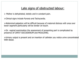
OBSTRUCTED
LABOUR
objectives
 By the end of this presentation the student shoulb be
able:
 Recap normal labor
 Define obstructed labour
 Manage obstructed labor
LABOR
 Normal labour: defined as regular painful uterine contractions which becomes
progressively stronger and more frequent accompanied by the effacement and progressive
dilatation of the cervix and the descent of the presenting part of the fetus.
 The labor is divided into 3 stages.
 The mechanism of labour is explained by the change in the position and the attitude of the
fetus, as the fetus initiates its way through the pelvis (birth canal).
 Mechanism includes:
 1) Engagement
 2) Descent
 3) Flexion
 4) Internal rotation
 5) Extension
 6) Restitution
 7) External rotation
X-TICS OF NORMAL LABOR
 Spontaneous onset
 Single cephalic presentation.
 37-42 weeks of gestation
 No artificial interventions.
 Unassisted spontaneous vaginal delivery.
 Duration of <12 hours in nulliparous women, and <8 hours in multiparous
women.
 A retrospective diagnosis.
 A labour which deviates from these features can be described as abnormal.
DEFINITION
 Is defined as failure of progressive descent of the presenting part,
despite adequate uterine contractions thus requiring surgical
intervention.
 As opposed to prolonged labor which involves powers and psyche in
addition to passage and passenger, obstructed labor is attributed to
mechanical obstruction resulting from abnormality in the passage
(pelvis) or the passenger (fetus).
 In obstructed labor there is abnormal fetal pelvic relationship.
PARTOGRAM SHOWING OBSTRUCTED LABOUR.
1 2 3 4 5 6 7 8 9 10 11 12 13 14 15 16 17 18 19 20 21 22 23 24
1
0
9
8
7
6
5
4
3
2
1
0
TIME
(HOURS)
LATENT
PHASE
ACTIVE PHASE
ALERT
ACTION
EPIDEMIOLOGY:
 One of the most common causes of maternal
morbidity and mortality in developing countries.
 Short stature
 8% of direct maternal deaths
 Common cause of complication e.g infections and
fistulas
 Fetal death from Asphyxia is common.
Risk factors:
• Maternal age: < 16 years
• Primigravidae
• Short stature (<150cm)
• Past obstetric history
– History of obstructed labor
– Delivery by cesarean section
– Difficult labor ending with VVF
– Ruptured uterus
• Maternal diseases:
– DM
– History suggestive of pelvic tumor
– History of cervical surgery
Causes:
Maternal causes:
a) Abnormalities of the passage (pelvic dystocia)
• Contracted pelvis: shape of pelvis normal but the diameters
are decreased. Causes of contracted pelvis can be genetic or
nutritional
• Pelvic deformities: fracture of pelvis, polio, osteomyelitis,
TB, SCD
• Abnormality of the pelvis: android, platypelloid or
anthropoid (many Tz women have Bantu pelvis)
 Cephalopelvic disproportion (Small pelvis, Contracted
pelvis, Big baby, Deformed pelvis)
b) Soft tissue obstruction of birth canal
• Uterus: double uterus, septate uterus
• Cervical stenosis: congenital or post-surgical
• Vaginal stenosis: congenital, surgical
• Myomas on the lower segment of the uterus or cervix
c) Abnormal placental location
• Low placentation i.e. placenta previa
Fetal causes:
• Fetal malpresentation:
– Brow
– Face
– Shoulder
– Breech
• Fetal apposition:
- Persistent occipital posterior
- Transverse lie
 Fetal macrosomia
 Compound presentation
 Locked twins
 Congenital malformations:
- Hydrocephalus
- Conjoined twins
- Hydrops fetalis
CLINICAL PICTURE (I)
Symptoms:
 Prolonged labor
 Frequent and strong uterine contractions
 Ruptured membranes
 Labor pains initially severe & frequent then becomes
mild.
 Pain is more on the back radiating to thighs due to
pressure over muscles and ligaments.
 The patient is exhausted by pain and the demands of
overworking the uterus
Signs of obstructed labor
 General condition:
 Maternal exhaustion: ketoacidosis, dehydration,
rapid pulse
 Shock in ruptured uterus
Abdominal examination
• Distended bladder
• Bowel distended with gas and can be palpated on
either side of the uterus.
• They give the sound of drums on tapping.
• Formation of Bandl’s ring
– This is visible at the level where the upper and lower
segments of the uterus meet, a firm ridge is formed running
obliquely across the uterine wall.
– There is increased thickening of the upper segment and
increased thinning of the lower segment
Late signs of obstructed labour:
 Mother is dehydrated, ketotic and in constant pain.
Clinical signs include Pyrexia and Tachycardia.
Abdominal palpation will be difficult because of maternal distress with area over
lower segment particularly will be tender on touch.
On vaginal examination the assessment of presenting part is complicated by
presence of CAPUT SUCCEDENUM and MOULDING.
Urinary output is present and on insertion of catheter you notice urine concentrated
With blood.
In untreated case the possible outcomes are:
 Secondary uterine inertia from uterine exhaustion
Generalized spasm or tonic contraction of the uterus,
where the uterus makes one last effort to overcome the
obstruction.
Rupture of the uterus , often as a result of tonic
contractions.
EVENTS DURING LABOUR (I)
 The upper segment actively contracts and retracts
while the lower segment is relatively passive
 Upper segment contracting almost instantly and
retracting makes uterus become hard and its walls
become very much thicker and shorter as it forces
fetus down and draws the lower segment and cervix
up
EVENTS DURING LABOUR (II)
 As time goes on, more and more of the fetus is driven
down into the relaxing lower segment which becomes
dangerously thin & will rupture if urgent action is not
taken
 Uterine contractions usually increase in force &
frequency, often accompanied by strong bearing
efforts
 Pains are severe & continuous
 The mother becomes exhausted & restless with dry &
discolored tongue & lips; Pulse rate rises
(≥120/minute); Temperature also rises
EVENTS DURING LABOUR (III)
 Obstruction always occurs in the cavity or just below
the pelvic brim; serious obstruction at the pelvic
outlet is uncommon. Death of the fetus results from
compression of the placental site circulation
 The vagina and vulva are oedematous and the birth
canal feels hot and dry. The oedematous cervix may
be felt below the presenting part and also a large
caput and marked moulding of cranial bones
Vaginal examination
• Excessive moulding of the fetal head
• Edema of the vulva, cervix
• Stained meconium
• Presence of caput succedaneum
• Membranes may or may not rupture, if they rupture
there is risk of infections.
Catheterization:
 Concentrated urine
 Urine mixed with blood (sometimes)
Others:
 Hypoglycaemia
 Acetonuria
 IUFD
In untreated case the possible outcomes are:
 Secondary uterine inertia from uterine exhaustion
(primigravidae)
Generalized spasm or tonic contraction of the
uterus, where the uterus makes one last effort to
overcome the obstruction.
Rupture of the uterus , often as a result of tonic
contractions (multigravidae)
Anticipation of obstructed labor:
• Close surveillance of a woman in labor will identify
obstruction before it has advanced to cause maternal
or fetal complications. The following parameters
should raise suspicion:
• Failure of progressive descent of the presenting part
and/or stagnation or slow dilatation of the cervix
• Cervix that is poorly applied to the presenting part
Cont..
 Incoordinate uterine contractions
 Early rupture of membranes
 Reduced pelvic measurements
 Diagonal conjugate of <11cm
 Flat sacral curve
 Prominent ischial spines
 Narrow subpubic arch
Maternal:
1.Trauma to the bladder
 The bladder walls get traumatized which may lead
to blood stained urine, a common finding in
obstructed labour.
The base of the bladder and urethra which are
compressed in between the presenting part and
symphisis pubis may undergo pressure necrosis.
This may later slough off resulting into genital
urinary fistula. Rectovaginal fistula may also develop as a
complication of neglected obstructed labour.
Complication of obstructed labour
2. Prolonged compression of the nerves thus
obstetric palsy (peroneal aspect of sciatic trunk
[supplies shin muscles] affected; patient presents with
foot drop).
3. Infections
– Genital sepsis is an invariably accompaniment especially after
rupture of the membranes with repeated vaginal examinations or
attempted manipulations outside.
– This may even result into intrauterine infections
4. Injury to the genital tract includes rupture of the
uterus which may be spontaneous in multipara or
may be traumatic following instrument deliver.
5. D.I.C: when the fetus dies in utero, it becomes
softened due to decay and triggers DIC and
maternal hge,shock and death.
6. Postpartum haemorrhage and shock
– May be due to isolated or combined effects of atonic uterus
or genital tract trauma
– Infections may also cause shock (septic shock)
7. Maternal death
– All these lead to increased maternal morbidity and
mortality.
– The deaths are mainly due to rupture of the uterus and
sepsis with metabolic change.
8.Others
• Anaemia
• Incontinence
Fetal:
Asphyxia.
Asphyxia results from tonic uterine contractions
which interfere with the uteroplacental circulation or
due to cord prolapsed especially in shoulder
presentation
Acidosis due to foetal hypoxia and maternal acidosis
Intracranial haemorrhage due to supermoulding of
the head leading to tentorial tear due to traumatic
delivery.
Ascending infection.
Fetal death.
MANAGEMENT OF
OBSTRUCTED LABOUR
• The main aim is to relieve the obstruction as
earliest as possible by a safe procedure
Initial assessment of the patient’s condition:
 Pallor, jaundice, pulse rate and respiratory rate
 Fundal height, foetal lie and presentation,
 Caput formation and moulding of foetal skull
bones
 Pelvic assessment for presence of infected liquor
 Assess urine for acetone, serum for electrolytes and
blood for gasses
 Blood grouping and cross-matching
In managing neglected obstructed labour
consider;
Resuscitation
Type of delivery
Preoperative care
Post operative care
1. Resuscitation
 Correct the dehydration, electrolyte imbalance and
hypoglycaemia by giving dextrose saline initially
then Ringers lactate - atleast 3L stat
 Empty bladder and stomach (catheterise and take;
urine for acetone bedside test)
 Blood grp & x- matching (in case pph follows)
Obstetric management:
• Before proceeding for definitive operation
treatment, rupture of the membrane must be
excluded.
• The balanced decision should be taken about the best
method of relieving the obstruction with least hazards
to the mother.
• There is no any place for ‘watch and wait’ neither
any scope of using oxytocin to stimulate uterine
contraction
2. Mode of delivery
• If the foetus is alive – do C/S
• If Dead – destructive procedure (craniotomy,
decapitation, cleidotomy)
• Vaginal delivery
The baby is invariably dead in most of the
neglected cases and destructive operation is the
best choice to relieve the obstruction
 If however the head is low down and vaginal
delivery is not risky, vacuum extraction may be
done in a living baby.
 After completion of the delivery and expulsion of
the placenta, exploration of the uterus and the
lower genital tract should be done to exclude
uterine rupture or tear
• Caesarean section
– This is the mainstay treatment.
– If the case is detected early with good foetal conditions, C/S
gives the best result.
– But in late neglected cases, even if the foetal heart sounds is
audible, desperate attempt to do C/S to save the baby may
lead to disastrous consequences. The baby may be delivered
stillborn or dies due to neonatal sepsis.
Other Methods of delivery:
Episiotomy:
 Works very well for P.G
 Especially if baby’s vertex is in an occipito-anterior
position.
Vacuum extraction: if baby is alive with 2/5 of the
head above the brim and only moderate molding.
Methods of delivery contd…
 In vaccuum extraction, use the rule of three pulls. 1st
dislodge the baby,then bring baby’s head to pelvic
floor, then deliver the baby.
 Contraindicated in: a dead baby, baby with 2/5 of
head above the brim, severe moulding and a
definitive CPD.
Methods of delivery contd…
 3. Symphisiotomy: indications; baby alive in cephalic
position, not more than 2/5 of head above the brim.
In case of soft tissue obstruction…
 Vaginal stricture(very common): from scar tissue
from previous delivery. If its thin divide it, if its
thick section and excise later when she isnt
pregnant.
 If thin, incise at 4 and 8 oclock positions after
vaginal delivery,suture the laceration.
 If there is a fibroid, leave it and remove it later.
Never remove a fibroid at C-section
3. Pre operative Care
Continue with IV fluids
Antibiotics; ampicillin 1g and
metronidazole 500 mg IV.
Consent of C/S
Should take nothing orally
4. Post-delivery care
• Continued monitoring of temperature, pulse, BP
and urine output & color
• Monitor abdominal distension
• Continue with antibiotics
• Bladder drainage for at least 10 days (in women
with blood in urine)
• Check for peroneal nerve damage (obstetric palsy)
and rehabilitate appropriately
• Bear in mind the possibility of PPH
• Counsel the patient in regard to future pregnancies
• Analgesics-pethidine 100mg 8 hrly x 24hrs
Primary prevention:
• Educate communities about the dangers of prolonged
labour and the need for speedy referral.
• Educate women who have had a caesarean section for
obstructed labour about the reasons for the operation
and what to do next time around
• Prevent malnutrition
• Accessible health systems
Prevention of obstructed labour:
Secondary Prevention:
 Proper ANC and guidance
 Assessment of risk factors
 Antenatal assessment- contracted pelvis?
Tertiary Prevention
 Proper Partogram usage especially in monitoring of
the mother and the fetus conditions (partogram
helps to prevent obstructed labour)
• Can be used in knowing time for operative
intervention and in improving the neonatal outcome.
• Can be highly effective in reducing complications
from prolonged labor for the mother (postpartum
hemorrhage, sepsis, uterine rupture, etc.) and for the
newborn (death, anoxia, infections, etc.).
Summary
 Obstructed labor and ruptured uterus are obstetrical
emergencies that must be given urgent attention
 Reference obstetrics by ten teachers , Williams text
book of obstetrics

10. Obstructed Labour-3.ppt

  • 1.
  • 2.
    objectives  By theend of this presentation the student shoulb be able:  Recap normal labor  Define obstructed labour  Manage obstructed labor
  • 3.
    LABOR  Normal labour:defined as regular painful uterine contractions which becomes progressively stronger and more frequent accompanied by the effacement and progressive dilatation of the cervix and the descent of the presenting part of the fetus.  The labor is divided into 3 stages.  The mechanism of labour is explained by the change in the position and the attitude of the fetus, as the fetus initiates its way through the pelvis (birth canal).  Mechanism includes:  1) Engagement  2) Descent  3) Flexion  4) Internal rotation  5) Extension  6) Restitution  7) External rotation
  • 4.
    X-TICS OF NORMALLABOR  Spontaneous onset  Single cephalic presentation.  37-42 weeks of gestation  No artificial interventions.  Unassisted spontaneous vaginal delivery.  Duration of <12 hours in nulliparous women, and <8 hours in multiparous women.  A retrospective diagnosis.  A labour which deviates from these features can be described as abnormal.
  • 5.
    DEFINITION  Is definedas failure of progressive descent of the presenting part, despite adequate uterine contractions thus requiring surgical intervention.  As opposed to prolonged labor which involves powers and psyche in addition to passage and passenger, obstructed labor is attributed to mechanical obstruction resulting from abnormality in the passage (pelvis) or the passenger (fetus).  In obstructed labor there is abnormal fetal pelvic relationship.
  • 6.
    PARTOGRAM SHOWING OBSTRUCTEDLABOUR. 1 2 3 4 5 6 7 8 9 10 11 12 13 14 15 16 17 18 19 20 21 22 23 24 1 0 9 8 7 6 5 4 3 2 1 0 TIME (HOURS) LATENT PHASE ACTIVE PHASE ALERT ACTION
  • 7.
    EPIDEMIOLOGY:  One ofthe most common causes of maternal morbidity and mortality in developing countries.  Short stature  8% of direct maternal deaths  Common cause of complication e.g infections and fistulas  Fetal death from Asphyxia is common.
  • 8.
    Risk factors: • Maternalage: < 16 years • Primigravidae • Short stature (<150cm) • Past obstetric history – History of obstructed labor – Delivery by cesarean section – Difficult labor ending with VVF – Ruptured uterus • Maternal diseases: – DM – History suggestive of pelvic tumor – History of cervical surgery
  • 9.
    Causes: Maternal causes: a) Abnormalitiesof the passage (pelvic dystocia) • Contracted pelvis: shape of pelvis normal but the diameters are decreased. Causes of contracted pelvis can be genetic or nutritional • Pelvic deformities: fracture of pelvis, polio, osteomyelitis, TB, SCD • Abnormality of the pelvis: android, platypelloid or anthropoid (many Tz women have Bantu pelvis)  Cephalopelvic disproportion (Small pelvis, Contracted pelvis, Big baby, Deformed pelvis)
  • 10.
    b) Soft tissueobstruction of birth canal • Uterus: double uterus, septate uterus • Cervical stenosis: congenital or post-surgical • Vaginal stenosis: congenital, surgical • Myomas on the lower segment of the uterus or cervix c) Abnormal placental location • Low placentation i.e. placenta previa
  • 11.
    Fetal causes: • Fetalmalpresentation: – Brow – Face – Shoulder – Breech • Fetal apposition: - Persistent occipital posterior - Transverse lie
  • 12.
     Fetal macrosomia Compound presentation  Locked twins  Congenital malformations: - Hydrocephalus - Conjoined twins - Hydrops fetalis
  • 13.
    CLINICAL PICTURE (I) Symptoms: Prolonged labor  Frequent and strong uterine contractions  Ruptured membranes  Labor pains initially severe & frequent then becomes mild.  Pain is more on the back radiating to thighs due to pressure over muscles and ligaments.  The patient is exhausted by pain and the demands of overworking the uterus
  • 14.
    Signs of obstructedlabor  General condition:  Maternal exhaustion: ketoacidosis, dehydration, rapid pulse  Shock in ruptured uterus
  • 15.
    Abdominal examination • Distendedbladder • Bowel distended with gas and can be palpated on either side of the uterus. • They give the sound of drums on tapping. • Formation of Bandl’s ring – This is visible at the level where the upper and lower segments of the uterus meet, a firm ridge is formed running obliquely across the uterine wall. – There is increased thickening of the upper segment and increased thinning of the lower segment
  • 16.
    Late signs ofobstructed labour:  Mother is dehydrated, ketotic and in constant pain. Clinical signs include Pyrexia and Tachycardia. Abdominal palpation will be difficult because of maternal distress with area over lower segment particularly will be tender on touch. On vaginal examination the assessment of presenting part is complicated by presence of CAPUT SUCCEDENUM and MOULDING. Urinary output is present and on insertion of catheter you notice urine concentrated With blood.
  • 17.
    In untreated casethe possible outcomes are:  Secondary uterine inertia from uterine exhaustion Generalized spasm or tonic contraction of the uterus, where the uterus makes one last effort to overcome the obstruction. Rupture of the uterus , often as a result of tonic contractions.
  • 18.
    EVENTS DURING LABOUR(I)  The upper segment actively contracts and retracts while the lower segment is relatively passive  Upper segment contracting almost instantly and retracting makes uterus become hard and its walls become very much thicker and shorter as it forces fetus down and draws the lower segment and cervix up
  • 19.
    EVENTS DURING LABOUR(II)  As time goes on, more and more of the fetus is driven down into the relaxing lower segment which becomes dangerously thin & will rupture if urgent action is not taken  Uterine contractions usually increase in force & frequency, often accompanied by strong bearing efforts  Pains are severe & continuous  The mother becomes exhausted & restless with dry & discolored tongue & lips; Pulse rate rises (≥120/minute); Temperature also rises
  • 20.
    EVENTS DURING LABOUR(III)  Obstruction always occurs in the cavity or just below the pelvic brim; serious obstruction at the pelvic outlet is uncommon. Death of the fetus results from compression of the placental site circulation  The vagina and vulva are oedematous and the birth canal feels hot and dry. The oedematous cervix may be felt below the presenting part and also a large caput and marked moulding of cranial bones
  • 21.
    Vaginal examination • Excessivemoulding of the fetal head • Edema of the vulva, cervix • Stained meconium • Presence of caput succedaneum • Membranes may or may not rupture, if they rupture there is risk of infections.
  • 22.
    Catheterization:  Concentrated urine Urine mixed with blood (sometimes) Others:  Hypoglycaemia  Acetonuria  IUFD
  • 23.
    In untreated casethe possible outcomes are:  Secondary uterine inertia from uterine exhaustion (primigravidae) Generalized spasm or tonic contraction of the uterus, where the uterus makes one last effort to overcome the obstruction. Rupture of the uterus , often as a result of tonic contractions (multigravidae)
  • 24.
    Anticipation of obstructedlabor: • Close surveillance of a woman in labor will identify obstruction before it has advanced to cause maternal or fetal complications. The following parameters should raise suspicion: • Failure of progressive descent of the presenting part and/or stagnation or slow dilatation of the cervix • Cervix that is poorly applied to the presenting part
  • 25.
    Cont..  Incoordinate uterinecontractions  Early rupture of membranes  Reduced pelvic measurements  Diagonal conjugate of <11cm  Flat sacral curve  Prominent ischial spines  Narrow subpubic arch
  • 26.
    Maternal: 1.Trauma to thebladder  The bladder walls get traumatized which may lead to blood stained urine, a common finding in obstructed labour. The base of the bladder and urethra which are compressed in between the presenting part and symphisis pubis may undergo pressure necrosis. This may later slough off resulting into genital urinary fistula. Rectovaginal fistula may also develop as a complication of neglected obstructed labour. Complication of obstructed labour
  • 27.
    2. Prolonged compressionof the nerves thus obstetric palsy (peroneal aspect of sciatic trunk [supplies shin muscles] affected; patient presents with foot drop). 3. Infections – Genital sepsis is an invariably accompaniment especially after rupture of the membranes with repeated vaginal examinations or attempted manipulations outside. – This may even result into intrauterine infections
  • 28.
    4. Injury tothe genital tract includes rupture of the uterus which may be spontaneous in multipara or may be traumatic following instrument deliver. 5. D.I.C: when the fetus dies in utero, it becomes softened due to decay and triggers DIC and maternal hge,shock and death. 6. Postpartum haemorrhage and shock – May be due to isolated or combined effects of atonic uterus or genital tract trauma – Infections may also cause shock (septic shock)
  • 29.
    7. Maternal death –All these lead to increased maternal morbidity and mortality. – The deaths are mainly due to rupture of the uterus and sepsis with metabolic change. 8.Others • Anaemia • Incontinence
  • 30.
    Fetal: Asphyxia. Asphyxia results fromtonic uterine contractions which interfere with the uteroplacental circulation or due to cord prolapsed especially in shoulder presentation Acidosis due to foetal hypoxia and maternal acidosis
  • 31.
    Intracranial haemorrhage dueto supermoulding of the head leading to tentorial tear due to traumatic delivery. Ascending infection. Fetal death.
  • 32.
  • 33.
    • The mainaim is to relieve the obstruction as earliest as possible by a safe procedure Initial assessment of the patient’s condition:  Pallor, jaundice, pulse rate and respiratory rate  Fundal height, foetal lie and presentation,  Caput formation and moulding of foetal skull bones  Pelvic assessment for presence of infected liquor  Assess urine for acetone, serum for electrolytes and blood for gasses  Blood grouping and cross-matching
  • 34.
    In managing neglectedobstructed labour consider; Resuscitation Type of delivery Preoperative care Post operative care
  • 35.
    1. Resuscitation  Correctthe dehydration, electrolyte imbalance and hypoglycaemia by giving dextrose saline initially then Ringers lactate - atleast 3L stat  Empty bladder and stomach (catheterise and take; urine for acetone bedside test)  Blood grp & x- matching (in case pph follows)
  • 36.
    Obstetric management: • Beforeproceeding for definitive operation treatment, rupture of the membrane must be excluded. • The balanced decision should be taken about the best method of relieving the obstruction with least hazards to the mother. • There is no any place for ‘watch and wait’ neither any scope of using oxytocin to stimulate uterine contraction
  • 37.
    2. Mode ofdelivery • If the foetus is alive – do C/S • If Dead – destructive procedure (craniotomy, decapitation, cleidotomy) • Vaginal delivery The baby is invariably dead in most of the neglected cases and destructive operation is the best choice to relieve the obstruction
  • 38.
     If howeverthe head is low down and vaginal delivery is not risky, vacuum extraction may be done in a living baby.  After completion of the delivery and expulsion of the placenta, exploration of the uterus and the lower genital tract should be done to exclude uterine rupture or tear
  • 39.
    • Caesarean section –This is the mainstay treatment. – If the case is detected early with good foetal conditions, C/S gives the best result. – But in late neglected cases, even if the foetal heart sounds is audible, desperate attempt to do C/S to save the baby may lead to disastrous consequences. The baby may be delivered stillborn or dies due to neonatal sepsis.
  • 40.
    Other Methods ofdelivery: Episiotomy:  Works very well for P.G  Especially if baby’s vertex is in an occipito-anterior position. Vacuum extraction: if baby is alive with 2/5 of the head above the brim and only moderate molding.
  • 41.
    Methods of deliverycontd…  In vaccuum extraction, use the rule of three pulls. 1st dislodge the baby,then bring baby’s head to pelvic floor, then deliver the baby.  Contraindicated in: a dead baby, baby with 2/5 of head above the brim, severe moulding and a definitive CPD.
  • 42.
    Methods of deliverycontd…  3. Symphisiotomy: indications; baby alive in cephalic position, not more than 2/5 of head above the brim.
  • 43.
    In case ofsoft tissue obstruction…  Vaginal stricture(very common): from scar tissue from previous delivery. If its thin divide it, if its thick section and excise later when she isnt pregnant.  If thin, incise at 4 and 8 oclock positions after vaginal delivery,suture the laceration.  If there is a fibroid, leave it and remove it later. Never remove a fibroid at C-section
  • 44.
    3. Pre operativeCare Continue with IV fluids Antibiotics; ampicillin 1g and metronidazole 500 mg IV. Consent of C/S Should take nothing orally
  • 45.
    4. Post-delivery care •Continued monitoring of temperature, pulse, BP and urine output & color • Monitor abdominal distension • Continue with antibiotics • Bladder drainage for at least 10 days (in women with blood in urine) • Check for peroneal nerve damage (obstetric palsy) and rehabilitate appropriately • Bear in mind the possibility of PPH • Counsel the patient in regard to future pregnancies • Analgesics-pethidine 100mg 8 hrly x 24hrs
  • 46.
    Primary prevention: • Educatecommunities about the dangers of prolonged labour and the need for speedy referral. • Educate women who have had a caesarean section for obstructed labour about the reasons for the operation and what to do next time around • Prevent malnutrition • Accessible health systems Prevention of obstructed labour:
  • 47.
    Secondary Prevention:  ProperANC and guidance  Assessment of risk factors  Antenatal assessment- contracted pelvis?
  • 48.
    Tertiary Prevention  ProperPartogram usage especially in monitoring of the mother and the fetus conditions (partogram helps to prevent obstructed labour)
  • 49.
    • Can beused in knowing time for operative intervention and in improving the neonatal outcome. • Can be highly effective in reducing complications from prolonged labor for the mother (postpartum hemorrhage, sepsis, uterine rupture, etc.) and for the newborn (death, anoxia, infections, etc.).
  • 50.
    Summary  Obstructed laborand ruptured uterus are obstetrical emergencies that must be given urgent attention
  • 51.
     Reference obstetricsby ten teachers , Williams text book of obstetrics