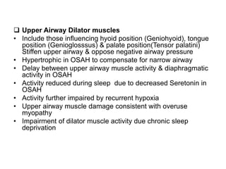
This document provides an overview of obstructive sleep apnea hypopnea (OSAH). It discusses the stages of normal sleep and how OSAH disrupts sleep. The pathogenesis of OSAH involves upper airway collapse due to imbalances between dilator muscle force and negative pressure. Symptoms include snoring, sleepiness, and gasping. Polysomnography is used to diagnose by measuring respiratory disturbances and oxygen levels. Treatments include weight loss, appliances like CPAP, and surgery to enlarge the airway.
2. physiology of deglutition and disorders of swallowingkrishnakoirala4
1. The document discusses the physiology of swallowing (deglutition) and disorders that can affect it. Swallowing involves coordinated muscle movements in the oral, pharyngeal, and esophageal stages to transport food to the stomach.
2. Common disorders of swallowing include reflux esophagitis, esophageal strictures, achalasia, hiatal hernia, and esophageal cancer. Investigations include barium swallow, endoscopy, and manometry.
3. Treatment depends on the underlying cause but may involve dilation, drugs, botulinum toxin injection, surgery, or chemoradiation for cancer. Careful evaluation is needed to identify structural issues or neurological problems affecting swallowing
This document discusses the physiology of phonation, or voice production. It defines phonation as the rapid opening and closing of the vocal cords due to the separation and apposition of the vocal folds, accompanied by breath under lung pressure, which creates vocal sound. It describes the anatomy involved in voice production including the lungs, diaphragm, larynx, throat, mouth and nose. It discusses theories of voice production and covers topics like pitch, volume, quality, vocal registers, vocal disorders, vocal injury, and video stroboscopy.
The document summarizes the physiology of deglutition (swallowing) in three stages: 1) oral stage where food is prepared in the mouth and propelled to the oropharynx, 2) pharyngeal stage where the nasopharyngeal and oropharyngeal isthmuses close and the larynx elevates to prevent aspiration as the food bolus is pushed down, and 3) esophageal stage where peristalsis propels the bolus through the esophagus and into the stomach. Key events in each stage include tongue movement, closure of the larynx, contraction of the pharyngeal constrictor muscles, and opening/closing of the cricopharyngeal sphinct
The document discusses auditory brainstem response (ABR) testing, which is used to evaluate hearing in newborns. ABR testing uses electrodes to measure electrical activity in the brainstem in response to auditory clicks or tones. It is an effective screening tool for detecting hearing loss, with a high sensitivity and specificity. ABR testing can identify abnormalities in the auditory nerve or brainstem that may indicate conditions like acoustic neuromas. It provides objective information about hearing thresholds and neural conduction in the auditory pathway.
Auditory neuropathy is a hearing disorder where sound enters the inner ear normally but transmission from the inner ear to the brain is impaired. It can be caused by genetic mutations, infections, or other conditions affecting the auditory nerve. On tests, people with auditory neuropathy have normal otoacoustic emissions but abnormal or absent auditory brainstem responses and acoustic reflexes. Treatment involves hearing aids, cochlear implants, or other assistive devices to provide auditory input and support language development.
VEMP testing provides a method to evaluate otolith function in the inner ear by measuring electromyographic responses from the sternocleidomastoid (cVEMP) and inferior oblique ocular muscles (oVEMP) elicited by sound stimulation. cVEMP assesses the saccule and vestibular nerve pathway while oVEMP assesses the utricle pathway. VEMP testing is useful in clinical diagnosis of various vestibular disorders including neuritis, Meniere's disease, vestibular schwannoma, and more. Standardization of stimulation and recording methods is still needed for VEMP to be effectively utilized in clinical practice.
This presentation gives some basic information regarding the definition , etiology and pathophysiology of " obstructive sleep apnea" which is a serious sleep disorder .Treatment methods are briefly reviewed with special emphasis on the role of the oral surgeon and orthodontist in the management of this medical condition .
The document discusses vocal resonance and the cavities involved in vocal resonance production. It describes the mouth, pharyngeal, and nasal cavities as the main resonators. The mouth cavity can be modified through positions of the lips, tongue, jaw, and soft palate. The pharyngeal cavity extends from the larynx to the soft palate and can change shape through movements of the tongue, soft palate, and larynx. The nasal cavity is also an important resonator that can be included or excluded through lowering or raising of the soft palate.
The use of voice is an integral part of communication; our voice is one of the defining features of our individuality, and it shares a lot of information about you, your voice tells others if you are happy or sad, healthy or unwell, young or old. Our voice can also reveal to others our background, such as the region of the world where we live, and even our social economic status, when a voice produced that perceived by others as unusual or strange and draws attention to the person who is speaking, it is quite likely the person is demonstrating a voice disorder.
So, I am happy to introduce this presentation about Pubertal voice disorders & Puberphonia, I would like this presentation to be useful and add a lot of information on this topic.
This document discusses testing of vestibular function. It begins by providing statistics on dizziness complaints. The rest of the document describes various office examinations and tests that can be used to evaluate vestibular function, including cranial nerve exams, positional tests like Dix-Hallpike and Fukuda stepping, and oculomotor function tests like head thrust and head shake nystagmus. It then reviews quantitative vestibular testing methods like electronystagmography (ENG), which can test individual labyrinths, and rotational chair testing, which is considered the gold standard for identifying bilateral vestibular lesions.
Auditory neuropathy spectrum disorder (ANSD) is characterized by normal outer hair cell function but abnormal or absent auditory brainstem response, despite mild to profound hearing loss. A 27-year-old female presented with right-sided hearing loss, vertigo, and tinnitus for several years. Testing found normal outer hair cell function but abnormal auditory brainstem responses, consistent with progressive ANSD. Treatment options for ANSD are limited but may include hearing aids, cochlear implants, or speech therapy depending on the severity and progression of the hearing loss.
HIS 120 Function of the Larynx and Speech ProductionRebecca Krouse
The larynx has two primary functions: biological functions like preventing substances from entering the lungs and non-biological functions like sound production. Sound production occurs when the vocal folds in the larynx vibrate from the air flow from the lungs. There are various theories about voice production, including the myoelastic-aerodynamic theory which states that the vocal folds vibrate due to air pressure from breathing, and the neurochronaxic theory which argues each vibration is initiated by a nerve impulse from the brain.
This document discusses voice disorders and their causes. It defines pitch, average fundamental frequencies, and how the larynx develops from birth through puberty. Inflection and how it affects pitch is explained. Vocal intensity and how it relates to loudness is covered. The five perceptual signs of voice disorders - pitch, loudness, quality, nonphonatory, and etiologies are summarized. Vocal abuse disorders like nodules and ulcers are defined. Medical conditions such as Parkinson's disease and organic diseases are listed as causes. Finally, psychiatric conditions like conversion disorders are mentioned as another potential cause of voice disorders.
The pterygopalatine fossa is a small pyramidal space located behind the maxilla and below the orbit. It contains the maxillary nerve, pterygopalatine ganglion, maxillary artery and veins. The fossa communicates with several areas through canals including the orbit, nasal cavity, infratemporal fossa and middle cranial fossa. It is an important distribution center for branches of the maxillary nerve and artery.
Cochlear Fluid is the one of the most important fluid not only for hearing sensation but also for the balance of human body. It is very important to know the embryology, anatomy, and physiology of cochlear fluid mechanism to know the various pathological conditions of inner ear.
Organic voice disorders include laryngeal reflux, congenital abnormalities, contact ulcers, leukoplakia, cancer, sulcus vocalis, and papilloma. Laryngeal reflux involves acid irritating the larynx and can cause hoarseness and throat clearing. Congenital abnormalities like laryngomalacia and subglottal stenosis can result in breathing and phonation difficulties. Contact ulcers may form from vocal abuse/misuse and can cause vocal fatigue and pain. Leukoplakia is a pre-cancerous whitish lesion on the vocal folds that impacts vocal quality and mass. Cancer is caused by factors like smoking and requires surgical treatment. Sulcus vocalis impairs
Obstructive sleep apnea (OSA) is a common breathing disorder in children characterized by pauses in breathing during sleep. The document summarizes epidemiology, pathophysiology, clinical features, diagnostic tests, and treatment options for paediatric OSA. The largest risk group is children with adenotonsillar hypertrophy. Polysomnography is the gold standard diagnostic test. Treatment includes weight loss, nasal steroids, adenotonsillectomy, CPAP, and jaw surgery. Complications of adenotonsillectomy include bleeding and respiratory issues in high risk children. Residual OSA occurs in some children following surgery.
The facial nerve originates from the pons and has both motor and sensory components. It passes through the internal acoustic meatus, facial canal within the temporal bone, and exits at the stylomastoid foramen. Within the facial canal it has several segments separated by genu. Important surgical landmarks along the facial canal include the geniculate ganglion, pyramidal eminence, and digastric ridge. The facial nerve gives off branches like the chorda tympani and greater petrosal nerve. It terminates in five branches that innervate muscles of facial expression. Knowledge of the facial nerve anatomy is important for otologic and parotid surgeries to avoid injury.
Videostroboscopy is a useful technique for evaluating the larynx. It uses synchronized flashing light passed through an endoscope to visualize vocal fold vibration in slow motion. This allows examination of vocal fold biomechanics, laryngeal mucosa, and mucosal vibration. Videostroboscopy can detect vocal fold lesions and other pathologies, helping to plan surgery and treatments for voice problems. The procedure involves calibrating a microphone, inserting a rigid or flexible endoscope, and having the patient phonate so vocal fold vibration can be observed. Common findings include vocal cysts, polyps, and nodules, which impact mucosal wave and glottic closure.
This document discusses vocal health and hygiene. It covers topics like the anatomy of the vocal folds, causes of voice disorders including abuse and misuse of the voice, common vocal disorders, and recommendations for vocal care. Some key recommendations include drinking plenty of water throughout the day, getting adequate rest, avoiding irritants like smoking and loud environments, and practicing vocal hygiene techniques like gargling with saline. It also notes that lack of sleep and certain drinks like sodas can negatively impact vocal health.
Physiology of larynx& theories of voice production(dr.ravindra daggupati)Ravindra Daggupati
The document discusses the physiology of the larynx and voice production. It covers the functions of the larynx including protection of the airways during swallowing, control of airflow and breathing, and sound generation. It describes the mechanics of phonation including the roles of the vocal folds, subglottic pressure, and the mucosal wave. Various theories of vocal fold vibration are presented, including the myoelastic aerodynamic theory and multi-mass models. The document also discusses laryngeal receptors, the neurology of speech, and vocal registers.
This document provides an overview of normal swallowing and anatomy related to swallowing. It discusses that swallowing is a complex phenomenon involving both voluntary and involuntary actions, with the average person swallowing around 600 times per day. It then details the anatomy involved in swallowing, including the 55 muscles, 5 cranial nerves, and centers in the central nervous system. The stages of swallowing are described as oral, pharyngeal, and esophageal. Dysphagia (impaired swallowing) can originate from disturbances in the mouth, pharynx, or esophagus. Approaches to evaluating dysphagia include history, clinical exams, and tests like the modified barium swallow and videoendoscopy.
Otoacoustic emissions (OAEs) are sounds produced by the inner ear that can be measured in the ear canal. There are different types of OAEs including spontaneous, stimulus frequency, transient evoked, and distortion product OAEs. OAEs are believed to be generated by outer hair cells in the cochlea. Brainstem auditory evoked response (BERA) involves recording electrical activity in the brainstem in response to auditory stimuli. BERA can help identify lesions in the auditory nerve or brainstem by analyzing latencies and amplitudes of waves I-V. Abnormal findings on OAEs or BERA can indicate conditions such as acoustic neuromas or other inner
TREATMENT OF RESONANCE DISORDERS: OPTIONS FOR THE NONMEDICAL SPEECH PATHOLOGISTLy Laane
This document discusses treatment options for resonance disorders, focusing on velopharyngeal dysfunction (VPD). It begins with an overview of anatomy related to resonance and clefting. Compensatory misarticulations that can occur with VPD are described. Treatment options include speech therapy for mild cases where the velopharyngeal port is almost but not quite closed, while surgery is recommended for more severe cases with a large gap. Surgical techniques like posterior pharyngeal flap and dynamic sphincter pharyngoplasty are outlined. Factors in determining the appropriate treatment are described.
This document discusses various congenital anomalies of the larynx that can occur due to errors in embryogenesis. It begins with an overview of laryngeal development from the 4th to 6th week of gestation. It then describes several supraglottic anomalies such as laryngomalacia, laryngeal or saccular cysts, and lymphangiomas. Glottic anomalies discussed include laryngeal webs, atresia, and vocal cord paralysis. Subglottic anomalies like congenital subglottic stenosis and subglottic hemangiomas are also covered. The document concludes with descriptions of genetic and central nervous system anomalies that can involve the larynx, such as Cri du Chat syndrome and
This document provides an overview of obstructive sleep apnea (OSA). It discusses the definition, signs and symptoms, epidemiology, diagnosis using polysomnography, and treatment options including continuous positive airway pressure therapy and upper airway surgery. The key points are that OSA involves recurrent collapse of the upper airway during sleep, leading to pauses in breathing and oxygen level drops. Diagnosis is via an overnight sleep study, and treatment focuses on eliminating airway obstructions through devices, weight loss, or surgery.
Obstructive sleep apnea (OSA) is a common disorder caused by collapse of the pharyngeal airway during sleep, resulting in oxygen desaturation and arousal from sleep. Significant advances have been made in evaluating and treating OSA, including polysomnography to diagnose the severity, and treatments such as CPAP, oral appliances, weight loss, and surgeries like UPPP to enlarge the airway. Surgical treatment is indicated for more severe cases or when other treatments are unsuccessful.
Surgical options for Obstructive sleep apnoea syndromeGirish S
OBSTRUCTIVE SLEEP APNEA SYNDROME- REVIEW AND VARIOUS SURGICAL OPTIONS IN DETAIL.. based on Cummings & Scott new edition.. MS OTORHINOLARYNGOLOGY...
complete and detailed review of each operations like uvulopalatoplasty,epiglottoplasty, pillar implantation, tongue base reduction, laser and coblation techniques.. .
The document discusses obstructive sleep apnea (OSA). It defines OSA as a sleep disorder involving cessation or decrease of airflow despite breathing effort. It describes the anatomy of the upper airway and the types of apnea, including central, obstructive, and mixed. Risk factors for OSA include obesity, male sex, and structural factors like a retrognathic jaw. Symptoms include snoring, sleep deprivation, and daytime sleepiness. Diagnosis involves polysomnography and upper airway imaging. Management options presented are lifestyle changes, oral appliances, surgery, and CPAP.
1) Obstructive sleep apnea (OSA) is a common sleep disorder characterized by recurrent collapse of the upper airway during sleep, resulting in pauses in breathing or reductions in air flow.
2) It is diagnosed through an overnight sleep study called a polysomnography that measures breathing, oxygen levels, and brain waves during sleep.
3) The standard treatment for moderate to severe OSA is nasal continuous positive airway pressure (CPAP) therapy, which prevents upper airway collapse and improves symptoms.
1. Obstructive sleep apnea (OSA) is caused by collapse of the upper airway during sleep, resulting in cessation of breathing. It is diagnosed through polysomnography and managed through lifestyle changes, oral devices, CPAP/BiPAP, or surgery.
2. Treatment options include weight loss, positional therapy, oral devices to advance the mandible or tongue, and CPAP/BiPAP which provide airway pressure to keep the airway open. Surgery is considered if other options fail or are not tolerated.
3. Surgical procedures aim to enlarge the airway space and include nasal surgery, uvulopalatophlasty, tonsillectomy, tongue base
This document provides an overview of obstructive sleep apnea (OSA). It discusses the history and definitions of OSA, pathogenesis involving anatomic and neural factors, epidemiology and risk factors such as obesity, and clinical features. The diagnosis of OSA involves screening, nocturnal oximetry, and polysomnography which is the gold standard test. Consequences of untreated OSA include neurocognitive, cardiovascular, and metabolic effects. Treatment options include positive airway pressure therapy, weight loss, oral appliances, surgery, and oxygen. Positive airway pressure therapy with CPAP is the standard treatment and involves titration to determine the optimal pressure level.
This document discusses obstructive sleep apnea syndrome (OSAS). It defines OSAS and describes the stages of sleep. OSAS is characterized by pauses in breathing or instances of reduced breathing during sleep. The severity is determined by the apnea-hypopnea index. Symptoms include daytime sleepiness and snoring. Risk factors include obesity, large tongue, and nasal obstruction. Treatment involves weight loss, sleep positioning, continuous positive airway pressure devices, and sometimes surgery to improve breathing during sleep. Complications of untreated OSAS include high blood pressure, heart disease, and stroke.
Slides prepared and compiled by highly experienced ENT teacher, Dr. Krishna Koirala from Nepal , for teaching undergraduate and postgraduate ENT students in the field of otorhinolaryngology.
A clear and concise explanation of the basic concepts in the subject matter concerned.
He is the Head of department with a sound knowledge in the field of ENT to teach both undergraduate and postgraduate ENT students
Obstructive sleep apnea /certified fixed orthodontic courses by Indian denta...Indian dental academy
The Indian Dental Academy is the Leader in continuing dental education , training dentists in all aspects of dentistry and offering a wide range of dental certified courses in different formats.
Indian dental academy provides dental crown & Bridge,rotary endodontics,fixed orthodontics,
Dental implants courses.for details pls visit www.indiandentalacademy.com ,or call
0091-9248678078
This document discusses sleep apnea and its diagnosis and treatment. It begins with an overview of sleep physiology and the different stages of sleep. It then discusses the different types of sleep apnea, most notably obstructive sleep apnea (OSA). The risk factors, symptoms, diagnostic tools and treatments for OSA are explained in detail. Key points include that OSA is caused by upper airway obstruction during sleep, and is diagnosed using polysomnography and treated initially through weight loss, sleep position changes, and CPAP therapy. Surgery may be considered if other treatments are unsuccessful.
Slides prepared and compiled by highly experienced ENT teacher, Dr. Krishna Koirala from Nepal, for teaching undergraduate and postgraduate ENT students in the field of otorhinolaryngology.
A clear and concise explanation of the basic concepts in the subject matter concerned.
He is the Head of department with a sound knowledge in the field of ENT to teach both undergraduate and postgraduate ENT students
This document discusses obstructive sleep apnea (OSA), including its causes, diagnosis, and treatment options. OSA is caused by a blockage of the airway during sleep, which can be diagnosed through tests measuring oxygen levels and sleep patterns. Treatment options include behavioral changes, devices like CPAP that open the airway, oral appliances that reposition the jaw or tongue, and in severe cases, surgery. Managing OSA requires identifying the underlying causes of airway blockage and treating them through non-invasive or surgical means.
1) Obstructive sleep apnea (OSA) occurs when the upper airway collapses during sleep, causing breathing to stop (apnea) or become shallow (hypopnea). This leads to hypoxia and fragmented sleep.
2) OSA can be obstructive, central, or mixed. Obstructive OSA is caused by collapse of the upper airway.
3) Left untreated, OSA can cause heart failure, arrhythmias, hypertension, and traffic accidents due to daytime sleepiness. Treatment includes weight loss, changing sleep position, oral appliances, and continuous positive airway pressure (CPAP). Surgery is considered if other treatments fail.
Obstructive sleep apnea is a common disorder characterized by recurrent collapse of the upper airway during sleep, causing disrupted breathing and fragmented sleep. It is diagnosed through a sleep study showing apneic episodes and oxygen desaturation. Risk factors include obesity, enlarged tonsils, and craniofacial abnormalities. Treatment involves lifestyle changes, oral appliances, CPAP, surgery, or a combination. Surgeries aim to enlarge the airway through procedures of the nose, palate, tongue, or jaw.
This document provides an overview of obstructive sleep apnea (OSA) and its implications for anesthesia. It defines OSA and discusses its causes, diagnosis, and physiological effects. It outlines risks of anesthesia for those with OSA, including difficult intubation and postoperative respiratory depression. It recommends preoperative screening and treatment with CPAP or weight loss. Intraoperatively, it advises securing the airway and avoiding sedatives that could cause collapse. Postoperatively, supplemental oxygen is important due to risk of apnea and respiratory depression upon waking.
This document discusses the evaluation and treatment of mouth breathing and snoring in children. It outlines the main causes of mouth breathing as allergic rhinitis, enlarged adenoids, enlarged tonsils, and deviated nasal septum. Clinical signs of mouth breathing include sleeping with an open mouth, snoring, nasal obstruction, and irritability. Evaluation involves taking a thorough history and performing physical exams to check for signs of enlarged adenoids or tonsils. Treatment options depend on the underlying cause, and may include removing enlarged adenoids or tonsils, treating allergies, or correcting structural issues.
Obstructive sleep apnea is characterized by recurrent collapse of the upper airway during sleep, causing intermittent cessation of breathing. Risk factors include obesity, a family history of sleep apnea, and retrognathia. Symptoms include loud snoring, witnessed breathing pauses, and excessive daytime sleepiness. Left untreated, obstructive sleep apnea can lead to hypertension, heart disease, stroke, and accidents from sleepiness. Treatment involves weight loss, CPAP therapy, oral appliances, and sometimes surgery. CPAP is the most effective treatment and requires education and follow-up support to ensure patient adherence.
Obstructive sleep apnea (OSA) is a common sleep disorder where the muscles in the back of the throat relax during sleep, blocking the airway and interrupting breathing. Symptoms include loud snoring, gasping or choking during sleep, and excessive daytime sleepiness. OSA is diagnosed through an overnight sleep study called a polysomnogram. Treatment options include lifestyle changes, oral appliances like mandibular advancement devices, and continuous positive airway pressure (CPAP) therapy. Oral appliances are effective for mild to moderate OSA while CPAP is the standard treatment for severe cases.
ANATOMY OF MID EAR and related structures.pptxJitenLad2
The middle ear consists of the tympanic cavity, Eustachian tube, and mastoid air cell system. The tympanic cavity contains the ossicles and is bounded by walls that form compartments. It communicates with the nasopharynx via the Eustachian tube and with the mastoid antrum. The mastoid antrum leads to extensive air cells within the temporal bone. Structures like the facial nerve canal pass through the walls and roof of the middle ear.
ANATOMY OF EXT EAR AND RELATED STRUCTURESJitenLad2
The external ear consists of the pinna (auricle), external auditory canal, and tympanic membrane. The pinna is made of elastic cartilage covered in skin and attached to muscles that allow movement. The external auditory canal is divided into an outer cartilaginous portion and inner bony portion, leading to the tympanic membrane. The tympanic membrane separates the external ear from the middle ear and is composed of an outer epithelial layer, inner mucosal layer, and middle fibrous layer that encloses the malleus handle.
BUCCAL CARCINOMA AND ITS COMPLICATIONS PPTJitenLad2
This document provides information on buccal carcinoma, including its location in the cheek, clinical presentation, staging, and treatment options. It notes that buccal carcinoma is the fifth most common oral cavity cancer. Risk factors include tobacco and betel nut use. Clinically, tumors may present as exophytic, ulcerative, or verrucous growths, causing pain, bleeding, or trismus. Treatment involves surgical resection such as segmental mandibulectomy or neck dissection, with the type of surgery dependent on the size and extent of tumor invasion.
MIDDLE CRANIAL FOSSA and structures passing throughJitenLad2
The middle cranial fossa is located between the anterior and posterior cranial fossae. It is shaped like a butterfly, being narrow and shallow in the middle and wide and deep on each side. It contains several important foramina including the optic canal, superior orbital fissure, foramen rotundum, foramen ovale, foramen spinosum, foramen laceration, and carotid canal. These foramina transmit nerves and blood vessels through the base of the skull.
This case presentation summarizes the forceps delivery of a primigravida woman. The 26-year-old woman, at 38 weeks gestation, was admitted with abdominal pain and found to be in latent labor. After progressing to full dilation, she was unable to bear down effectively. An outlet forceps delivery was performed under local anesthesia due to maternal exhaustion. A baby girl was delivered in good condition, and the placenta was removed manually with an intact membrane. Both mother and baby recovered well.
CASE PRESENTATION ON occipito fronto presentation JitenLad2
This document presents a case study of a 26-year-old primigravida woman at 38 weeks and 4 days of twin pregnancy who presented with abdominal pain. On examination, she was found to be in active labor with an occiput posterior fetal position. She underwent an emergency lower segment cesarean section due to lack of descent of the fetal head after full cervical dilation. The procedure was uncomplicated and she was discharged on postpartum day 4 in stable condition.
The temporal bone contributes to the base and lateral wall of the skull. It is divisible into four parts - squamous, mastoid, petrous, and tympanic. The squamous part forms the temporal fossa and articulates with other bones. The mastoid part projects backward and houses air cells. The petrous part is pyramid-shaped and contains structures like the internal acoustic meatus. The tympanic part forms parts of the external acoustic meatus and tympanic cavity. The temporal bone articulates with surrounding bones and contains numerous structures important for hearing and cranial nerve transmission.
This document discusses bone histology and physiology. It contains information on the three main types of bone cells - osteoblasts, osteocytes, and osteoclasts - and their functions in bone formation and resorption. It also describes the structure of bone tissue and the skeletal system's roles in support, protection, movement, mineral homeostasis, blood cell production, and triglyceride storage. Key aspects of bone physiology covered include mineral storage and regulation of calcium and phosphate levels in the body.
The vagus nerve is the 10th cranial nerve that originates in the brainstem and has the longest course of all cranial nerves, extending from the head to the abdomen. It provides both sensory and motor functions, including taste sensation to the epiglottis and root of the tongue, motor innervation to the pharynx and larynx muscles, and parasympathetic innervation to the heart and gastrointestinal tract. Anatomically, it exits the cranium through the jugular foramen and has numerous branches in the neck before extending into the thorax and abdomen.
This study aims to assess the correlation between cord blood albumin levels and the need for phototherapy in term and preterm neonates. The study will include 400 newborns and measure their cord blood albumin levels at birth using the bromocresol green method. It will also record their serum bilirubin levels and whether they require phototherapy, based on American Academy of Pediatrics guidelines. Appropriate statistical analysis will be used to determine if lower cord blood albumin predicts higher rates of phototherapy need. The researchers hope this study provides useful data on screening for neonatal jaundice in the local population based on cord blood albumin levels.
The document provides an anatomical overview of the nose, including both external and internal structures. It describes the osteocartilaginous framework and muscles that make up the external nose. Internally, it details the nasal cavity, nasal valves, nasal septum, lateral nasal walls consisting of the inferior, middle, and superior turbinates, as well as other structures such as the bulla ethmoidalis, uncinate process, and osteomeatal unit. The summary outlines the key internal and external nasal structures.
The document describes the anatomy of the axilla. The axilla is a pyramidal-shaped space located between the upper limb and trunk, with the apex directed upward towards the root of the neck. It contains fat, lymph nodes, the brachial plexus nerves and cords, axillary vessels, and other structures. The walls of the axilla are formed by bones and muscles, including the clavicle, scapula, ribs, and pectoral and latissimus muscles. Conditions affecting the axilla include shoulder dislocations, lymphadenitis, and thoracic outlet syndrome.
radiological anatomy of liver segments (1).pptxJitenLad2
The document summarizes the normal radiological anatomy and segmentation of the liver. It describes:
- The liver can be divided into three lobes: right, left, and caudate. The right lobe is separated from the left by Cantlie's line, which contains the middle hepatic vein.
- Couinaud classification divides the liver into eight segments based on vascular inflow and outflow. Each segment has its own branch of the portal vein, hepatic artery and bile duct.
- Common pathologies seen on imaging include cirrhosis, cysts, abscesses, hemangiomas, focal nodular hyperplasia, and hepatocellular carcinoma.
The document outlines the objectives and activities of the Pradhan Mantri Surakshit Matritva Abhiyan (PMSMA) in India, which aims to provide fixed-day assured and comprehensive antenatal care services on the 9th of every month. It discusses forming district coordination committees, conducting orientation workshops, community mobilization activities, and monitoring and evaluation. It also details the operationalization of PMSMA facilities, including required human resources and medical equipment. An IT ecosystem including a portal, SMS, toll-free helpline, and mobile app is described to enable volunteer registration and recognition for the initiative.
1. The document provides information about the roles, referrals, and linkages of Integrated Counselling and Testing Centres (ICTCs) in India.
2. ICTCs aim to reduce HIV transmission by increasing access to voluntary HIV testing and counselling. They provide counselling, testing, and link people to medical, psychological, and social support services.
3. ICTCs are located in various public and private health facilities. They require infrastructure like counselling rooms and laboratories, as well as staff like counsellors, medical officers, and laboratory technicians.
The document provides information on various modes of mechanical ventilation and strategies for weaning patients off ventilators. It discusses negative pressure ventilation techniques like iron lungs as well as modern positive pressure modes like pressure control ventilation, synchronized intermittent mandatory ventilation (SIMV), and proportional assist ventilation (PAV). The goals of mechanical ventilation are to maintain ventilation and tissue oxygenation while decreasing the work of breathing. Modes are selected based on the level of support needed and to facilitate eventual weaning from the ventilator.
Rhinomanometry is a form of manometry used in evaluation of the nasal cavity. Rhinomanometry is a standard diagnostic tool aiming to objectively evaluate the respiratory function of the nose. It measures pressure and flow during normal inspiration and expiration through the nose. Increased pressure during respiration is a result of increased resistance to airflow through nasal passages (nasal blockage), while increased flow, which means the speed of airstream, is related to better patency. Nasal obstruction leads to increased values of nasal resistance. Rhinomanometry may be used to measure only one nostril at a time (anterior rhinomanometry) or both nostrils simultaneously (posterior rhinomanometry).
rhinimanometryRhinomanometry is a form of manometry used in evaluation of the nasal cavity. Rhinomanometry is a standard diagnostic tool aiming to objectively evaluate the respiratory function of the nose. It measures pressure and flow during normal inspiration and expiration through the nose. Increased pressure during respiration is a result of increased resistance to airflow through nasal passages (nasal blockage), while increased flow, which means the speed of airstream, is related to better patency. Nasal obstruction leads to increased values of nasal resistance. Rhinomanometry may be used to measure only one nostril at a time (anterior rhinomanometry) or both nostrils simultaneously (posterior rhinomanometry).
Solution manual for managerial accounting 18th edition by ray garrison eric n...rightmanforbloodline
Solution manual for managerial accounting 18th edition by ray garrison eric noreen and peter brewer_compressed
Satisfying Spa Massage Experience at Just 99 AED - Malayali Kerala Spa AjmanMalayali Kerala Spa Ajman
Our Spa Massage Center Ajman prioritizes efficiency to ensure a satisfying massage experience for our clients at Malayali Kerala Spa Ajman. We offer a hassle-free appointment system, effective health issue identification, and precise massage techniques.
Our Spa in Ajman stands out for its effectiveness in enhancing wellness. Our therapists focus on treating the root cause of issues, providing tailored treatments for each client. We take pride in offering the most satisfying Pakistani Spa service, adjusting treatment plans based on client feedback.
For the most result-oriented Russian Spa treatment in Ajman, visit our Massage Center. Our Russian therapists are skilled in various techniques to address health concerns. Our body-to-body massage is efficient due to individualized care and high-grade massage oils.
This particular slides consist of- what is hypotension,what are it's causes and it's effect on body, risk factors, symptoms,complications, diagnosis and role of physiotherapy in it.
This slide is very helpful for physiotherapy students and also for other medical and healthcare students.
Here is the summary of hypotension:
Hypotension, or low blood pressure, is when the pressure of blood circulating in the body is lower than normal or expected. It's only a problem if it negatively impacts the body and causes symptoms. Normal blood pressure is usually between 90/60 mmHg and 120/80 mmHg, but pressures below 90/60 are generally considered hypotensive.
Health Tech Market Intelligence Prelim Questions -Gokul Rangarajan
The Ultimate Guide to Setting up Market Research in Health Tech part -1
How to effectively start market research in the health tech industry by defining objectives, crafting problem statements, selecting methods, identifying data collection sources, and setting clear timelines. This guide covers all the preliminary steps needed to lay a strong foundation for your research.
This lays foundation of scoping research project what are the
Before embarking on a research project, especially one aimed at scoping and defining parameters like the one described for health tech IT, several crucial considerations should be addressed. Here’s a comprehensive guide covering key aspects to ensure a well-structured and successful research initiative:
1. Define Research Objectives and Scope
Clear Objectives: Define specific goals such as understanding market needs, identifying new opportunities, assessing risks, or refining pricing strategies.
Scope Definition: Clearly outline the boundaries of the research in terms of geographical focus, target demographics (e.g., age, socio-economic status), and industry sectors (e.g., healthcare IT).
3. Review Existing Literature and Resources
Literature Review: Conduct a thorough review of existing research, market reports, and relevant literature to build foundational knowledge.
Gap Analysis: Identify gaps in existing knowledge or areas where further exploration is needed.
4. Select Research Methodology and Tools
Methodological Approach: Choose appropriate research methods such as surveys, interviews, focus groups, or data analytics.
Tools and Resources: Select tools like Google Forms for surveys, analytics platforms (e.g., SimilarWeb, Statista), and expert consultations.
5. Ethical Considerations and Compliance
Ethical Approval: Ensure compliance with ethical guidelines for research involving human subjects.
Data Privacy: Implement measures to protect participant confidentiality and adhere to data protection regulations (e.g., GDPR, HIPAA).
6. Budget and Resource Allocation
Resource Planning: Allocate resources including time, budget, and personnel required for each phase of the research.
Contingency Planning: Anticipate and plan for unforeseen challenges or adjustments to the research plan.
7. Develop Research Instruments
Survey Design: Create well-structured surveys using tools like Google Forms to gather quantitative data.
Interview and Focus Group Guides: Prepare detailed scripts and discussion points for qualitative data collection.
8. Sampling Strategy
Sampling Design: Define the sampling frame, size, and method (e.g., random sampling, stratified sampling) to ensure representation of target demographics.
Participant Recruitment: Plan recruitment strategies to reach and engage the intended participant groups effectively.
9. Data Collection and Analysis Plan
Data Collection: Implement methods for data gathering, ensuring consistency and validity.
Analysis Techniques: Decide on analytical approaches (e.g., statistical
Test bank clinical nursing skills a concept based approach 4e pearson educati...rightmanforbloodline
Test bank clinical nursing skills a concept based approach 4e pearson education
Mental Health and well-being Presentation. Exploring innovative approaches and strategies for enhancing mental well-being. Discover cutting-edge research, effective strategies, and practical methods for fostering mental well-being.
At Malayali Kerala Spa Ajman we providing the top quality massage services for our customers.
Our massage center prioritizes efficiency to ensure a quality massage experience for our clients at Malayali Kerala Spa Ajman. We offer a convenient appointment system and precise massage services.
Reach us at Villa No 7, Near Ammar Bin Yasir Street Al Rashidiya 2 - Ajman - United Arab Emirates.
Phone : +971 529818279
NURSING MANAGEMENT OF PATIENT WITH EMPHYSEMA .PPTblessyjannu21
Prepared by Prof. BLESSY THOMAS, VICE PRINCIPAL, FNCON, SPN.
Emphysema is a disease condition of respiratory system.
Emphysema is an abnormal permanent enlargement of the air spaces distal to terminal bronchioles, accompanied by destruction of their walls and without obvious fibrosis.
Emphysema of lung is defined as hyper inflation of the lung ais spaces due to obstruction of non respiratory bronchioles as due to loss of elasticity of alveoli.
It is a type of chronic obstructive
pulmonary disease.
It is a progressive disease of lungs.
nursing management of patient with Empyema pptblessyjannu21
prepared by Prof. BLESSY THOMAS, SPN
Empyema is a disease of respiratory system It is defines as the accumulation of thick, purulent fluid within the pleural space, often with fibrin development.
Empyema is also called pyothorax or purulent pleuritis.
It’s a condition in which pus gathers in the area between the lungs and the inner surface of the chest wall. This area is known as the pleural space.
Pus is a fluid that’s filled with immune cells, dead cells, and bacteria.
Pus in the pleural space can’t be coughed out. Instead, it needs to be drained by a needle or surgery.
Empyema usually develops after pneumonia, which is an infection of the lung tissue. it is mainly caused due in infectious micro-organisms. It can be treated with medications and other measures.
This particular slides consist of- what is Pneumothorax,what are it's causes and it's effect on body, risk factors, symptoms,complications, diagnosis and role of physiotherapy in it.
This slide is very helpful for physiotherapy students and also for other medical and healthcare students.
Here is a summary of Pneumothorax:
Pneumothorax, also known as a collapsed lung, is a condition that occurs when air leaks into the space between the lung and chest wall. This air buildup puts pressure on the lung, preventing it from expanding fully when you breathe. A pneumothorax can cause a complete or partial collapse of the lung.
2. INTRODUCTION
• Good sleep hygiene is critical for one’s overall physical and
mental health.
• Normally it should take about 10 - 15 minutes to fall asleep after
going to bed.
• If you are asleep in less than 5 minutes, that could be a sign of
excessive sleepiness.
• Sleep is a temporary state of unconciousness that can be
interrupted by external stimuli
• Divided into 2 phases i.e. “Quiet” or non rapid eye movement
sleep (NREM) & “Active” or rapid eye movement sleep (REM)
3. NREM
EEG waves progressively slow down & increase in amplitude
Stage I & II- Low voltage & mixed frequency
• Stage I & II at the sleep initiation
• Stage II – Half of adult’s sleep
Stage III & IV- High voltage & low frequency
• Aka “Slow Wave Sleep” or “Delta Sleep”
• Gradual progression to deeper “Slow Wave Sleep”
• % decreases with age
• About 40 - 50% in children to total absence by age 40 - 50
4.
5. REM Sleep
Low voltage & high frequency waves
Periods of rapid eye movements & twitching of face as well as
limbs
Occurrence of dreams
Increased autonomic activity with marked fluctuations in heart
rate, respiratory rate & blood pressure
90 minutes after onset of sleep
Recurrence between 4 to 6 times during a night’s sleep
Longer periods with sleep progression
25 % of total sleeptime
8. Apnoea
Cessation of airflow at the nostrils and mouth for atleast
10 seconds
Hypopnoea (reduction in tidal volume)
50% reduction in airflow, lasting for 10 seconds in the
presence of continued respiratory effort
Respiratory Disturbance Index (RDI) or Apnoea
Hypopnoea index (AHI)
Number of apnoeas and hypopnoeas per hour of
sleep
10. RDI O2 desaturation Day time
sleepiness
Primary
snoring
UARS
OSAS
< 5 / hr
< 5 / hr
> 5 / hr
SaO2 > 90%
SaO2 > or = 90%
SaO2 < 90%
No
Yes
Yes
SLEEP-RELATED UPPER
AIRWAY OBSTRUCTION
11. SLEEP APNOEA - TYPES
• Obstructive sleep apnoea - cessation of airflow in the
presence of continued respiratory effort
• Central sleep apnoea - no airflow at the nose or mouth
associated with a cessation of all respiratory effort
• Mixed apnoea - begins initially as central apnoea, then
becomes obstructive
12. • Intrinsic dyssomnia characterized by recurrent episodes
of upper airway collapse and obstruction during sleep
• Associated with recurrent oxyhemoglobin desaturation
and arousal from sleep
• Both anatomic and neuromuscular factors are important
OBSTRUCTIVE SLEEPAPNOEA
13. Pcrit for airway collapse
• Balance between force of dilator muscles & negative pressure
of inspiratory muscles
• Normal Negative, OSAH Positive
PATHOGENESIS
14. Narrow upper airways
• Fat deposition around airway & in related soft tissues
• Increasing age
• Individual variations in mandibular, tongue & soft palate size
(Genetically determined)
• Adenotonsillar hypertrophy during childhood
15. Upper Airway Dilator muscles
• Include those influencing hyoid position (Geniohyoid), tongue
position (Genioglosssus) & palate position(Tensor palatini)
Stiffen upper airway & oppose negative airway pressure
• Hypertrophic in OSAH to compensate for narrow airway
• Delay between upper airway muscle activity & diaphragmatic
activity in OSAH
• Activity reduced during sleep due to decreased Seretonin in
OSAH
• Activity further impaired by recurrent hypoxia
• Upper airway muscle damage consistent with overuse
myopathy
• Impairment of dilator muscle activity due chronic sleep
deprivation
16. Nonmuscular Factors
• Sleep Position (Posterior displacement of tongue & mandible in
supine position, more in REM)
• Low end expiratory volumes because of abdominal weight
leading to small upper airway
• More oval airway due to fat deposition in lateral wall leading to
impaired dilator muscle function
• Upper airway edema due to chronic vascular over perfusion &
mechanical trauma associated with snoring and recurrent upper
airway obstruction
17. • Hypoxemia, hypercapnia, increased respiratory effort & negative
airway pressure cause arousal by direct stimulation of
respiratory centre & RAS
• Apnoea termination associated with brief cortical or subcortical
arousal
• Increased upper airway muscle activity at the time of arousal
associated with loud snort and a short period of compensatory
hyperventilation.
22. HISTORY
• Snoring: Heard in same room, anywhere in the house
or by neighbours as well
• Drug History: Sedatives, antipsychotic drugs
• H/O of cardiac abnormalities
• H/O thyroid problems
• H/O alcohol intake, quantity per day
• H/O smoking, number per day
• Current weight & whether it is increasing
• Sleep latency: Increased in patients with OSAH
24. Face
Maxillary Retrusion
• Subnasale posterior to vertical line drawn from soft tissue
nasion (point of deepest depression at root of nose) to
subnasale (junction of columella & upper lip)
Mandible Retrusion:
• Anterior prominence of chin is > 2mm behind the vertical
line drawn from vermilion border of lower lip
• Buccal groove of mandibular first molar is posterior to
buccal cusp of maxillary first molar
ENT & HEAD AND NECK EXAMINATION
25. Oral Cavity & Pharynx
Tongue
• Macroglossia/Microglossia
• Freidman Tongue Position
I - Tonsils, tonsillar pillars & uvula seen
II - Uvula seen but not tonsils
III - Soft palate seen
IV - Only hard palate seen
26. • Hard Palate :- High arched
• Soft Palate: Low lying, Thick, Webbed
(Redundant posterior pillars)
27. Uvula
• Long/Wide/Thick/Embedded in
soft tissue
Tonsil
• Grade I - < 25% obliteration of airway
• GradeII - 25-50% obliteration of airway
• GradeIII - 50-75% obliteration of airway
• GradeIV - >75% obliteration of airway
Pharynx
• Nasopharyngeal tumor/ enlarged adenoids/
palatal & lingual tonsils/ retropharyngeal mass
28. • Deformities of ext. nose
• Soft tissue asymmetry
• Nasal valve collapse
• Polyps
• Stenosis
• DNS
• Turbinate hypertrophy
• Nasal masses
Larynx
Tumors/ Oedema/ Laryngotracheomalacia
Nose
Increased nasal resistance leads to negative pressure in
the pharynx
29. OVERNIGHT POLYSOMNOGRAPHY
• EEG- Division of NREM into Stages 1 to 4
• Submental EMG- Differentiates between REM & arousal
• EOG- Detection of REM stage
• O2 Saturation- pO2 < 92% & pCO2 > 53% is diagnostic
• ECG/Holter for Apnoea induced arrythmia
• Nasal/Oral Airflow
• Chest/Abdominal Movememt- Elastic belt around chest &
abdomen differentiates between Central & Obstructive Sleep
Apnoea
• ET CO2 - >50 % for more than 9 % of sleep / > 53 % peak level
• Oesophageal Baloon Manometer- Measurement of respiratory
effort instead of respiratory movement
• Anterior Tibial EMG – Periodic movement during sleep
• Arterial Blood Gas – Adequacy of alveolar ventilation, Severity
of hypoxia & Acid-base balance status
• Rhinomanometry to measure amount of nasal resistance
30. MULLER’s MANEOUVRE
Flexible nasopharyngoscopy in awake patients
Swallowing with closed mouth & nostrils
Lateral narrowing > AP Narrowing
Doesn’t satisfactorily predict the level of airway obstuction
during sleep
Evaluates only airway lumen
Grade I- Minimal collpase
Grade II- 50% collpase
Grade III- 75% collpase
GradeIV- Obliteration of airway
31. CEPHALOMETRY
Lateral X-ray Head & Neck
Length of Soft Palate: Males-41mm, Females-37.3mm
Posterior Airway Space: Males-14.5mm, Females-13.7mm
Position of hyoid relative to mandibular plane (MPH)
Males-19.5mm, Females-15.8mm
MPH>24mm & PAS <5mm Increased chances of OSAH
FLUOROSCOPY
Detection of upper closure during sleep in patients with OSAH
Not sensitive & significant
Radiation exposure
IMAGING STUDIES
32. COMPUTED TOMOGRAPHY
• Narrowing in the retropalatal region during wakefulness & sleep
• Large tongue volume & small airway in obese patients with sleep
apnoea
• Dynamic Imaging with Electon Beam CT can analyse changes in
upper airway during inspiraton and expiration in wakefullness
• Gives an accurate assesssment of upper airway cross sectional
area
• Poor resolution of upper airway adipose tissue
Phase I- Increased upper airway area (Onset of Inspiration)
Phase II- Constant upper airway area (During Inspiration)
Phase III- Largest airway calibre (Onset of expiration)
Phase IV- Least upper airway area (End of expiration)
33. MAGNETIC RESONANCE IMAGING
Excellent upper airway soft tissue resolution including adipose
tissue
Accurate determination of cross sectional area & volume
Contraindicated in patients with ferromagnetic clips or
pacemakers
34. Grade
I
II
III
IV
V
VI
Obstruction
Simple palatal flutter
Single level palatal obstruction
Palatal level obstruction with intermittent
oropharyngeal involvement
Sustained multisegmental obstruction
Tongue base level obstruction
Isolated epiglottic involvement
SLEEP NASENDOSCOPY
Sedation with Propofol to a level of sleep sufficient to induce
snoring
Examination of upper aerodigestive tract in supine position
with a nasendoscope, to determine the levels of obstruction
35. TREATMENT
•
•
•
•
Medical
Appliances - nasal splint, mandibular positioning device,
tongue retaining device
Surgical
If anatomic obstruction is present, corrective surgery should
be done
NONSURGICAL TREATMENT
•
•
•
•
Weight loss
Treatment of systemic disorders
Alcohol advice
Drugs review
36. MEDICAL TREATMENT
Drug treatment
•
•
•
• Protryptiline (increases the neuromuscular activity of upper
airway & decreases REM sleep)
• Theophylline
• Progesterone
• Modafinil (improves wakefulness by decreasing GABA
mediated neurotransmission)
37. APPLIANCES
• Mandibular positioning device – in non obese patients with
micrognathia / retrognathia, advances the mandible and
increases posterior airway space, has success rate of 50 % &
compliance rate of 25%
• Tongue retaining device
• Positional devices
• Nasal splints
• Nasal CPAP & Nasal BiPAP
39. Continuous positive airway pressure
treatment
• The CPAP is a machine composed of a flow generator, a
hose, and an interface
• The flow generator provides airflow, the interface is a
mask placed on the person’s face, and the hose connects
the two
• The user is provided with a constant stream of
compressed air
• The pressure from the air keeps the airway open &
preventing the number of sleep apnea episodes
• It is the pressure, not the actual airflow that forces the
airway to stay open
• The air pressure is measured in cm per water, and most
patients use a range from 6 to 14 cm per water
40.
41. Disadvantages of CPAP
• Many are reluctant to try this method at first due to the
inconvenience of the mask, hose, and machine
• The air pressure also causes some patients to experience
nasal congestion or a runny nose, and it may take a few
weeks to adjust to the machine
• The disadvantages are mainly because of comfort reasons,
and there are really no serious side effects
42. SURGICAL TREATMENT
Indications
Oxygen saturation < 85%
Apnoeic Index > 40
Significant day time sleep
Heroic sleep
Significant cardiac arrythmia during sleep
Failed medical treatment
43. NASAL PROCEDURES
• The main goal is to relive the obstruction as an
adjunctive measure to improve the outcomes of
continuous positive airway pressure (CPAP) by
reducing CPAP pressure requirements, an oral
appliance, or other surgery.
• Although nasal surgery in isolation does not have a
consistent effect on the apnea-hypopnea index in
OSA patients.
Adv:- improving snoring
Improve subjective sleep quality
Reduce daytime sleepiness
44. Turbinate reductions reduce the obstruction
caused by inferior turbinate.
• Submucosal resection combined with lateral
displacement is the most effective at decreasing nasal
obstruction caused by inferior turbinate hypertrophy.
• In patients with turbinate hypertrophy from allergic and
nonallergic rhinitis, medical therapy is the primary
treatment.
• Include intranasal corticosteroid sprays, antihistamines,
decongestants, cromolyn, sympathomimetics,
immunotherapy, and even direct injection of the
turbinates with corticosteroids
45. • Patients with mucosal redundancy or bony
turbinate hypertrophy will not respond to
medical therapy and are surgical candidates.
• Partial inferior turbinectomy; turbinoplasty;
laser-assisted turbinoplasty; cryosurgery;
treatment with infrared light; chemical or
electrical coagulation; and radiofrequency
volume turbinate reduction.
46. • Preserving mucociliary clearance is one of the most
important factors for maintaining functionality of
the turbinates.
• The surgeon must also keep in mind that techniques
that involve a laser, radiofrequency device, or other
devices add additional cost and time to the surgery
47. 1) Turbinectomy :-
A total turbinectomy should not be
performed because it predisposes to the
empty nose syndrome and causes
excessive drying and crusting of the nose.
It is done with the help of nasal endoscope
and microdebrider ,is used to shave away
the extra tissue and suction it out.
Done under GA or LA .
48. MODIFIFIED MABRY TECHNIQUE
• To resect the redundant mucosa and conchal
bone while preserving adequate turbinate to
maintain function and prevent empty nose
syndrome.
• Disruption in the remaining turbinate is
minimal so as to preserve mucociliary
clearance.
49. Modifified Mabry technique
An incision is made on the undersurface of the inferior turbinate (blue arrow) and the
mucosal flap is elevated medially (black arrow). The Knight scissors are then used to
conservatively resect the lateral mucosal flap in continuity with excess bone (white
arrow). Then, the mucosa is reapproximated laterally .
50. 2) Turbinoplasty :-
Also called outfracture technique.
It is done in order to change the position of the
turbinate.
Depending on the severity of nasal obstruction,
some turbinate tisssue may be shaved off.
Microdebrider assisted or ultrasound turbinate
reduction.
Done under GA or LA.
51. 3) Radiofrequency ablation :-
Radiofrequency energy is used to shrink
down the turbinates.
Most minimally invasive technique.
Can be done under LA.
If the turbinate grows back, this procedure
can be repeated later.
52. Needle like instrumnt inserted into the turbinate
Energy is transmitted to the tissue to cause a
controlled damage
So by the time healing process occurs
Turbinate will be reduced
Allowing improved airflow through the nose
54. Septoplasty straightening a deformity of
the nasal septum.
Rhinoplasty corrects any anatomical
deformities that compromise the nasal
airway.
55. Nasal valve surgery improves the airflow
in patient with nasal valve obstruction.
• The harvested septal cartilage can easily be
fashioned into spreader grafts for stenting of
the internal nasal valves or batten grafts for
bolstering the valve area.
• Ear carilage can also be used.
57. Internal nasal valve
correction :-
• Starting from the
hemitransfixion incision,
extramucosal
skeletonization of the
endonasal section of the
middle vault allowed for
the exposure of supero-
medial, superior and
lateral aspect of the valve
area.
58. • Deformities of the caudal margin of the upper
lateral cartilages (ULCs) were corrected, as
well as thickening or deflections of the
superior segment of the septum.
• The septum-upper lateral cartilage complex
was contoured to restore a normal anatomic
relationship and a physiologic valve angle.
59. External nasal valve correction :-
• Morphologic columellar anomalies are corrected by
columelloplasty.
• The acute naso-labial angle is adjusted by placing an
intracolumellar strut.
• Intrcolumellar strut graft is usually constructed from autologous
septal cartilage.
60. UPPER PHARYNGEAL PROCEDURES
Tonsillectomy:--
• The extent to which tonsillar hypertrophy contributes to
OSA in adults remains unclear.
• Tonsillectomy with adenoidectomy is the first line
treatment in pediatric patients with severe OSA and
adenotonsillar hypertrophy.
• It also showed substantial improvement in AHI severity,
oxyhemoglobin saturation and sleep quality in obese
patient with OSA .
• Patients who undergo tonsillectomy often experience
significant reduction in the CPAP pressure required.
62. Uvulopalatopharyngoplasty
• First surgical procedure specifically
• Most commonly performed surgical procedure to
treat OSA.
• There are multiple approaches have been introducing to
address the narrowing or collapse of the retropalatal
region.
• It traditionally involved removal of the uvula, a portion of
the soft palate, tonsils and closure of the tonsillar pillars.
• All the new techniques increase the dimension of the
pharyngeal airway to reduce obstruction.
63. LATERAL PHARYNGOPLASTY
• Done under GA.
• This procedure combined with tonsillectomy
and involve combination of tissue removal
and tissue repositioning of the soft palate as
well as the lateral pharyngeal wall.
64. • Goal :- Increase the size of the airway
without affecting breathing, speaking, and
swallowing.
Drawback :-
• Require more dissection
• More difficult to perform
• Longer recovery time
65. Microdissection of the superior pharyngeal constrictor muscle within the tonsillar
fossa, sectioning of this muscle
66. Suturing of the created laterally based flap of that muscle to the
same side palatoglossus muscle
67. Zetapalatopharyngoplasty
(Z-palatoplasty)
• Done under GA.
• Criteria :- Friedman Stage 2 and 3
:- Appearance of obstruction at the level
of soft palate contributing to OSAH
• Widen the space between the palate and
posterior pharyngeal wall, between the palate
and tongue base, and either to maintain or even
widen the lateral dimensions of the pharynx.
69. Mucosa over the palatal flap is
removed, exposing the palatal
musculature
Uvula and palate are split in the midline
with cold knife
70. Uvular flap along with the soft palate
are reflected back laterally over the
soft palate
Two layered closure of the palatal
flaps
71. Lateral view of the soft palate and
the uvula after the mucosa is
excised. Note the uvula and palate
are hanging close to the posterior
pharyngeal wall, narrowing the
retropharyngeal space.
Lateral view showing the
widening of the nasopharynx
after midline palatoplasty.
72. • After the ZPPP, the anterolateral direction of pull
on the soft palate widens the retropalatal space.
74. Expansion sphincter pharyngoplasty
• Also known as functional expansion
pharyngoplasty.
• Done under GA.
• Better result than UPPP.
• Used to treat OSA and is combined with
tonsillectomy in patient who have tonsils that
have not previously been removed.
75. • Goal :- Increase the size of the airway without
affecting breathing, speaking, and swallowing.
• Indication :- Moderate or severe OSA with
lateral pharyngeal wall collapse.
• Creates more powerful tension in the lateral
pharyngeal wall and reduces the bulk of lateral
pharyngeal soft tissue by isolating and rotating
the palatopharyngeus muscle
superoanterolaterally.
76.
77. After completion of tonsillectomy, the right
palatopharyngeus muscle (asterisk) is identified
78. A horizontal incision is made to divide the inferior
end of the palatopharyngeus muscle (asterisk),
with care taken to leave its fascia attachments to
the deeper horizontal constrictor muscle.
79. Absorbable suture is used to fix the
palatopharyngeus muscle to the pterygo
mandibular raphe (white arrowhead) through a
mucosal tunnel (yellow arrowhead) at the anterior
pillar.
80. Mucosal closure is performed, and ESP
creates tension in the lateral
pharyngeal wall.
81.
82.
83. Palatal advancement
• Also known as Transpalatal advancement
phayngoplasty or Palatal advancement
pharyngoplasty.
• Done under GA.
• It is performed less often than other type
of palatal surgery.
84. • Goal :- Increase the size of the airway without
affecting breathing, speaking, and swallowing.
• Soft palate is elevated by advancing it towards
the hard palate.
• Advantage :- Less pain than other palatal
surgery
85.
86.
87.
88. ADVERSE EFFECT OF PALATAL
SURGERY
• Bleeding
• Oronasal fistula :- Generally heal without any
treatment , certain patient require mouthpiece to
covering the healing area
• Infection
• Difficult swallowing
• Change in speech
• Throat dryness
• Tooth injury
• Continued snoring:-This surgery eliminate OSA
but some patient have loud snoring.
89. PALATAL RADIOFREQUENCY
• GA or sedation not required.
• Criteria :- Typically reserved for snoring or
Upper airway resistant syndrome patient
• Required 2 to 3 treatment session.
90. •Energy is delivered at 3-5 locations with a
specially designed probe to heat the soft tissue in
a controlled fashion and create a certain amount
of damage.
93. •To determine the likelihood for successful resolution of
OSA after UPPP, a staging system was developed based
on tonsil size, tongue-palate position, and BMI.
Stage Friedman palate
position
Tonsil size BMI
I 1-2 3-4 <40
II 1-2
3-4
0-1-2
3-4
<40
III 3-4 0-1-2 <40
IV any any >40
94. Success rate of UPPP :-
• Stage I - 80%
• Stage II - 37.9%
• Stage III - 8.7%
95. UPPP
• Procedure addresses the obstruction at the soft
palate area only, without improving the airway at
the base of the tongue (hypopharyngeal area) or
nasal cavity.
• In addition, scar contracture at the posterior
border of the soft palate can create a “curtain”
effect, pulling the soft palate downward against
the tongue and causing significant transverse
narrowing between the posterior faucial pillars,
further contributing to OSA.
96. ADVERSE EFFECTS OF UPPP
• Severe transient throat pain and chronic subjective
dysphagia.(MOST COMMON)
• Pharyngonasal reflux (nasal regurgitation)
• Trouble with smell and taste
• Pharyngeal dryness
• Voice change(Hyoernasal speech)
• Palatal stenosis
• The new technique used in UPPP like radiofrequency
tissue volume reduction (RFTVR) is safer and less
painful than resection technique.
98. TONGUE REDUCTION PROCEDURES
1) Radiofrequency tissue ablation :-
• Also known as tongue coblation,
Somnoplasty.
• It is a minimal invasive procedure.
• Procedure can be done in outpatient clinic
without requiring sedation or GA.
• Usually 2-3 treatment sessions required.
103. 3) Partial Midline Glossectomy:-
• Resection of the midline tongue base tissue.
• Done unde GA.
• Adverse effect :- More painful so abandoned.
• It is performed by working through the open mouth
without any external incisions except a small one on
the neck for a plastic drain placed at the time of
surgery
104.
105.
106. ADVERSE EFFECT OF TONGUE
REDUCTION
• Bleeding
• Infection
• Change of voice :-
Due to hypoglossal nerve damage which change the
movement of tongue
107. Tongue numbness or tingling :-
Radiofrequency treatment involves heating part of the tongue,
and this can produce lingual nerve damage.
This nerve is located along the sides of the tongue deep
down in the tongue, and therefore the treatments are
primarily given to the central part of the tongue and the
superficial areas on the sides of the tongue.
If nerve damage or these symptoms do occur, usually the
damage is temporary with recovery over the course of days or
weeks, but the damage can take months to recover or be
permanent.
Tongue weakness or trouble swallowing:-
Due to Hypoglossal nerve involvement
108. TONGUE ADVANCEMENT
1. Tongue-base suspension :-
• Done under GA.
• Stabilize the tongue and prevent
retrolingual collapse by placing a suture
to the anterior mandible to create a
tongue base sling.
109. • The mouth was opened by a
jaw retractor and the
operation was performed
using the repose bone screw
system , which consists of a
bone screw, which is a
sharp-tipped small-diameter
titanium screw with two
prolene monofilament
number 1 sutures crimped
into its base.
110. • The floor of the mouth at
the frenulum was
anesthetized locally using a
vasoconstrictor.
• An incision of about 1.5cm
was made in the midline of
the frenulum, between the
two Wharton ducts, taking
care not to injure the ductal
orifices.
111. • The inner periosteum was
then lifted by a curved
hemostat that was passed
through the incision into
the floor of the mouth.
• Inserter is activated to
drive the screw to the
inner table of mandible.
112. • One of the 2 prolene
sutures is passed to the
tongue base about 1.5 cm
lateral to the midline on the
right side by the suture
passer.
113. • Second prolene suture is
passed to the opposite
side of the tongue base
about 1.5 cm lateral to the
midline on the left side by
the suture passer.
114. • The Mayo needle is passed
submucosally to the
opposite side.
117. 2. Genioglossus advancement :-
• Genioglossus is the largest muscle of the
tongue.
• Advancing the genial tubercle of the
anterior mandible forward and create an
osteotomy around it.
• Done under GA.
118. • An incision is made inside
the lower lip.
• The chin muscle and other
soft tissues are cleared away
to expose the central part of
the lower jaw.
• Small rectangular cuts
(roughly 1 x 2 cm) are made
in the lower jaw below the
lower front teeth to capture
the area of attachment of
the genioglossus muscle.
• This rectangle of bone is
moved forward and turned
slightly.
119. • A small titanium screw is
used to hold the bone
fragment in place by securing
it to the remainder of the
lower jawbone.
• The chin muscle and soft
tissues are replaced, and
stitches are used to close the
incision.
• The teeth and lower jawbone
are not moved.
120. 3. Hyoid suspension :-
• Suspend the hyoid bone to the thyroid cartilage
by using permanent suture.
• Done under GA.
• A small (usually 7-8 cm or 3 inches) skin
incision is made, ideally in a natural neck skin
crease to camouflage the scar.
121. • Four stitches are placed
around the hyoid bone
and the upper portion of
the thyroid cartilage to
stabilize the hyoid bone.
122. • The skin incision is
closed with stitches,
and usually a small
drain is placed for 1-2
days to allow any fluid
to escape without being
trapped underneath the
skin.
123. Maxillomandibular advancement
• The maxilla and the mandible are advanced
together with both upper and lower teeth to
widen the retrolingual and the retropalatal
segments of the upper airway.
• It is beneficial mainly for patients with
craniofacial issues.
124. • The maxilla is moved by a Le fort I osteotomy
and the mandible by a sagittal split
osteotomy.
• It showed a significant increase in the
pharyngeal airway dimensions and decrease
AHI score below the threshold of 20.
125.
126. HYPOGLOSSAL NERVE STIMULATION
• New treatment for OSA by Implantable
neurostimulator device was approved by US
Food and Drug Administration in 2014.
• It keeps the lower pharyngeal airway open
during sleep by activates the protrusion
muscles of the tongue via the hypoglossal
nerve.
127. • Selective hypoglossal nerve
stimulation with respiratory
sensing uses an intercostal
sensor for the detection of the
patient’s breathing cycle.
• This information is processed
in the subclavian impulse
generator, which then delivers
an impulse to the stimulation
electrode at the lateral neck.
• The submentally positioned
cuff of the electrode activates
protrusor branches of the
distal hypoglossal nerve and
herewith, opens the upper
airway.
128. • For optimal positioning of
the stimulation electrode,
neuromonitoring guidance is
recommended to identify the
protrusor branches of the
hypoglossal nerve.
• The system is activated four
weeks after implantation
with another four weeks to
allow acclimatization.
• About eight weeks after
implantation, a
polysomnographic therapy
adjustment is performed in a
sleep laboratory to optimize
OSA therapy.
129. • Later, a regular annual
follow-up appointment is
recommended.
• When the sensing lead
detects inspiration is
occurring, the impulse
generator sends a signal
via the stimulation lead to
the hypoglossal nerve,
which results in slight
forward displacement of
the stiffened tongue.
130. OPERATIVE TECHNIQUE
• Plan neck incision:- 4-6 cm in
length, 1 cm from midline, 2 cm
below mandible
• Divide skin and platysma, small
inferior subplatysma flap identify
and retract digastric tendon and
submandibular gland, identify
mylohyoid and divide ranine vein
as necessary
• Hypoglossal nerve identification:
place vessel loop to isolate nerve
131.
132. • Plan implantable pulse generator
(IPG) pocket: 5 cm inferior to
clavicle, 5.5 cm in length.
• Divide skin and subcutaneous
tissue. Bluntly dissect directly
down to pectoralis fascia,Size
pocket to 3 finger breadths.
133. • Sensor lead placement:-
• Incision at inferior margin of
right pectoralis major, inferior to
nipple.
• Choose intercostal space that is
superior to the xiphoid.
• Divide skin and serratus muscle.
• Identify external intercostals
(traversing inferior-medial).
• Place 1 cm bent malleable
retractor under external
intercostal to identify internal
intercostal muscle.
134. • Retractor should meet no
resistance at it enters
correct the correct plane
between internal and
external intercostal
muscles.
• Carefully place sensor lead
between intercostals and
secure with anchor
sutures.
135. Eligibility criteria include:
• Age > = 21 years old
• Moderate or severe OSA (AHI > 20 but less
than 65 events per hour)
• Predominantly obstructive events (central and
mixed apneas <= 25 percent of AHI).
• Unable to tolerate CPAP
• DISE shows no concentric velopharyngeal
collapse or any other anatomical findings.
• BMI < 32 kg/m2
136. • Hypoglossal Nerve Stimulation showed 68% decrease in
AHI score, oxygen desaturation index score decreased
by 70%, and improved quality of life.
• Adverse efffect :-
• Bleeding
• Infection
• Tongue weakness
• Tongue numbness
• Facial weakness
• Change in voice
• Limitation on MRI examination
• Pneumothorax (Collapsed lung)
137. TRACHEOSTOMY
• It is recommended primarily for patient with
sever and life threating OSA who failed all the
other treatment options and in morbid obese
patients.
• The most immediate, effective and definitive
treatment for OSA is placing a permanent
cannula in the neck to bypassing the upper
pharyngeal airway.
138. BARIATRIC SURGERY
• OSA is seen in about 45% of bariatric patients .
• Surgically induced weight loss showed
significantly improves obesity-related sleep
apnea.
139. MULTILEVEL SURGERY
• Patient with OSA could have multiple locations of
collapse in upper and lower pharyngeal tracts.
• Those patients would benefit from multilevel
surgery.
• DISE is now a standard procedure during the
presurgical evaluation which gives the surgeon
personalized anatomical information.
• A combination of multilevel procedures improved
the outcome compare to single-site procedure.