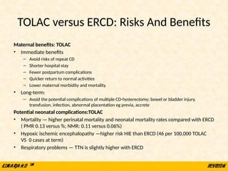
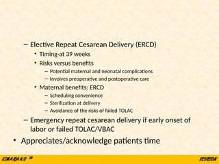
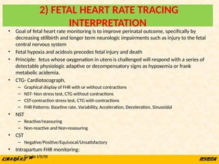
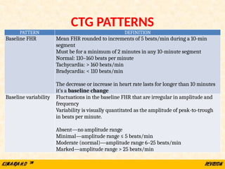
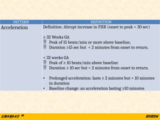
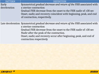
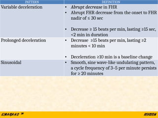
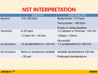
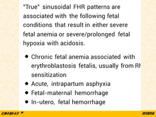
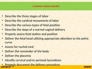
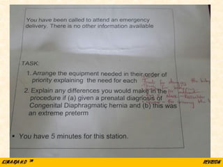
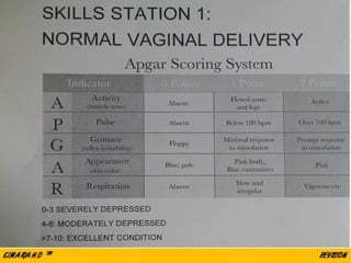
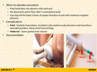
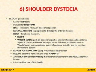
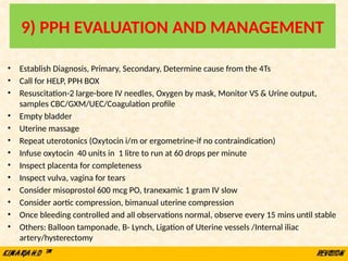
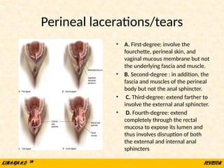
BASIC OBSTETRICS SKILLS
MBChB IV
Prof Eunice Cheserem
&
Dr Alfred Osoti
&
Dr Rose J. Kosgei
10 OBSTETRICS SKILL STATIONS
1) Counseling in Obstetrics-1 Previous Scar
2) Fetal heart rate tracing interpretation – normal and abnormal tracings
3) Normal vaginal delivery
4) Breech Vaginal Delivery
5) Assisted vaginal delivery-vacuum extraction
6) Shoulder dystocia
7) Sterile speculum examination
8) Digital vaginal examination
9) PPH evaluation and management
10) Perineal tear & episiotomy repair
11) Leopold’s maneuver
12) Partograph –Obstructed labour
1) COUNSELING IN OBSTETRICS
1 PREVIOUS SCAR
• Greet patient, Introduces self, Sits squarely, Open Posture, Leans
Forward, Eye Contact, Relax
• Demonstrate understanding of the underlying condition and
management
– Explain the diagnosis-1 previous scar
• Review previous scar- Indications as non-recurrent, outcome, complications
– Elective delivery options - ERCD vs TOLAC/VBAC
• ERCD-Elective Repeat Cesarean Delivery
• TOLAC-Trial Of Labor After Previous Cesarean Delivery
• VBAC-Vaginal Birth After Cesarean Delivery
• TOLAC/VBAC
– Current pregnancy-confirm no contraindication to TOLAC/VBAC
– TOLAC/VBAC -Success rate
– Potential maternal/neonatal complications
APPROPRIATE CANDIDATES
FOR TOLAC
• One prior low transverse uterine
incision
– Success rate of TOLAC 60 - 70 %
– Uterine rupture rate of about 0.7 %
– Success rates are higher if prior vaginal
delivery
• Clinically adequate pelvis
• Appropriate fetal weight < 3.5 kg
• No other uterine scars or previous
rupture
• Interpregnancy interval of >18 months
• Physicians and facility immediately
available throughout active labor
capable of monitoring labor and
performing an emergency cesarean
delivery
CONTRAINDICATIONS TO
TOLAC
• High risk for uterine rupture
– Previous classical or J or T-shaped
incision, or extensive transfundal
uterine surgery or myoma resections
– Previous uterine rupture
– ≥2 previous scars
• Medical complications that
preclude vaginal delivery
• Obstetric indications of cesarean
delivery e.g. placenta previa,
footling breech presentation
• Lack of an appropriate
facility — surgical, anesthesia,
nursing, pediatric staff and
laboratory resources to provide
multiple transfusions
• Antepartum predictors of success
– Indication for prior cesarean delivery
– Fetal malpresentation (75%)
– Non-reassuring fetal heart rate pattern (60%)
– Failure to progress or CPD if non-recurrent cause (54%)
– History of prior vaginal delivery
– Before or after previous CS (OR prior vaginal delivery
3.90; prior VBAC 4.76)
• Demographic factors
– Hispanic, African American, and Asian women more
likely to pursue TOLAC, but are less likely to have a
successful VBAC versus non-Hispanic and white women
– Increasing maternal age, single marital status, and less
than 12 years of education have reduced likelihood
– Age >35 less likely to have a successful VBAC and more
likely to experience complications
– Success increases with increasing maternal height
– Success lower in obese women
• Interpregnancy interval of less than six
months risk factor for uterine rupture
and maternal morbidity
• Maternal medical disease
– Hypertension, diabetes, asthma,
renal disease, and heart disease
may reduce likelihood
• Intrapartum factors
– Admission labor status:
• Spontaneous labor or high
bishop score more likely to
succeed than induced labor or
low Bishop score
– Fetal macrosomia
• Wt. > 3.5 kg reduces likelihood
TOLAC/VBAC -Success rate
TOLAC versus ERCD: Risks And Benefits
Maternal benefits: TOLAC
• Immediate benefits
– Avoid risks of repeat CD
– Shorter hospital stay
– Fewer postpartum complications
– Quicker return to normal activities
– Lower maternal morbidity and mortality.
• Long-term:
– Avoid the potential complications of multiple CD-hysterectomy; bowel or bladder injury,
transfusion, infection, abnormal placentation eg previa, accrete
Potential neonatal complications:TOLAC
• Mortality — higher perinatal mortality and neonatal mortality rates compared with ERCD
( PMR 0.13 versus %; NMR: 0.11 versus 0.06%)
• Hypoxic ischemic encephalopathy —higher risk HIE than ERCD (46 per 100,000 TOLAC
VS 0 cases at term)
• Respiratory problems — TTN is slightly higher with ERCD
– Elective Repeat Cesarean Delivery (ERCD)
• Timing-at 39 weeks
• Risks versus benefits
– Potential maternal and neonatal complications
– Involves preoperative and postoperative care
• Maternal benefits: ERCD
– Scheduling convenience
– Sterilization at delivery
– Avoidance of the risks of failed TOLAC
– Emergency repeat cesarean delivery if early onset of
labor or failed TOLAC/VBAC
• Appreciates/acknowledge patients time
2) FETAL HEART RATE TRACING
INTERPRETATION
• Goal of fetal heart rate monitoring is to improve perinatal outcome, specifically by
decreasing stillbirth and longer term neurologic impairments such as injury to the fetal
central nervous system
• Fetal hypoxia and acidosis precedes fetal injury and death
• Principle: fetus whose oxygenation in utero is challenged will respond with a series of
detectable physiologic adaptive or decompensatory signs as hypoxemia or frank
metabolic acidemia.
• CTG- Cardiotocograph,
– Graphical display of FHR with or without contractions
– NST- Non stress test, CTG without contractions
– CST-contraction stress test, CTG with contractions
– FHR Patterns: Baseline rate, Variability, Acceleration, Deceleration, Sinusoidal
• NST
– Reactive/reassuring
– Non-reactive and Non-reassuring
• CST
– Negative/Positive/Equivocal/Unsatisfactory
• Intrapartum FHR monitoring:
– Grade I/II/III
CTG PATTERNS
PATTERN DEFINITION
Baseline FHR Mean FHR rounded to increments of 5 beats/min during a 10-min
segment
Must be for a minimum of 2 minutes in any 10-minute segment
Normal: 110–160 beats per minute
Tachycardia: > 160 beats/min
Bradycardia: < 110 beats/min
The decrease or increase in heart rate lasts for longer than 10 minutes
it’s a baseline change
Baseline variability Fluctuations in the baseline FHR that are irregular in amplitude and
frequency
Variability is visually quantitated as the amplitude of peak-to-trough
in beats per minute.
Absent—no amplitude range
Minimal—amplitude range ≤ 5 beats/min
Moderate (normal)—amplitude range 6–25 beats/min
Marked—amplitude range > 25 beats/min
PATTERN DEFINITION
Acceleration Definition: Abrupt increase in FHR (onset to peak < 30 sec)
≥ 32 Weeks GA
 Peak of 15 beats/min or more above baseline,
 Duration ≥15 sec but < 2 minutes from onset to return.
< 32 weeks GA
 Peak of ≥ 10 beats/min above baseline
 Duration ≥ 10 sec but < 2 minutes from onset to return.
• Prolonged acceleration: lasts ≥ 2 minutes but < 10 minutes
in duration
• Baseline change: an acceleration lasting ≥10 minutes
PATTERN DEFINITION
Early
deceleration
Symmetrical gradual decrease and return of the FHR associated with
a uterine contraction
Gradual FHR decrease from the onset to the FHR nadir of ≥30 sec
Onset, nadir, and recovery coincident with beginning, peak, and end
of contraction, respectively.
Late deceleration Symmetrical gradual decrease and return of the FHR associated with
a uterine contraction
Gradual FHR decrease from the onset to the FHR nadir of ≥30 sec
Nadir after the peak of the contraction.
Onset, nadir, and recovery occur after beginning, peak, and end of
contraction respectively
PATTERN DEFINITION
Variable deceleration • Abrupt decrease in FHR
• Abrupt FHR decrease from the onset to FHR
nadir of ≤ 30 sec
• Decrease ≥ 15 beats per min, lasting ≥15 sec,
<2 min in duration
Prolonged deceleration • Decrease ≥15 beats per min, lasting ≥2
minutes < 10 min
• Deceleration ≥10 min is a baseline change
Sinusoidal • Smooth, sine wave-like undulating pattern,
a cycle frequency of 3–5 per minute persists
for ≥ 20 minutes
NST INTERPRETATION
REACTIVE NON-REACTIVE
Baseline 110–160 bpm Bradycardia <110 bpm
Tachycardia > 160 bpm
Erratic or rising baseline
Variability 6–25 bpm
£ 5 bpm for < 40 min
£ 5 (absent or minimal) >40 min
>25bpm >10min.
Sinusoidal
Accelerations ³2 accelerations in < 40 min < 2 accelerations in ≥40 min
Decelerations None or occasional variable
< 30 sec
Variable decelerations ≥30 sec
Prolonged decelerations
CST-INTERPRETATION
Negative: no late or significant variable decelerations
Positive: late decelerations after ≥50% of contractions
Equivocal–suspicious: intermittent late decelerations or significant
variable decelerations
Equivocal: FHR decelerations in the presence of contractions more
frequent than every 2 minutes or lasting longer than 90 seconds
Unsatisfactory: fewer than three contractions in 10 minutes or an
uninterpretable tracing
Three-Tiered Intrapartum Fetal Heart Rate
Interpretation System
Category I
• Baseline rate: 110–160 bpm
• Baseline FHR variability: moderate
• No Late or variable decelerations
• Early decelerations present or absent
• Accelerations: present or absent
Category II
• FHR tracings does not meet criteria for either category as Category I or Category III
and is considered indeterminate
Category III: -Predictive of abnormal fetal acid status at time of observation
• Absent baseline FHR variability and any of:
– Recurrent late decelerations
– Recurrent variable decelerations
– Bradycardia
• Sinusoidal pattern
• Make preparations for immediate delivery while initiating resuscitative measures
3) NORMAL VAGINAL DELIVERY
• Describe the three stages of labor
• Describe the cardinal movements of labor
• Describe the various types of fetal position
• Describe the steps of a normal vaginal delivery
• Properly assess fetal station and position
• Deliver the fetal head utilizing appropriate attention to the pelvic
curve
• Assess for nuchal cord
• Deliver the remainder of the body
• Deliver the placenta
• Identify cervical and/or perineal lacerations
• Properly document the delivery procedure
4) BREECH VAGINAL DELIVERY
• TERM BREECH TRIAL (Lancet, 2000) planned CS is associated with improved neonatal outcomes
compared to planned vaginal birth (poor neonatal outcomes of 1.6% -v- .5.0%). However
patients may present in second stage necessitating vaginal breech delivery
• Procedure
1) Confirm diagnosis and no contraindications to vaginal delivery - Leopold maneuvers
o Identify fetal pole in the fundus. Breech gives sensation of large nodular mass.
o Palms on the sides of abdomen. Identify (hard smooth) back on one side and irregular mobile parts on the other
side
o Using thumb and fingers grasp presenting part (diagnose whether engagement has occurred)
o Facing mothers feet, use fingertips to palpate presenting parts.
2) Call Consultant/staff experienced in Breech Delivery
3) Consider episiotomy
4) “Hands off” – do not assist in the delivery of the baby until maternal efforts have resulted in expulsion
of the fetus at least to the scapulae
5) Pull down a small loop of cord to prevent traction on the cord
6) Wrap body in a towel to allow for support and grip
7) Gentle rotation will usually allow delivery of arms. If not use the Lovsett’s maneuver. Follow humerus
down and rotate each arm across the chest and out.
8) The aftercoming head may deliver spontaneously. If not, use the Mariceau-smellie-veit maneuver. The
trunk of the baby lies on the operator’s right forearm. The head is flexed by applying pressure to the
cheek bones and upper lip while gentle traction is applied on the shoulders with the left hand.
9) If the head is entrapped, uterine relaxation in theatre with GA (halothane) may allow delivery.
• Complications
– Cord prolapse
– Birth trauma as a result of extended arm or head,
incomplete dilatation of the cervix or CPD
– Asphyxia from cord prolapse, cord compression, placental
detachment or arrested head
– Damage to abdominal organs
– Broken neck
• Documentation: detailed, clear and comprehensive
including details of counseling and the identity of all
those involved in the procedures.
5) ASSISTED VAGINAL DELIVERY-VACUUM
EXTRACTION
• ABCDEFGHI
• Prerequisites:
– ≥ 36 weeks
– Cephalic, Vertex, Full cervical dilation, Fetal head at station 0, Descent not more than 1/5
– Mother conscious & cooperative
• Procedure:
– Obtain verbal informed consent, Put on sterile gown and sterile gloves, Confirm
equipment is working
– Position the mother in semi-lithotomy, Empty bladder, Confirm full cervical dilation,
Identify posterior fontanel, Identify sagittal suture, Assess for episiotomy
– Place center of cup 2 cm anterior to the posterior fontanel/flexion point on the sagittal
suture
– Stabilize the suction cup with two fingers
– Check and free maternal tissues
– Create vacuum of 0.2kg/sq.cm (yellow)
– To pull increase vacuum pressure to 0.8kg/sq.cm (green)
– Apply traction during uterine contraction only perpendicular to the cup
• When to abandon procedure
– Fetal head does not advance with each pull
– No descend to pelvic floor after 3 contractions/pulls
– Cup slips off the head 3 times at proper direction of pull with maximum negative
pressure
• Complications
– Fetal –Cephalo-haematoma, localized scalp oedema scalp abrasions and lacerations,
Neonatal jaundice, Intracranial haemorrhage
– Maternal – lower genital tract injuries
• Documentation
6) SHOULDER DYSTOCIA
• Definition: need for additional obstetric maneuvers to effect delivery of fetal shoulders
at the time of vaginal delivery
• Pathophysiology: impaction of anterior fetal shoulder on maternal pubic bone causing
delay in delivery of the shoulder after head has been delivered
• Risk Factors: Mainly due to increased fetal birth weight: Maternal: High BMI,
Multiparity, Advanced Maternal Age, Diabetes, Postterm Pregnancy, Previous
macrosomic infant, excessive weight gain in pregnancy, Maternal birth weight over
4000 g.
• Warning Signs: -prolonged second stage, “turtling” (head retracts into perineum after
delivery)
• Management: goal  release anterior shoulder from entrapment, Prevent fetal
asphyxia and permanent Erb's palsy, Avoid physical injury (eg, bone fractures,
maternal trauma).
• Complications:
– Maternal: PPH, cervical/vaginal lacerations
– Fetal:clavicle/humerus fractures, brachial plexus injuries, birth asphyxia, death
PATHOPHYSIOLOGY
• Normal:
– The fetal biacromial diameter enters the pelvis at an oblique angle
with the posterior shoulder ahead of the anterior one, rotating to the
anterior-posterior position at the pelvic outlet with external rotation
of the fetal head.
– The anterior shoulder can then slide under the symphysis pubis for
delivery
• Abnormal/shoulder dystocia:
– The fetal shoulders remain in an anterior-posterior position during
descent OR descend simultaneously rather than sequentially into the
pelvic inlet
– THE anterior shoulder becomes impacted behind the symphysis pubis
– AND the sacral promontory may obstruct the posterior shoulder.
6) SHOULDER DYSTOCIA
• HELPERR (pneumonic):
– Call for HELP-team
– Evaluate for EPISOTOMY
– LEGS – McRoberts Manuver –knee chest position
– EXTERNAL PRESSURE (suprapubic)-to dislodge the anterior shoulder
– ENTER – Rotational manuvers,
• RUBINS
• WOOD’S SCREW: push on posterior aspect of anterior shoulder and on anterior
aspect of posterior shoulder and try to rotate shoulders to oblique, Reverse
Wood’s Screw: push on anterior aspect of posterior shoulder and try to rotate
shoulders to oblique
– REMOVE POSTERIOR ARM - grasp foetal elbow, not shoulder
– ROLL the patient to her hands and knee (GASKIN’S)
– Zavanelli/Gunn-Zavanelli-O'Leary maneuver : Replacement of fetal head, Abdominal
Rescue
– Intentional fracture of the clavicle
• Call for HELP AND PREPARE
– Nursing, anesthesia, obstetric, and pediatric staff
should be called to the room to provide assistance as
needed
– Instructions given in a clear and calm manner to
gravida and personnel
– No pushing while preparations are made and
maneuvers are undertaken to reposition the fetus
– Patient positioned with her buttocks flush with the
edge of the bed to provide optimal access for
executing maneuvers to affect delivery
– Empty bladder
• Evaluate for EPISIOTOMY
LEGS – McROBERTS MANUVER
– Hyperflexion and abduction of the hips causing cephalad rotation of the symphysis pubis
and flattening of the lumbar lordosis that frees the impacted shoulder
– Two assistants, each grasps a maternal leg and sharply flexes the thigh back against the
abdomen
– Relieves shoulder dystocia via
• Marked cephalad rotation of the symphysis pubis and subsequent flattening of the sacrum, thus
removing the sacral promontory as an obstruction site
• By bringing the pelvic inlet into the plane perpendicular to the maximum expulsive force, pushing
efficiency improves significantly
– NO change in diameter, McRoberts maneuver does not change the actual dimensions of
the maternal pelvis
EXTERNAL PRESSURE (SUPRAPUBIC)
– An assistant applies pressure suprapubically with the palm or fist, directing the
pressure on the anterior shoulder both downward (to below the pubic bone) and
laterally (toward the baby's face or sternum)
– Performed in conjunction with McRoberts maneuver
– Adducts the shoulders or bring them into an oblique plane, the oblique diameter is the
widest diameter of the maternal pelvis
– Useful in mild cases and those from impacted anterior shoulder
ENTER – Rotational maneuvers
• REMOVE POSTERIOR ARM
– Introduce a hand into the vagina to locate the posterior shoulder and
arm : If the fetal abdomen faces the maternal right, the operator's left
hand should be used; if the fetal abdomen faces the maternal left, the
right hand is used.
– The posterior arm should be identified and followed to the elbow. If the
elbow is flexed, the operator can grasp the forearm and hand and pull out
the arm. If it is extended, pressure is applied in the antecubital fossa. This
flexes the elbow across the fetal chest and allows the forearm or hand to
be grasped.
– The arm is then pulled out of the vagina, which brings the posterior
shoulder into the pelvis and reduces the shoulder diameter by 2 to 3 cm
– If the anterior shoulder cannot be delivered at this point, the fetus can be
rotated and the procedure repeated for the anterior (now posterior) arm.
• RUBIN MANEUVER
– Adducts fetal shoulder so that the shoulders are displaced from the
anteroposterior diameter of the inlet, thereby allowing the posterior
arm to enter the pelvis
– Requires adequate anesthesia
• One hand placed in the vagina and on the back surface of the posterior fetal
shoulder, and then rotate it anteriorly (towards the fetal face). If the fetal spine
is on the maternal left, the operator's right hand is used; the left hand is used if
the fetal spine is on the maternal right
• Alternatively, hand placed on the back surface of the anterior fetal shoulder, if
it is more accessible.
• WOODS SCREW MANEUVER
• Rotates the fetus by exerting pressure on the anterior, clavicular surface of the
posterior fetal shoulder to turn the fetus until the anterior shoulder emerges
from behind the maternal symphysis
• If the fetal spine is on the maternal left, the operator uses the left hand to push
on the clavicle of the posterior arm and rotate the baby 180 degrees in a
counterclockwise direction
• ROLL THE PATIENT TO HER HANDS AND KNEES, GASKIN ALL-FOURS
– Places the mother on her hands and knees, OR racing start or ‘sprinter’
position NOT knee-chest position
– Delivery achieved by gentle downward traction on the posterior shoulder
(the shoulder against the maternal sacrum) or upward traction on the
anterior shoulder (the shoulder against the maternal symphysis)
• ZAVANELLI/GUNN-ZAVANELLI-O'LEARY MANEUVER
• Replacement of the fetal head in the pelvis, then cesarean delivery
– Administer tocolytic eg terbutaline (0.25 mg SC/nifedipine/nitroglycerin)
– Rotate the head back to an occiput anterior position (reversal of
restitution)
– Flex head from its extended position and push it as far cephalad as
possible using firm pressure with palm of one hand.
– The other hand may be used to depress the perineum
– Proceed with cesarean delivery.
Others
• Abdominal rescue
– A low transverse hysterotomy, then transabdominal manual rotation of
the anterior shoulder to the oblique diameter fetus delivered vaginally
• Intentional clavicular fracture
– Intentional-to shorten the biacromial diameter by pulling the anterior
clavicle outward
– Difficult; can lead to injury of underlying vascular and pulmonary
structures
• Documentation : Clear and complete documentation in the
medical record is critical
Rotation of posterior shoulder-Woods
MANUAL REMOVAL OF POSTERIOR
SHOULDER GASKIN
RUBIN ZAVANELLI
7) STERILE SPECULUM EXAMINATION
• Review indications-e.g. preterm premature rupture of fetal membranes
• Thoroughly explain the procedure to the patient
• Sterile gloving
• Report findings on inspection of external genitalia
• Swabs vulva -5 swab technique
• Application of sterile towels
• Lubricates speculum
• Separates the labia and exposes introitus with non non-dominant hand and
introduces lubricated speculum with dominant hand
• Position speculum to asses cervix
• Report on status of vaginal walls, Cervix (dilatation, position, defects,
inflammation)
• May report on cord, colour of liquor, pooling on valsalva , any other
discharge/blood
8) DIGITAL VAGINAL EXAMINATION
• Thoroughly explain the procedure to the patient
• Assess the patient between contractions when most relaxed
• Sterile technique: 5 sterile gloves, lubricates the first two fingers of the
dominant hand
• Use the non dominant hand to spread the labia minora and the dominant
‐
hand to assess the cervix
• Assess cervical dilation, effacement, consistency, position
• Identify the presenting part, palpate for fontanelles and sutures
• Rule out common malpresentations: breech, shoulder, brow, face,
compound
• Assess for the presence of a prolapsed umbilical cord
• Dispose the contaminated equipment
• Document on a partograph
• Institute appropriate management
9) PPH EVALUATION AND MANAGEMENT
• Establish Diagnosis, Primary, Secondary, Determine cause from the 4Ts
• Call for HELP, PPH BOX
• Resuscitation-2 large-bore IV needles, Oxygen by mask, Monitor VS & Urine output,
samples CBC/GXM/UEC/Coagulation profile
• Empty bladder
• Uterine massage
• Repeat uterotonics (Oxytocin i/m or ergometrine-if no contraindication)
• Infuse oxytocin 40 units in 1 litre to run at 60 drops per minute
• Inspect placenta for completeness
• Inspect vulva, vagina for tears
• Consider misoprostol 600 mcg PO, tranexamic 1 gram IV slow
• Consider aortic compression, bimanual uterine compression
• Once bleeding controlled and all observations normal, observe every 15 mins until stable
• Others: Balloon tamponade, B- Lynch, Ligation of Uterine vessels /Internal iliac
artery/hysterectomy
Inflate Condom with
water till no further
bleeding is occuring
(usually about 300-
500 mls )
UTERUS
Foleys Catheter
Condom
String
Apply clamp to keep water within
Condom after inflation
Giving set
Water/NS
OR
syringe
THE CONDOM TAMPONADE
Clean
water
Steps
1. Place condom over balloon end of Foleys catheter
2. Tie lower end of condom snugly below level of the
balloon using suture / string. Tie should be tight
enough to prevent leakage of water but should not
strangulate catheter and prevent inflow of water into
condom. Check for leakage by inflating ballon with
about 20cc water.
3. Aseptically place the condom end high into
uterine cavity by digital manipulation or with aid of
speculum and forceps
4. Inflate CT by connecting open/outlet end of
catheter to giving set connected to infusion bag or
use clean water with aid of large syringe. (cut the
giving set at level of rubber to enable it fit into
catheter)
5. Inflate condom with water or saline to about
300- 500 mls (or to amount at which no further
bleeding is observed).
6. Clamp catheter when desired volume is
achieved and bleeding is controlled.
7. Maintain In-situ for 24 hours if bleeding
controlled and patient is stable.
8. Give Broad spectrum antibiotic cover
9. Monitor patient closely, resuscitate and/or treat
complications e.g. shock, coagulopathy
10. When patient is stable (after 24 hours) slowly deflate
condom by letting out 50 mls of water/saline every hour.
11. Re-inflate to previous level if bleeding reoccurs whilst
deflating.
12. CT may be kept in place for up to 24 hours
13. If Bleeding is not controlled within 15 mins of initial
insertion of CT abandon procedure and seek surgical
intervention immediately.
The B-Lynch Suture
Step 1: Using Absorbale large suture.
In-out-over…In-out-over…In-out-tie
B-Lynch Suture #2
Courtesy: Lynch BC, Coker A, Laval AH et al. The B_Lynch technique for control of Masive PPH,
An Alternative to Hysterectomy. Five Cases Reported. Br. J. Obstet Gynecol 1997, 104 327-376
B-Lynch Suture #3
10) PERINEAL TEAR & EPISIOTOMY
REPAIR
• Episiotomy
• is a surgical incision of the perineum performed to widen the
vaginal opening to facilitate the delivery of an infant
• May be associated with increased risk of extension to severe
perineal lacerations, dyspareunia, and future pelvic floor
dysfunction: Types: Midline and mediolateral
• Tears
• Are spontaneous perineal lacerations arising from perineal
trauma at delivery. Degrees 1-4
• Repair in layers
• Postoperative care
• Pain control, perineal hygiene, sitz baths
Technique
• Place an anchor stitch above the wound apex
– Close the vaginal mucosa and submucosa with continuous
interlocking stitches to close the vaginal incision and
reapproximate the cut margins of the hymenal ring using
an absorbable 2–0 or 3–0 suture
• Close the fascia and muscles to restore the perineal
body using a continuous absorbable 2–0 or 3–0 suture
• Carry the continuous suture upward as a subcuticular
stitch
• Tie the final knot proximal to the hymenal ring.
Perineal lacerations/tears
• A. First-degree: involve the
fourchette, perineal skin, and
vaginal mucous membrane but not
the underlying fascia and muscle.
• B. Second-degree : in addition, the
fascia and muscles of the perineal
body but not the anal sphincter.
• C. Third-degree: extend farther to
involve the external anal sphincter.
• D. Fourth-degree: extend
completely through the rectal
mucosa to expose its lumen and
thus involves disruption of both
the external and internal anal
sphincters
Obs OSCE dry Run (Five minutes)
SCENARIO
You are provided with a model and equipment.
(pelvic model and a Kiwi vacuum extractor)
• Question
1. Using the model provided, demonstrate the
correct placement of the cup
• End

OBSTETRICS SKILLS. PRESENTATION pptx

  • 1.
    BASIC OBSTETRICS SKILLS MBChBIV Prof Eunice Cheserem & Dr Alfred Osoti & Dr Rose J. Kosgei
  • 2.
    10 OBSTETRICS SKILLSTATIONS 1) Counseling in Obstetrics-1 Previous Scar 2) Fetal heart rate tracing interpretation – normal and abnormal tracings 3) Normal vaginal delivery 4) Breech Vaginal Delivery 5) Assisted vaginal delivery-vacuum extraction 6) Shoulder dystocia 7) Sterile speculum examination 8) Digital vaginal examination 9) PPH evaluation and management 10) Perineal tear & episiotomy repair 11) Leopold’s maneuver 12) Partograph –Obstructed labour
  • 3.
    1) COUNSELING INOBSTETRICS 1 PREVIOUS SCAR • Greet patient, Introduces self, Sits squarely, Open Posture, Leans Forward, Eye Contact, Relax • Demonstrate understanding of the underlying condition and management – Explain the diagnosis-1 previous scar • Review previous scar- Indications as non-recurrent, outcome, complications – Elective delivery options - ERCD vs TOLAC/VBAC • ERCD-Elective Repeat Cesarean Delivery • TOLAC-Trial Of Labor After Previous Cesarean Delivery • VBAC-Vaginal Birth After Cesarean Delivery • TOLAC/VBAC – Current pregnancy-confirm no contraindication to TOLAC/VBAC – TOLAC/VBAC -Success rate – Potential maternal/neonatal complications
  • 4.
    APPROPRIATE CANDIDATES FOR TOLAC •One prior low transverse uterine incision – Success rate of TOLAC 60 - 70 % – Uterine rupture rate of about 0.7 % – Success rates are higher if prior vaginal delivery • Clinically adequate pelvis • Appropriate fetal weight < 3.5 kg • No other uterine scars or previous rupture • Interpregnancy interval of >18 months • Physicians and facility immediately available throughout active labor capable of monitoring labor and performing an emergency cesarean delivery CONTRAINDICATIONS TO TOLAC • High risk for uterine rupture – Previous classical or J or T-shaped incision, or extensive transfundal uterine surgery or myoma resections – Previous uterine rupture – ≥2 previous scars • Medical complications that preclude vaginal delivery • Obstetric indications of cesarean delivery e.g. placenta previa, footling breech presentation • Lack of an appropriate facility — surgical, anesthesia, nursing, pediatric staff and laboratory resources to provide multiple transfusions
  • 5.
    • Antepartum predictorsof success – Indication for prior cesarean delivery – Fetal malpresentation (75%) – Non-reassuring fetal heart rate pattern (60%) – Failure to progress or CPD if non-recurrent cause (54%) – History of prior vaginal delivery – Before or after previous CS (OR prior vaginal delivery 3.90; prior VBAC 4.76) • Demographic factors – Hispanic, African American, and Asian women more likely to pursue TOLAC, but are less likely to have a successful VBAC versus non-Hispanic and white women – Increasing maternal age, single marital status, and less than 12 years of education have reduced likelihood – Age >35 less likely to have a successful VBAC and more likely to experience complications – Success increases with increasing maternal height – Success lower in obese women • Interpregnancy interval of less than six months risk factor for uterine rupture and maternal morbidity • Maternal medical disease – Hypertension, diabetes, asthma, renal disease, and heart disease may reduce likelihood • Intrapartum factors – Admission labor status: • Spontaneous labor or high bishop score more likely to succeed than induced labor or low Bishop score – Fetal macrosomia • Wt. > 3.5 kg reduces likelihood TOLAC/VBAC -Success rate
  • 6.
    TOLAC versus ERCD:Risks And Benefits Maternal benefits: TOLAC • Immediate benefits – Avoid risks of repeat CD – Shorter hospital stay – Fewer postpartum complications – Quicker return to normal activities – Lower maternal morbidity and mortality. • Long-term: – Avoid the potential complications of multiple CD-hysterectomy; bowel or bladder injury, transfusion, infection, abnormal placentation eg previa, accrete Potential neonatal complications:TOLAC • Mortality — higher perinatal mortality and neonatal mortality rates compared with ERCD ( PMR 0.13 versus %; NMR: 0.11 versus 0.06%) • Hypoxic ischemic encephalopathy —higher risk HIE than ERCD (46 per 100,000 TOLAC VS 0 cases at term) • Respiratory problems — TTN is slightly higher with ERCD
  • 7.
    – Elective RepeatCesarean Delivery (ERCD) • Timing-at 39 weeks • Risks versus benefits – Potential maternal and neonatal complications – Involves preoperative and postoperative care • Maternal benefits: ERCD – Scheduling convenience – Sterilization at delivery – Avoidance of the risks of failed TOLAC – Emergency repeat cesarean delivery if early onset of labor or failed TOLAC/VBAC • Appreciates/acknowledge patients time
  • 8.
    2) FETAL HEARTRATE TRACING INTERPRETATION • Goal of fetal heart rate monitoring is to improve perinatal outcome, specifically by decreasing stillbirth and longer term neurologic impairments such as injury to the fetal central nervous system • Fetal hypoxia and acidosis precedes fetal injury and death • Principle: fetus whose oxygenation in utero is challenged will respond with a series of detectable physiologic adaptive or decompensatory signs as hypoxemia or frank metabolic acidemia. • CTG- Cardiotocograph, – Graphical display of FHR with or without contractions – NST- Non stress test, CTG without contractions – CST-contraction stress test, CTG with contractions – FHR Patterns: Baseline rate, Variability, Acceleration, Deceleration, Sinusoidal • NST – Reactive/reassuring – Non-reactive and Non-reassuring • CST – Negative/Positive/Equivocal/Unsatisfactory • Intrapartum FHR monitoring: – Grade I/II/III
  • 9.
    CTG PATTERNS PATTERN DEFINITION BaselineFHR Mean FHR rounded to increments of 5 beats/min during a 10-min segment Must be for a minimum of 2 minutes in any 10-minute segment Normal: 110–160 beats per minute Tachycardia: > 160 beats/min Bradycardia: < 110 beats/min The decrease or increase in heart rate lasts for longer than 10 minutes it’s a baseline change Baseline variability Fluctuations in the baseline FHR that are irregular in amplitude and frequency Variability is visually quantitated as the amplitude of peak-to-trough in beats per minute. Absent—no amplitude range Minimal—amplitude range ≤ 5 beats/min Moderate (normal)—amplitude range 6–25 beats/min Marked—amplitude range > 25 beats/min
  • 10.
    PATTERN DEFINITION Acceleration Definition:Abrupt increase in FHR (onset to peak < 30 sec) ≥ 32 Weeks GA  Peak of 15 beats/min or more above baseline,  Duration ≥15 sec but < 2 minutes from onset to return. < 32 weeks GA  Peak of ≥ 10 beats/min above baseline  Duration ≥ 10 sec but < 2 minutes from onset to return. • Prolonged acceleration: lasts ≥ 2 minutes but < 10 minutes in duration • Baseline change: an acceleration lasting ≥10 minutes
  • 11.
    PATTERN DEFINITION Early deceleration Symmetrical gradualdecrease and return of the FHR associated with a uterine contraction Gradual FHR decrease from the onset to the FHR nadir of ≥30 sec Onset, nadir, and recovery coincident with beginning, peak, and end of contraction, respectively. Late deceleration Symmetrical gradual decrease and return of the FHR associated with a uterine contraction Gradual FHR decrease from the onset to the FHR nadir of ≥30 sec Nadir after the peak of the contraction. Onset, nadir, and recovery occur after beginning, peak, and end of contraction respectively
  • 12.
    PATTERN DEFINITION Variable deceleration• Abrupt decrease in FHR • Abrupt FHR decrease from the onset to FHR nadir of ≤ 30 sec • Decrease ≥ 15 beats per min, lasting ≥15 sec, <2 min in duration Prolonged deceleration • Decrease ≥15 beats per min, lasting ≥2 minutes < 10 min • Deceleration ≥10 min is a baseline change Sinusoidal • Smooth, sine wave-like undulating pattern, a cycle frequency of 3–5 per minute persists for ≥ 20 minutes
  • 13.
    NST INTERPRETATION REACTIVE NON-REACTIVE Baseline110–160 bpm Bradycardia <110 bpm Tachycardia > 160 bpm Erratic or rising baseline Variability 6–25 bpm £ 5 bpm for < 40 min £ 5 (absent or minimal) >40 min >25bpm >10min. Sinusoidal Accelerations ³2 accelerations in < 40 min < 2 accelerations in ≥40 min Decelerations None or occasional variable < 30 sec Variable decelerations ≥30 sec Prolonged decelerations
  • 14.
    CST-INTERPRETATION Negative: no lateor significant variable decelerations Positive: late decelerations after ≥50% of contractions Equivocal–suspicious: intermittent late decelerations or significant variable decelerations Equivocal: FHR decelerations in the presence of contractions more frequent than every 2 minutes or lasting longer than 90 seconds Unsatisfactory: fewer than three contractions in 10 minutes or an uninterpretable tracing
  • 15.
    Three-Tiered Intrapartum FetalHeart Rate Interpretation System Category I • Baseline rate: 110–160 bpm • Baseline FHR variability: moderate • No Late or variable decelerations • Early decelerations present or absent • Accelerations: present or absent Category II • FHR tracings does not meet criteria for either category as Category I or Category III and is considered indeterminate Category III: -Predictive of abnormal fetal acid status at time of observation • Absent baseline FHR variability and any of: – Recurrent late decelerations – Recurrent variable decelerations – Bradycardia • Sinusoidal pattern • Make preparations for immediate delivery while initiating resuscitative measures
  • 19.
    3) NORMAL VAGINALDELIVERY • Describe the three stages of labor • Describe the cardinal movements of labor • Describe the various types of fetal position • Describe the steps of a normal vaginal delivery • Properly assess fetal station and position • Deliver the fetal head utilizing appropriate attention to the pelvic curve • Assess for nuchal cord • Deliver the remainder of the body • Deliver the placenta • Identify cervical and/or perineal lacerations • Properly document the delivery procedure
  • 22.
    4) BREECH VAGINALDELIVERY • TERM BREECH TRIAL (Lancet, 2000) planned CS is associated with improved neonatal outcomes compared to planned vaginal birth (poor neonatal outcomes of 1.6% -v- .5.0%). However patients may present in second stage necessitating vaginal breech delivery • Procedure 1) Confirm diagnosis and no contraindications to vaginal delivery - Leopold maneuvers o Identify fetal pole in the fundus. Breech gives sensation of large nodular mass. o Palms on the sides of abdomen. Identify (hard smooth) back on one side and irregular mobile parts on the other side o Using thumb and fingers grasp presenting part (diagnose whether engagement has occurred) o Facing mothers feet, use fingertips to palpate presenting parts. 2) Call Consultant/staff experienced in Breech Delivery 3) Consider episiotomy 4) “Hands off” – do not assist in the delivery of the baby until maternal efforts have resulted in expulsion of the fetus at least to the scapulae 5) Pull down a small loop of cord to prevent traction on the cord 6) Wrap body in a towel to allow for support and grip 7) Gentle rotation will usually allow delivery of arms. If not use the Lovsett’s maneuver. Follow humerus down and rotate each arm across the chest and out. 8) The aftercoming head may deliver spontaneously. If not, use the Mariceau-smellie-veit maneuver. The trunk of the baby lies on the operator’s right forearm. The head is flexed by applying pressure to the cheek bones and upper lip while gentle traction is applied on the shoulders with the left hand. 9) If the head is entrapped, uterine relaxation in theatre with GA (halothane) may allow delivery.
  • 23.
    • Complications – Cordprolapse – Birth trauma as a result of extended arm or head, incomplete dilatation of the cervix or CPD – Asphyxia from cord prolapse, cord compression, placental detachment or arrested head – Damage to abdominal organs – Broken neck • Documentation: detailed, clear and comprehensive including details of counseling and the identity of all those involved in the procedures.
  • 25.
    5) ASSISTED VAGINALDELIVERY-VACUUM EXTRACTION • ABCDEFGHI • Prerequisites: – ≥ 36 weeks – Cephalic, Vertex, Full cervical dilation, Fetal head at station 0, Descent not more than 1/5 – Mother conscious & cooperative • Procedure: – Obtain verbal informed consent, Put on sterile gown and sterile gloves, Confirm equipment is working – Position the mother in semi-lithotomy, Empty bladder, Confirm full cervical dilation, Identify posterior fontanel, Identify sagittal suture, Assess for episiotomy – Place center of cup 2 cm anterior to the posterior fontanel/flexion point on the sagittal suture – Stabilize the suction cup with two fingers – Check and free maternal tissues – Create vacuum of 0.2kg/sq.cm (yellow) – To pull increase vacuum pressure to 0.8kg/sq.cm (green) – Apply traction during uterine contraction only perpendicular to the cup
  • 26.
    • When toabandon procedure – Fetal head does not advance with each pull – No descend to pelvic floor after 3 contractions/pulls – Cup slips off the head 3 times at proper direction of pull with maximum negative pressure • Complications – Fetal –Cephalo-haematoma, localized scalp oedema scalp abrasions and lacerations, Neonatal jaundice, Intracranial haemorrhage – Maternal – lower genital tract injuries • Documentation
  • 27.
    6) SHOULDER DYSTOCIA •Definition: need for additional obstetric maneuvers to effect delivery of fetal shoulders at the time of vaginal delivery • Pathophysiology: impaction of anterior fetal shoulder on maternal pubic bone causing delay in delivery of the shoulder after head has been delivered • Risk Factors: Mainly due to increased fetal birth weight: Maternal: High BMI, Multiparity, Advanced Maternal Age, Diabetes, Postterm Pregnancy, Previous macrosomic infant, excessive weight gain in pregnancy, Maternal birth weight over 4000 g. • Warning Signs: -prolonged second stage, “turtling” (head retracts into perineum after delivery) • Management: goal  release anterior shoulder from entrapment, Prevent fetal asphyxia and permanent Erb's palsy, Avoid physical injury (eg, bone fractures, maternal trauma). • Complications: – Maternal: PPH, cervical/vaginal lacerations – Fetal:clavicle/humerus fractures, brachial plexus injuries, birth asphyxia, death
  • 28.
    PATHOPHYSIOLOGY • Normal: – Thefetal biacromial diameter enters the pelvis at an oblique angle with the posterior shoulder ahead of the anterior one, rotating to the anterior-posterior position at the pelvic outlet with external rotation of the fetal head. – The anterior shoulder can then slide under the symphysis pubis for delivery • Abnormal/shoulder dystocia: – The fetal shoulders remain in an anterior-posterior position during descent OR descend simultaneously rather than sequentially into the pelvic inlet – THE anterior shoulder becomes impacted behind the symphysis pubis – AND the sacral promontory may obstruct the posterior shoulder.
  • 29.
    6) SHOULDER DYSTOCIA •HELPERR (pneumonic): – Call for HELP-team – Evaluate for EPISOTOMY – LEGS – McRoberts Manuver –knee chest position – EXTERNAL PRESSURE (suprapubic)-to dislodge the anterior shoulder – ENTER – Rotational manuvers, • RUBINS • WOOD’S SCREW: push on posterior aspect of anterior shoulder and on anterior aspect of posterior shoulder and try to rotate shoulders to oblique, Reverse Wood’s Screw: push on anterior aspect of posterior shoulder and try to rotate shoulders to oblique – REMOVE POSTERIOR ARM - grasp foetal elbow, not shoulder – ROLL the patient to her hands and knee (GASKIN’S) – Zavanelli/Gunn-Zavanelli-O'Leary maneuver : Replacement of fetal head, Abdominal Rescue – Intentional fracture of the clavicle
  • 30.
    • Call forHELP AND PREPARE – Nursing, anesthesia, obstetric, and pediatric staff should be called to the room to provide assistance as needed – Instructions given in a clear and calm manner to gravida and personnel – No pushing while preparations are made and maneuvers are undertaken to reposition the fetus – Patient positioned with her buttocks flush with the edge of the bed to provide optimal access for executing maneuvers to affect delivery – Empty bladder • Evaluate for EPISIOTOMY
  • 31.
    LEGS – McROBERTSMANUVER – Hyperflexion and abduction of the hips causing cephalad rotation of the symphysis pubis and flattening of the lumbar lordosis that frees the impacted shoulder – Two assistants, each grasps a maternal leg and sharply flexes the thigh back against the abdomen – Relieves shoulder dystocia via • Marked cephalad rotation of the symphysis pubis and subsequent flattening of the sacrum, thus removing the sacral promontory as an obstruction site • By bringing the pelvic inlet into the plane perpendicular to the maximum expulsive force, pushing efficiency improves significantly – NO change in diameter, McRoberts maneuver does not change the actual dimensions of the maternal pelvis EXTERNAL PRESSURE (SUPRAPUBIC) – An assistant applies pressure suprapubically with the palm or fist, directing the pressure on the anterior shoulder both downward (to below the pubic bone) and laterally (toward the baby's face or sternum) – Performed in conjunction with McRoberts maneuver – Adducts the shoulders or bring them into an oblique plane, the oblique diameter is the widest diameter of the maternal pelvis – Useful in mild cases and those from impacted anterior shoulder
  • 32.
    ENTER – Rotationalmaneuvers • REMOVE POSTERIOR ARM – Introduce a hand into the vagina to locate the posterior shoulder and arm : If the fetal abdomen faces the maternal right, the operator's left hand should be used; if the fetal abdomen faces the maternal left, the right hand is used. – The posterior arm should be identified and followed to the elbow. If the elbow is flexed, the operator can grasp the forearm and hand and pull out the arm. If it is extended, pressure is applied in the antecubital fossa. This flexes the elbow across the fetal chest and allows the forearm or hand to be grasped. – The arm is then pulled out of the vagina, which brings the posterior shoulder into the pelvis and reduces the shoulder diameter by 2 to 3 cm – If the anterior shoulder cannot be delivered at this point, the fetus can be rotated and the procedure repeated for the anterior (now posterior) arm.
  • 33.
    • RUBIN MANEUVER –Adducts fetal shoulder so that the shoulders are displaced from the anteroposterior diameter of the inlet, thereby allowing the posterior arm to enter the pelvis – Requires adequate anesthesia • One hand placed in the vagina and on the back surface of the posterior fetal shoulder, and then rotate it anteriorly (towards the fetal face). If the fetal spine is on the maternal left, the operator's right hand is used; the left hand is used if the fetal spine is on the maternal right • Alternatively, hand placed on the back surface of the anterior fetal shoulder, if it is more accessible. • WOODS SCREW MANEUVER • Rotates the fetus by exerting pressure on the anterior, clavicular surface of the posterior fetal shoulder to turn the fetus until the anterior shoulder emerges from behind the maternal symphysis • If the fetal spine is on the maternal left, the operator uses the left hand to push on the clavicle of the posterior arm and rotate the baby 180 degrees in a counterclockwise direction
  • 34.
    • ROLL THEPATIENT TO HER HANDS AND KNEES, GASKIN ALL-FOURS – Places the mother on her hands and knees, OR racing start or ‘sprinter’ position NOT knee-chest position – Delivery achieved by gentle downward traction on the posterior shoulder (the shoulder against the maternal sacrum) or upward traction on the anterior shoulder (the shoulder against the maternal symphysis) • ZAVANELLI/GUNN-ZAVANELLI-O'LEARY MANEUVER • Replacement of the fetal head in the pelvis, then cesarean delivery – Administer tocolytic eg terbutaline (0.25 mg SC/nifedipine/nitroglycerin) – Rotate the head back to an occiput anterior position (reversal of restitution) – Flex head from its extended position and push it as far cephalad as possible using firm pressure with palm of one hand. – The other hand may be used to depress the perineum – Proceed with cesarean delivery.
  • 35.
    Others • Abdominal rescue –A low transverse hysterotomy, then transabdominal manual rotation of the anterior shoulder to the oblique diameter fetus delivered vaginally • Intentional clavicular fracture – Intentional-to shorten the biacromial diameter by pulling the anterior clavicle outward – Difficult; can lead to injury of underlying vascular and pulmonary structures • Documentation : Clear and complete documentation in the medical record is critical
  • 36.
    Rotation of posteriorshoulder-Woods
  • 37.
    MANUAL REMOVAL OFPOSTERIOR SHOULDER GASKIN
  • 38.
  • 39.
    7) STERILE SPECULUMEXAMINATION • Review indications-e.g. preterm premature rupture of fetal membranes • Thoroughly explain the procedure to the patient • Sterile gloving • Report findings on inspection of external genitalia • Swabs vulva -5 swab technique • Application of sterile towels • Lubricates speculum • Separates the labia and exposes introitus with non non-dominant hand and introduces lubricated speculum with dominant hand • Position speculum to asses cervix • Report on status of vaginal walls, Cervix (dilatation, position, defects, inflammation) • May report on cord, colour of liquor, pooling on valsalva , any other discharge/blood
  • 40.
    8) DIGITAL VAGINALEXAMINATION • Thoroughly explain the procedure to the patient • Assess the patient between contractions when most relaxed • Sterile technique: 5 sterile gloves, lubricates the first two fingers of the dominant hand • Use the non dominant hand to spread the labia minora and the dominant ‐ hand to assess the cervix • Assess cervical dilation, effacement, consistency, position • Identify the presenting part, palpate for fontanelles and sutures • Rule out common malpresentations: breech, shoulder, brow, face, compound • Assess for the presence of a prolapsed umbilical cord • Dispose the contaminated equipment • Document on a partograph • Institute appropriate management
  • 41.
    9) PPH EVALUATIONAND MANAGEMENT • Establish Diagnosis, Primary, Secondary, Determine cause from the 4Ts • Call for HELP, PPH BOX • Resuscitation-2 large-bore IV needles, Oxygen by mask, Monitor VS & Urine output, samples CBC/GXM/UEC/Coagulation profile • Empty bladder • Uterine massage • Repeat uterotonics (Oxytocin i/m or ergometrine-if no contraindication) • Infuse oxytocin 40 units in 1 litre to run at 60 drops per minute • Inspect placenta for completeness • Inspect vulva, vagina for tears • Consider misoprostol 600 mcg PO, tranexamic 1 gram IV slow • Consider aortic compression, bimanual uterine compression • Once bleeding controlled and all observations normal, observe every 15 mins until stable • Others: Balloon tamponade, B- Lynch, Ligation of Uterine vessels /Internal iliac artery/hysterectomy
  • 42.
    Inflate Condom with watertill no further bleeding is occuring (usually about 300- 500 mls ) UTERUS Foleys Catheter Condom String Apply clamp to keep water within Condom after inflation Giving set Water/NS OR syringe THE CONDOM TAMPONADE Clean water
  • 43.
    Steps 1. Place condomover balloon end of Foleys catheter 2. Tie lower end of condom snugly below level of the balloon using suture / string. Tie should be tight enough to prevent leakage of water but should not strangulate catheter and prevent inflow of water into condom. Check for leakage by inflating ballon with about 20cc water. 3. Aseptically place the condom end high into uterine cavity by digital manipulation or with aid of speculum and forceps 4. Inflate CT by connecting open/outlet end of catheter to giving set connected to infusion bag or use clean water with aid of large syringe. (cut the giving set at level of rubber to enable it fit into catheter) 5. Inflate condom with water or saline to about 300- 500 mls (or to amount at which no further bleeding is observed). 6. Clamp catheter when desired volume is achieved and bleeding is controlled. 7. Maintain In-situ for 24 hours if bleeding controlled and patient is stable. 8. Give Broad spectrum antibiotic cover 9. Monitor patient closely, resuscitate and/or treat complications e.g. shock, coagulopathy
  • 44.
    10. When patientis stable (after 24 hours) slowly deflate condom by letting out 50 mls of water/saline every hour. 11. Re-inflate to previous level if bleeding reoccurs whilst deflating. 12. CT may be kept in place for up to 24 hours 13. If Bleeding is not controlled within 15 mins of initial insertion of CT abandon procedure and seek surgical intervention immediately.
  • 45.
    The B-Lynch Suture Step1: Using Absorbale large suture. In-out-over…In-out-over…In-out-tie B-Lynch Suture #2 Courtesy: Lynch BC, Coker A, Laval AH et al. The B_Lynch technique for control of Masive PPH, An Alternative to Hysterectomy. Five Cases Reported. Br. J. Obstet Gynecol 1997, 104 327-376
  • 46.
  • 47.
    10) PERINEAL TEAR& EPISIOTOMY REPAIR • Episiotomy • is a surgical incision of the perineum performed to widen the vaginal opening to facilitate the delivery of an infant • May be associated with increased risk of extension to severe perineal lacerations, dyspareunia, and future pelvic floor dysfunction: Types: Midline and mediolateral • Tears • Are spontaneous perineal lacerations arising from perineal trauma at delivery. Degrees 1-4 • Repair in layers • Postoperative care • Pain control, perineal hygiene, sitz baths
  • 48.
    Technique • Place ananchor stitch above the wound apex – Close the vaginal mucosa and submucosa with continuous interlocking stitches to close the vaginal incision and reapproximate the cut margins of the hymenal ring using an absorbable 2–0 or 3–0 suture • Close the fascia and muscles to restore the perineal body using a continuous absorbable 2–0 or 3–0 suture • Carry the continuous suture upward as a subcuticular stitch • Tie the final knot proximal to the hymenal ring.
  • 51.
    Perineal lacerations/tears • A.First-degree: involve the fourchette, perineal skin, and vaginal mucous membrane but not the underlying fascia and muscle. • B. Second-degree : in addition, the fascia and muscles of the perineal body but not the anal sphincter. • C. Third-degree: extend farther to involve the external anal sphincter. • D. Fourth-degree: extend completely through the rectal mucosa to expose its lumen and thus involves disruption of both the external and internal anal sphincters
  • 52.
    Obs OSCE dryRun (Five minutes) SCENARIO You are provided with a model and equipment. (pelvic model and a Kiwi vacuum extractor) • Question 1. Using the model provided, demonstrate the correct placement of the cup • End