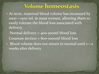
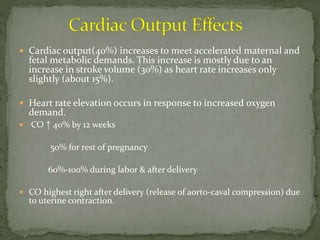
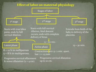
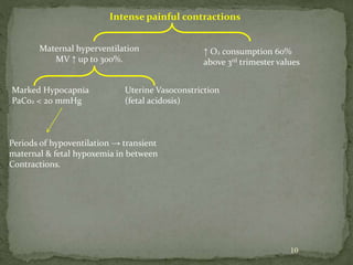
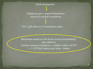
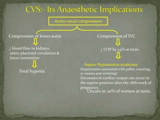
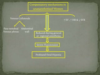
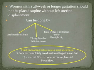
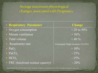
This document summarizes several physiological changes that occur during pregnancy. Key changes include increased blood volume, cardiac output, and respiratory rate. Hormonal changes lead to decreased sensitivity to local anesthetics and inhalational agents. The supine position can cause issues late in pregnancy due to compression of the inferior vena cava and aorta. Regional techniques require lower doses of local anesthetics during pregnancy. Overall, pregnancy results in significant cardiovascular and respiratory adaptations to meet increased metabolic demands of the mother and fetus.
A 71 year old man with peripheral edema pleural effusion ascites and breathle...Md. Shahidul Islam
A 71 year old man with peripheral edema, pleural effusion, ascites and breathlessness. See how one thing led to another, and the final diagnosis and treatment
A 71 year old man with peripheral edema pleural effusion ascites and breathle...Md. Shahidul Islam
A 71 year old man with peripheral edema, pleural effusion, ascites and breathlessness. See how one thing led to another, and the final diagnosis and treatment
Pregnant patients are admitted in ICU with a number of pregnancy related problems. Some of them are really life threatening. Identification and prompt action is the key to save lives.
Anatomical & physiological changes in pregnancy & their clinical implications...alka mukherjee
• Women undergo several changes during pregnancy, including cardiovascular, hematologic, metabolic, renal, and respiratory changes that provide adequate nutrition and gas exchange for the developing fetus.
• Progesterone and estrogen levels rise continually through pregnancy, together with blood sugar, breathing rate, and cardiac output.
• The body’s posture changes during pregnancy to accommodate the growing fetus and the mother will experience weight gain.
• Breasts grow and change in preparation for lactation once the infant is born. Once lactation begins, the woman’s breasts swell significantly and can feel achy, lumpy, and
heavy (engorgement). This is relieved by nursing the infant.
• Plasma and blood volume increase over the course of the pregnancy and lead to changes in heart rate and blood pressure. Women may also have a higher risk of blood clots, especially in the weeks following labor.
• During pregnancy, both protein metabolism and carbohydrate metabolism are affected. One kilogram of extra protein is deposited, with half going to the fetus and placenta, and another half going to uterine contractile proteins, breast glandular tissue, plasma protein, and hemoglobin.
• Circulatory Changes
• Plasma and blood volume slowly increase by 40–50% over the course of the pregnancy (due to increased aldosterone) to accommodate the changes, resulting in an increase in heart rate (15 beats/min more than usual), stroke volume, and cardiac output. Cardiac output increases by about 50%, primarily during the first trimester.
• The systemic vascular resistance also drops due to the smooth muscle relaxation and overall vasodilation caused by elevated progesterone, leading to a fall in blood pressure. Diastolic blood pressure consequently decreases between 12–26 weeks, and increases again to pre-pregnancy levels by 36 weeks.
• Edema (swelling) of the feet is common during pregnancy, partly because the enlarging uterus compresses veins and lymphatic drainage from the legs.
• The platelet count tends to fall progressively during normal pregnancy, although it usually remains within normal limits. In a proportion of women (5–10%), the count will reach levels of 100–150 × 109 cells/l by term and this occurs in the absence of any pathological process. In practice, therefore, a woman is not considered to be thrombocytopenic in pregnancy until the platelet count is less than 100 × 109 cells/l.
• Pregnancy causes a two- to three-fold increase in the requirement for iron, not only for haemoglobin synthesis but also for for the foetus and the production of certain enzymes. There is a 10- to 20-fold increase in folate requirements and a two-fold increase in the requirement for vitamin B12.
Changes in the coagulation system during pregnancy produce a physiological hypercoagulable state (in preparation for haemostasis following delivery).
TEST BANK for Operations Management, 14th Edition by William J. Stevenson, Ve...kevinkariuki227
TEST BANK for Operations Management, 14th Edition by William J. Stevenson, Verified Chapters 1 - 19, Complete Newest Version.pdf
- Video recording of this lecture in English language: https://youtu.be/lK81BzxMqdo
- Video recording of this lecture in Arabic language: https://youtu.be/Ve4P0COk9OI
- Link to download the book free: https://nephrotube.blogspot.com/p/nephrotube-nephrology-books.html
- Link to NephroTube website: www.NephroTube.com
- Link to NephroTube social media accounts: https://nephrotube.blogspot.com/p/join-nephrotube-on-social-media.html
ARTIFICIAL INTELLIGENCE IN HEALTHCARE.pdfAnujkumaranit
Artificial intelligence (AI) refers to the simulation of human intelligence processes by machines, especially computer systems. It encompasses tasks such as learning, reasoning, problem-solving, perception, and language understanding. AI technologies are revolutionizing various fields, from healthcare to finance, by enabling machines to perform tasks that typically require human intelligence.
Title: Sense of Smell
Presenter: Dr. Faiza, Assistant Professor of Physiology
Qualifications:
MBBS (Best Graduate, AIMC Lahore)
FCPS Physiology
ICMT, CHPE, DHPE (STMU)
MPH (GC University, Faisalabad)
MBA (Virtual University of Pakistan)
Learning Objectives:
Describe the primary categories of smells and the concept of odor blindness.
Explain the structure and location of the olfactory membrane and mucosa, including the types and roles of cells involved in olfaction.
Describe the pathway and mechanisms of olfactory signal transmission from the olfactory receptors to the brain.
Illustrate the biochemical cascade triggered by odorant binding to olfactory receptors, including the role of G-proteins and second messengers in generating an action potential.
Identify different types of olfactory disorders such as anosmia, hyposmia, hyperosmia, and dysosmia, including their potential causes.
Key Topics:
Olfactory Genes:
3% of the human genome accounts for olfactory genes.
400 genes for odorant receptors.
Olfactory Membrane:
Located in the superior part of the nasal cavity.
Medially: Folds downward along the superior septum.
Laterally: Folds over the superior turbinate and upper surface of the middle turbinate.
Total surface area: 5-10 square centimeters.
Olfactory Mucosa:
Olfactory Cells: Bipolar nerve cells derived from the CNS (100 million), with 4-25 olfactory cilia per cell.
Sustentacular Cells: Produce mucus and maintain ionic and molecular environment.
Basal Cells: Replace worn-out olfactory cells with an average lifespan of 1-2 months.
Bowman’s Gland: Secretes mucus.
Stimulation of Olfactory Cells:
Odorant dissolves in mucus and attaches to receptors on olfactory cilia.
Involves a cascade effect through G-proteins and second messengers, leading to depolarization and action potential generation in the olfactory nerve.
Quality of a Good Odorant:
Small (3-20 Carbon atoms), volatile, water-soluble, and lipid-soluble.
Facilitated by odorant-binding proteins in mucus.
Membrane Potential and Action Potential:
Resting membrane potential: -55mV.
Action potential frequency in the olfactory nerve increases with odorant strength.
Adaptation Towards the Sense of Smell:
Rapid adaptation within the first second, with further slow adaptation.
Psychological adaptation greater than receptor adaptation, involving feedback inhibition from the central nervous system.
Primary Sensations of Smell:
Camphoraceous, Musky, Floral, Pepperminty, Ethereal, Pungent, Putrid.
Odor Detection Threshold:
Examples: Hydrogen sulfide (0.0005 ppm), Methyl-mercaptan (0.002 ppm).
Some toxic substances are odorless at lethal concentrations.
Characteristics of Smell:
Odor blindness for single substances due to lack of appropriate receptor protein.
Behavioral and emotional influences of smell.
Transmission of Olfactory Signals:
From olfactory cells to glomeruli in the olfactory bulb, involving lateral inhibition.
Primitive, less old, and new olfactory systems with different path
MANAGEMENT OF ATRIOVENTRICULAR CONDUCTION BLOCK.pdfJim Jacob Roy
Cardiac conduction defects can occur due to various causes.
Atrioventricular conduction blocks ( AV blocks ) are classified into 3 types.
This document describes the acute management of AV block.
Ozempic: Preoperative Management of Patients on GLP-1 Receptor Agonists Saeid Safari
Preoperative Management of Patients on GLP-1 Receptor Agonists like Ozempic and Semiglutide
ASA GUIDELINE
NYSORA Guideline
2 Case Reports of Gastric Ultrasound
These simplified slides by Dr. Sidra Arshad present an overview of the non-respiratory functions of the respiratory tract.
Learning objectives:
1. Enlist the non-respiratory functions of the respiratory tract
2. Briefly explain how these functions are carried out
3. Discuss the significance of dead space
4. Differentiate between minute ventilation and alveolar ventilation
5. Describe the cough and sneeze reflexes
Study Resources:
1. Chapter 39, Guyton and Hall Textbook of Medical Physiology, 14th edition
2. Chapter 34, Ganong’s Review of Medical Physiology, 26th edition
3. Chapter 17, Human Physiology by Lauralee Sherwood, 9th edition
4. Non-respiratory functions of the lungs https://academic.oup.com/bjaed/article/13/3/98/278874
New Directions in Targeted Therapeutic Approaches for Older Adults With Mantl...i3 Health
i3 Health is pleased to make the speaker slides from this activity available for use as a non-accredited self-study or teaching resource.
This slide deck presented by Dr. Kami Maddocks, Professor-Clinical in the Division of Hematology and
Associate Division Director for Ambulatory Operations
The Ohio State University Comprehensive Cancer Center, will provide insight into new directions in targeted therapeutic approaches for older adults with mantle cell lymphoma.
STATEMENT OF NEED
Mantle cell lymphoma (MCL) is a rare, aggressive B-cell non-Hodgkin lymphoma (NHL) accounting for 5% to 7% of all lymphomas. Its prognosis ranges from indolent disease that does not require treatment for years to very aggressive disease, which is associated with poor survival (Silkenstedt et al, 2021). Typically, MCL is diagnosed at advanced stage and in older patients who cannot tolerate intensive therapy (NCCN, 2022). Although recent advances have slightly increased remission rates, recurrence and relapse remain very common, leading to a median overall survival between 3 and 6 years (LLS, 2021). Though there are several effective options, progress is still needed towards establishing an accepted frontline approach for MCL (Castellino et al, 2022). Treatment selection and management of MCL are complicated by the heterogeneity of prognosis, advanced age and comorbidities of patients, and lack of an established standard approach for treatment, making it vital that clinicians be familiar with the latest research and advances in this area. In this activity chaired by Michael Wang, MD, Professor in the Department of Lymphoma & Myeloma at MD Anderson Cancer Center, expert faculty will discuss prognostic factors informing treatment, the promising results of recent trials in new therapeutic approaches, and the implications of treatment resistance in therapeutic selection for MCL.
Target Audience
Hematology/oncology fellows, attending faculty, and other health care professionals involved in the treatment of patients with mantle cell lymphoma (MCL).
Learning Objectives
1.) Identify clinical and biological prognostic factors that can guide treatment decision making for older adults with MCL
2.) Evaluate emerging data on targeted therapeutic approaches for treatment-naive and relapsed/refractory MCL and their applicability to older adults
3.) Assess mechanisms of resistance to targeted therapies for MCL and their implications for treatment selection
2. Pregnancy produces profound physiological
changes(adaptive to stress) that become more significant
as pregnancy progresses in duration.
This changes can be due to:-
1) Hormonal alteration,
2) Mechanical effect of gravid uterus,
3) Increased oxygen & metabolic requirement,
4) Haemodynamical alteration.
In addition unique challenges - two patients are cared for
simultaneously, failure to take care can be disastrous for
one or both of them.
4. Fluid retention is the most fundamental systemic change of
normal pregnancy.
The total plasma volume is increased during pregnancy
45%. The most marked expansion occurs in extra cellular
volume (ECV) with some increase in intra cellular water.
The factors contributing –
Increase sodium retention.(RAAS)
Decrease in thirst threshold.
Decrease in plasma oncotic pressure(↓ plasma
albumin levels)
5. At term, maternal blood volume has increased by
1000—1500 mL in most women, allowing them to
easily tolerate the blood loss associated with
delivery.
Normal delivery = 400-500ml blood loss
Cesarean section = 800-1000ml blood loss
Blood volume does not return to normal until 1—2
weeks after delivery
6. Cardiac output(40%) increases to meet accelerated maternal and
fetal metabolic demands. This increase is mostly due to an
increase in stroke volume (30%) as heart rate increases only
slightly (about 15%).
Heart rate elevation occurs in response to increased oxygen
demand.
CO ↑ 40% by 12 weeks
50% for rest of pregnancy
60%-100% during labor & after delivery
CO highest right after delivery (release of aorto-caval compression) due
to uterine contraction.
7. Peripheral vascular resistance (VR) decreases due to
the vasodilatory effects of progesterone and the
proliferation of low resistance vascular beds in the
inter-villous spaces of the placenta.
Because of the decrease in peripheral vascular
resistance(in spite of increased CO), arterial blood
pressure does not significantly change or may show a
slight fall in an uncomplicated pregnancy.
Diastolic(20%) > Systolic(8%)
The response to adrenergic agents and vasoconstrictors is
thus blunted.
8. Despite the increase in blood volume, there is no
change in the central venous pressure(CVP) during
pregnancy. This is most likely due to dilated systemic
and pulmonary circulations.
Cardiac chambers enlarge and myocardial hypertrophy is
often noted on echocardiography.(Eccentric due to
activation of RAAS).
Also show variable ECG,
ECHO, CXR Changes.
9. 9
Effect of labor on maternal physiology
Stages of labor
1st stage 2nd stage 3rd stage
Starts with true labor
pains, ends by full
cervical dilation.
Starts with full cervical
dilation, fetal descent
occurs, ends with complete
delivery of fetus.
Extends from birth of the
baby to delivery of the
placenta.
Latent phase
Active phase
Progressive cervical effacement
& minor dilatation (2 – 4 cm).
Progressive cervical dilatation
up to 10 cm.
8 – 12 h in nulliparous
5 – 8 h in multiparous.
Contractions are 1.5- 2 min apart,
last 1 – 1.5 min
15 – 120 min.
15 – 30 min.
10. 10
Intense painful contractions
Maternal hyperventilation
MV ↑ up to 300%.
↑ O2 consumption 60%
above 3rd trimester values
Marked Hypocapnia
PaCo2 < 20 mmHg
Uterine Vasoconstriction
(fetal acidosis)
Periods of hypoventilation → transient
maternal & fetal hypoxemia in between
Contractions.
11. 11
Each contraction
Displaces 300 – 500ml blood from
uterus to central circulation.
CO ↑ 45% above 3rd trimesteric value.
Maximum strain on the heart occurs immediately
after delivery.
Uterine intense involution→ sudden relieve of IVC
→ ↑ COP 80% above pre-labor values.
12. Effect of Pregnancy on Cardiovascular Investigations
Investigation Findings
Chest radiography Apparent cardiomegaly
Enlarged left atrium (lateral views)
Increased vascular markings
Straightening of left-sided heart border
Postpartum pleural effusion
ECG Right-axis deviation
Right bundle branch block
ST-segment depression on left precordial leads
Q waves in lead III
T-wave inversion in leads III, V2, and V3
Rotation of 15 degrees(QRS axis)
ECHO Trivial tricuspid regurgitation
Pulmonary regurgitation (up to 94% at term)
Increased left atrial size by 12%-14%
Increased left ventricle end-diastolic dimensions by 6%-10%
Inconsistent increase in left ventricle thickness
Mitral regurgitation (28% at term)
Pleural effusion (40% postpartum)
13. Occurs in 20% of women at term.
Aorto-caval compression
Compression of IVCCompression of lower aorta
↓ COP by 24% at term.↓ blood flow to kidneys,
utero-placental circulation &
lower extremities
Supine Hypotension syndrome
(hypotension associated with pallor, sweating,
or nausea and vomiting)
Decreases in cardiac output can occur in
the supine position after the 28th week of
pregnancy.
Fetal hypoxia
14. Compensatory mechanisms in
unanaesthetised Women
Venous Collaterals
↑ SV , ↑ HR & ↓ SVR
Para vertebral
Venous plexus
Abdominal
wall
Reduced during general
or regional anesthesia.
Severe Hypotension
Profound Fetal Hypoxia
15. Women with a 28-week or longer gestation should
not be placed supine without left uterine
displacement.
Can be done by
Left lateral decubitus
Tilting the table
Left side down
Rigid wedge (>15 degree)
under
The right hip
Fluid preloading before neuro-axial anesthesia
It does not completely avoid maternal hypotension but
It ↑ maternal CO → preserve utero-placental
blood flow.
16. Physical examination of the term pregnant woman
may also be abnormal with auscultation commonly
revealing a wide, loud, split first heart sound, an S3
sound, and a soft systolic ejection murmur.
Hence it is essential to differentiate the abnormal
cardiovascular changes from normal physiological
changes of pregnancy.
17. • Criteria to diagnose cardiac disease during pregnancy:
1) Presence of diastolic murmurs.
2) Systolic murmurs of severe intensity (grade 3).
3) Unequivocal enlargement of heart (X-ray).
4) Presence of severe arrhythmias, atrial fibrillation or
flutter
18. Changes in the respiratory system during pregnancy
involve the upper airways, minute ventilation, lung
volumes, and oxygen consumption.
Major physiological changes occur in the respiratory
system during pregnancy due to a combination of both
hormonal and mechanical factors.
Dyspnoea is a common complaint in pregnancy
affecting over half of women at some stage.
19. Respiratory Parameter Change
Oxygen consumption + 20 to 50%
Minute ventilation + 50%
Tidal volume + 40 %
Respiratory rate Unchanged/ Slight increase(+10-15%)
PaO2 + 10%
PaCO2 - 15%
HCO3 - 15%
FRC (functional residual capacity) - 20%
From Birnbach DJ, Gatt SP, Datta S (eds): Textbook of Obstetric Anaesthesia. New York, Churchill Livingstone, 2000, p 35.
20. Due to increased metabolic demands, Oxygen
consumption (+ 20 - 50%) and minute ventilation
(+40 - 50%) progressively increase during pregnancy.
(Increased progesterone sensitizes the central
respiratory center to carbon dioxide – directly
stimulating ventilation)
The pregnant woman thus takes larger Tidal
volumes(40-50%) to eliminate carbon dioxide.
PaCO2 decreases to(28—32 mm Hg); significant
respiratory alkalosis is prevented by a compensatory
↓ in plasma HCO3 concentration.
21. Hyperventilation may also increase PaO2.
Elevated levels of 2,3-diphosphoglycerate offset
the effect of hyperventilation (↓PaCO2) on
hemoglobin affinity for oxygen.
The combination of increased 2,3-DPG with
increase in cardiac output enhances oxygen
delivery to tissues.
22.
23. The maternal respiratory pattern changes as the
uterus enlarges
Diaphragm rises up(4cm) compensatory increases in
Antero-posterior diameters
Diaphragm motion not restricted
Chest wall motion limitation
Thoracic breathing favored over Abdominal
20% decrease in FUNCTIONAL RESIDUAL CAPACITY(FRC).
* No change in CLOSING CAPACICITY (CC) & VITAL CAPACITY
24. ↓ FRC + ↑O2 Consumption + Unchanged CC
Rapid O2 de-saturation during
periods of apnea (diminished capacity to
tolerate apnea).
Supine Position & Regional Block
further diminishes FRC
Pre-oxygenation prior to induction of general
anesthesia should be given to avoid hypoxemia in
pregnant patients.
Rapid development of hypoxemia
25. Rapid gaseous induction
The decrease in FRC coupled with the increase in minute
ventilation accelerates the uptake of all inhalational
anesthetics.
↓FRC → less dilution
↑MV → rapid deep depth
26. 26
Hormonal Changes Capillary engorgement of
respiratory tract mucosa
1) ↑ Incidence of difficult intubation.
2) Trauma and bleeding during
endotracheal intubation.
☼ Repeated attempts at laryngoscopy must be
minimized
☼ Use a small ETT (6 – 7 mm) during GA
27. 1) Progressive decrease in MAC .
40% at term
(Returns to normal by 3rd day postpartum).
Progesterone increases
20 times normal
level at term
β- endorphin surge during
labor & delivery
28. LA requirements for subarachnoid or epidural anaesthesia
are reduced in pregnancy (30%)
a) ↑ diffusion of LA to the receptor site.
b) ↑ sensitivity of nerve fibers to LA (Lower concentration
needed).
c) engorged epidural venous plexus.
Spinal ligaments including ligament flavum SOFT hence
loss of resistance technique used for regional block may be
masked
CNS : its anaesthetic importance
29. IVC obstruction by enlarging
uterus
Engorged Epidural
Venous Plexus
1) ↓CSF Volume
2) ↓Volume of
Epidural Space
3) ↑Epidural space
Pressure
1,2: This enhances the cephalad spread of LA
during regional blocks.
3 : Predisposes to higher incidence of Dural puncture &
intravascular injection
30. The parturient should be considered a full stomach patient
during most of gestation
☼ Upward & ant. displacement of the stomach by the
uterus → Incompetence of gastro-esophageal
sphincter → Gastro-esophageal reflux & aspiration.
☼ ↑ Progesterone → ↓ tone of gastro-esophageal sphincter.
☼ Placental Gastrin → Hyper-secretion of gastric acid.
☼ Gastric emptying → Delayed with labor.
31. Narcotics and anti-cholinergic reduce lower
oesophageal sphincter pressure (used with precaution)
For GA:
Pharmacological prophylaxis against aspiration.
Supine position with lateral tilt
No positive pressure ventilation before intubation
Rapid sequence induction.
Sellick’s maneuver
32. Renal vasodilatation increases renal blood flow
early during pregnancy.
↑ Cardiac output(CO) ↑ GFR & ↑ RPF(renal
plasma flow) by 50%.
↑ Renin & Aldosterone level promotes Na+ retention
leading to volume overload.
↓ Renal tubular threshold for glucose & amino acids →
mild glycosuria & proteinuria (< 300mg/d).
33. ↑ GFR ↑ clearance of urea, uric acid and
creatinine
↓ plasma concentrations of
sr. Creatinine & BUN
BUN and Creatinine levels that would be considered
marginally elevated in pre-pregnant patients are usually
indicative of severe renal impairment in pregnancy.
34. Hepatic function and blood flow are unchanged.
A mild ↓ in serum albumin is due to an expanded
plasma volume. Thus, the free fraction of albumin-
bound medications is increased.
A 25—30% decrease in serum pseudo cholinesterase
activity is also present at term, but it rarely produces
significant prolongation of NMB action.
Increased cholesterol gall stone
formation(progesterone).
35. ↑ Blood Volume ( up to 90ml/Kg)
↑ by 1000 – 1500 ml at term.
↑ Plasma Volume(45%) > ↑ RBC mass(30%)
Dilutional anemia & ↓ blood viscosity
Facilitates maternal & fetal
exchange of respiratory gases,
nutrients & metabolites
↓ Impact of maternal blood
loss at delivery
36. • Pregnancy leads to a hypercoagulable state, due to,
a) factors VII, VIII, X, XII ,IX ( only factor XI )
b) fibrinogen and FDP's
c) fibrinolytic activity - levels of plasminogen activators
d) antithrombin III
Probably a protective adaptation to lessen the risks
associated with the acute haemorrhage that occurs at
delivery.
Increased risk of thromboembolic disease (Post-Anaesthesia
ambulation )
37. Leucocytosis up to 13,000/µL.
↑ ESR
10-20% ↓ in platelet count.(Mild thrombocytopenia not a
contra-indication for neuraxial block).
Marked ↓ cell mediated immunity→ ↑ susceptibility to
viral infection.
38. Pregnancy is Diabetogenic
Human Placental lactogen(HPL) → relative insulin resistance.
• Biochemically Starvation like state
↓ Blood Glucose & Amino Acid levels.
↑ Free Fatty Acids, Ketones & triglycerides.
To promote fetal growth.
Maternal Insulin Levels Steadily Rise During Pregnancy
↓
39. Secretion of HCG and elevated levels of estrogens promote
hypertrophy of the thyroid gland.
Hyperplasia of Pancreatic ß Cells occurs in response to
increased demand for insulin secretions.
↑ TBG (↑ T3 & ↑ T4 ) → Free T3, T4 & TSH remain
normal.
40. • Increased level of relaxin - Softening Of Cervix
- Relax Symphysis Pubis
- Pelvic Joints
- Chest Wall Changes
Increased risk of back pain(lax ligaments)
42. 42
Due to maximum dilatation of uterine vasculature its auto regulation is lost
Uterine Blood Flow
Directly proportional to difference between
uterine arterial and venous pressure.
Inversely proportional to uterine
vascular resistance.
Uterine vasculature has abundant α-adrenergic & some β-adrenergic receptors.
Previously , vasoconstrictor agents with predominant β-adrenergic activity
(e.g. Ephedrine) were of choice for hypotension during pregnancy.
Recent studies show that α-adrenergic drugs (e.g.Phenylephrine) have
similar effects causing less fetal acidosis.
44. 44
Placental transfer of anaesthetic agents
Placental transfer of drugs depends on:
1) Molecular weight : < 1000 Da cross easily.
2) Protein binding– inversely proportional
3) Lipid solubility: Highly ionized substances have poor lipid solubility.
4) Maternal & fetal pH : affect ionization of the drug.
5) Maternal drug concentration: affected by dose given
and route of administration.
6) Timing of administration.
45. 45
Limited effects if <
1MAC & delivery within
10 min. of induction
Cross placenta
freely
Inhalational Agents
Intravenous Agents:
Thiopental, ketamine
& propofol
Limited fetal effects in
usual induction doses
(drug distribution,
metabolism & placental
uptake)
Variable effects.Cross placenta freelyOpioids
Most significant respiratory depressant effectsMorphine
Significant respiratory depression peaking 1- 3
hr after administration.
Pethidine
Minimal effect if < 1µg/Kg.Fentanyl
Minimal effects on
fetus.
The highly ionized
property impedes
placental transfer.
Muscle Relaxants
46. 46
Local anesthetics → Placental transfer depends on:
1) pKa.
2) Maternal & fetal pH : Fetal acidosis → higher fetal to maternal
drug ratios . Binding of hydrogen ions to the nonionized form → trapping
of local anesthetic in fetal circulation
3) Degree of protein binding : highly protein bound agents
diffuse poorly across the placenta.
Chloroprocaine has the least placental transfer as it is rapidly
broken down by plasma cholinesterase in the maternal circulation.
47. Summary
To illustrate how all these changes may affect anaesthetic
management, lets imagine performing a general anaesthetic for
caesarean section and list some key points
1) Careful attention to the assessment of the airway and any
necessary preparation to deal with a potentially difficult airway
in the pre operative period.
2)When positioning the patient on the table, remember to use
either a left tilt of 15 – 30 degrees on the table or a wedge
under the right buttock to minimize aorto-caval compression.
3)Venous access often easier due to engorgement of the venous
system
48. 4) Pre oxygenation is essential and should be with a tight fitting
mask for at least 3 minutes.
5) Rapid sequence induction with the application of cricoid
pressure is mandatory. Intubation may be difficult and so
adjuncts for difficult intubation should be available.
6) Once the airway is secured, ventilation should be aimed to
keep the PCO2 in the normal range for pregnancy.
7) The MAC of volatile anaesthetic is slightly reduced.
8) There is decreased sensitivity to endogenous and exogenous
catecholamines and so if vasopressors are required to maintain
adequate blood pressure, the amounts needed may be greater.
10) Extubation should be done with the patient awake and on
their side to reduce the risk of aspiration of gastric contents.