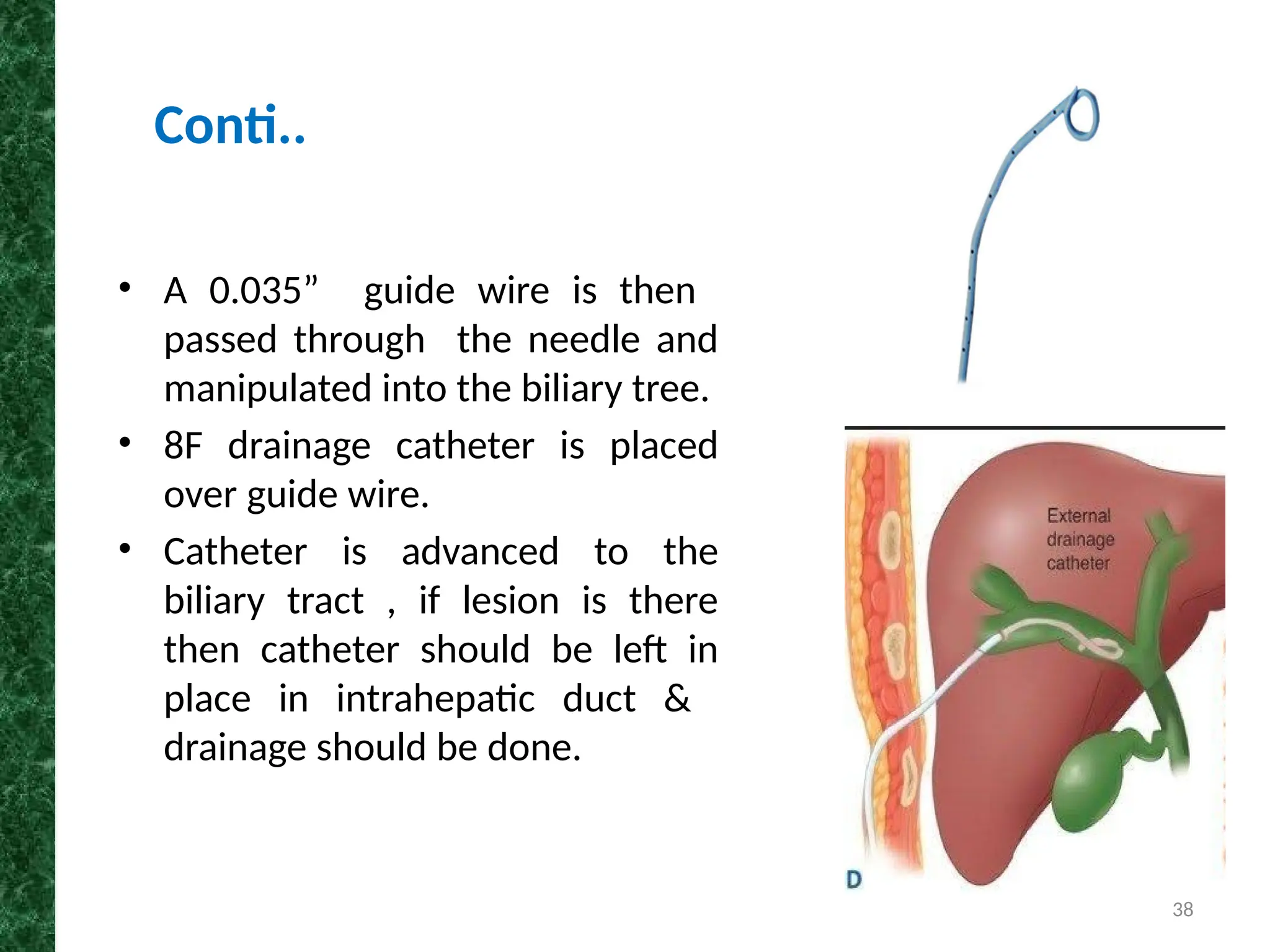
1
Diagnostic and Therapeutic Non- Vascular
Interventional Studies
JOGINDER SINGH
RADIOLOGICAL TECHNOLOGIST
DEPARTMENT OF RADIODIAGNOSIS AND IMAGING
PGIMER, CHANDIGARH
2
• Biopsy/FNAC
• Percutaneous Fluid Drainage
• PTC
• PTBD
• ERCP
• Ablation Therapy
• Vertebroplasty
Contents
3
• Intervention is the sub- specialty of radiology which
deals with minimally invasive image guided
procedures to diagnose & treat disease in nearly
every organ of the body.
• Done for catheterization or stenting of organ ductal
system.
• For biopsy or to drain fluids out of an organ.
• Done for tumor ablations in many organs of the body.
Introduction
4
Diagnostic procedures Therapeutic procedures
• Biopsy
• FNAC
• PTC
• T Tube Cholangiogram
• ERCP
• PTBD
• ERCP
• RFA
• MWA
• Crayoablation
• IRE
• Vertebroplasty
Non-Vascular Procedures
5
Imaging Modalities for
Non Vascular Intervention
6
• A biopsy is the removal of tissue from any part of the
body to examine it for disease .
• The tissue is removed by placing a needle through the
skin to the area of abnormality.
Percutaneous Biopsy/FNAC
7
• Diagnosis of any malignancy , metastatic
disease
• Tumor staging
• Recurrent disease after treatment.
• Diagnosis of inflammatory or infectious
processes, abnormal fluid collections.
Indications
8
• Uncorrectable coagulation abnormalities.
• Lack of a safe percutaneous pathway to the
lesion.
• An uncooperative patient in whom motion may
increase the risk of bleeding.
• Biopsy usually is not indicated in a patient who
will undergo surgery.
Contraindications
9
• Disposable plastic syringes
• Betadine
• Gloves
• Drapes and sheets
• Local anesthesia
• Specican
Tools Required
10
Needles
Aspiration
LP needle
Cheeba
needle
Cutting
needle
Side cutting
needle
Manual
Semi
automatic
Automatic
End cutting
needle
11
• The Chiba needle is one of the most
commonly used
biopsy/percutaneous access
needles.
• It is a two-part hollow needle with a
beveled tip angled at 30 degrees.
The inner stylet is removable.
• It is typically between 18-22 G.
Chiba needle
12
Side-cutting biopsy needle (left) and end-cutting
biopsy needle (right)
13
Side-cutting needles: manual (top),
semiautomated (middle), and
automated (bottom).
14
Mechanism of a side-cutting biopsy needle.
Mechanism of an automated end-cutting biopsy needle.
15
Comparison of specimens obtained from side-
cutting and end-cutting needles.
16
• Biopsy : In a core biopsy, a large-bore needle is
inserted and used to pull out a chunk of soft tissue or
bone.
• Fine Needle Aspiration: In fine needle aspiration , a
smaller needle is inserted and a syringe is used to
create a vacuum, sucking a sample up into the
needle.
Technique
17
• The patient can be in supine, prone or oblique
position as required . The shortest path to the
lesion is preferred.
• During needle advancement and biopsy, the
patient should suspend respiration to minimize
inadvertent motion of the needle.
Procedure
18
• The position of the needle tip can be observed
continuously during biopsy so that different sectors of
the lesion are sampled to increase the diagnostic yield.
Conti..
19
• Biopsy can be done in any part of the body .e.g.
• Liver biopsy
• Renal biopsy
• Lung biopsy
• Pancreas biopsy
• Thyroid biopsy
• Breast biopsy
Conti..
20
Complications
• Bleeding
• Organ injury
After Care
• Blood counts should be checked 4 hours after the
biopsy to exclude significant bleeding.
• If the patient and blood count are stable, no further
evaluation other than follow-up with the biopsy
results.
21
• PFD uses imaging guidance to place a needle or
catheter through the skin into the abscess to remove or
drain the infected fluid.
• PFD provides definitive treatment of most sterile and
infected collections in the chest, abdomen, pelvis, and
musculoskeletal system.
Percutaneous Fluid Drainage
22
Indications
Chest:
Pleural effusion
Pulmonary abscess
Abdomen:
Post-traumatic collection
Pancreatic fluid collection
Abscess due to crohn’s
disease
23
Patient Preparation
• Coagulopathies must be corrected before the
procedure.
• Patients with suspected infected fluid collections
usually are receiving antibiotics by the time drainage
is requested.
• Cephalosporin (e.g., 1 g of cefazolin IV) is given
within 1 hour of the procedure.
• PFD is performed with intravenous conscious
sedation and local anesthesia.
• Informed consent is must.
24
Technique
• Sonography and fluoroscopy is preferable because
they allow real-time monitoring and less radiation
to the patient than with CT.
• The shortest distance between the skin and the
collection without interposing vessels is chosen for
needle entry and catheter insertion.
• The area is sterilized, and local anesthesia is
applied to the skin and soft tissues where the
needle and catheter will be inserted.
25
Procedure
• Puncture the site with 18G needle .
• Follow needle till its tip is well inside the fluid/abscess cavity.
• Remove stylet of needle.
• Aspirate 2 to 5 ml of fluid.
• Store in sterile vial for microbiological analysis or completely aspirate
all fluid as a single stage therapeutic procedure.
26
• A 0.035“ guide wire is inserted through an 18G Chiba needle.
• Remove the needle.
• Place catheter in abscess cavity over guide wire.
• Remove the guide wire.
• Connect catheter to negative suction collection system.
• Suture catheter & skin wound.
• Secure catheter to skin with adhesive tape.
Conti..
27
Complications
• The major complications of PFD are bleeding,
bowel or bladder perforation, and sepsis.
• Hemorrhage is more likely to occur in a patient
with an uncorrected coagulopathy or when a sub-
optimal access route was used for catheter
insertion.
28
• The biliary system consists of the
organs and structures that secrete
and transport bile, including the liver,
gallbladder, and bile ducts.
• Right and left hepatic ducts unite to
form common hepatic duct.
• Common hepatic and cystic duct from
GB unite to form common bile duct .
This runs from liver to the duodenum.
Biliary System
29
• Percutaneous trans-hepatic cholangiography (PTC) is
a way of examining the bile duct system .
• This procedure is done under local anesthesia by a
radiologist.
• During the exam , a thin needle is inserted through
the skin and liver into a bile duct.
• The contrast is injected & the bile duct system is
outlined.
Percutaneous trans-hepatic
cholangiography (PTC)
30
Indications
• To see the position of bile duct.
• Prior to biliary drainage procedures/stenting
• To define the level of obstruction in patients
with obstructive biliopathy.
• To evaluate for presence of suspected
choledocholithiasis.
31
Contraindications
• Coagulopathy
• Low PTI count
• Sensitivity to Contrast media
• Severe jaundice
• Ascites
• Biliary tract sepsis
32
Technique
• Patient is lying down in supine position on the fluoroscopy table,
upper abdomen is cleaned and draped.
• The skin puncture site is anesthetized with local anesthetic.
• A fine (22G) Chiba needle is advanced into the liver.
• After the stylet is removed the needle is slowly withdrawn while
injecting a small amount of contrast.
• When contrast is seen to opacify the biliary tree, cholangiography
performed to delineate the biliary anatomy.
33
Precautions:
• No high injection flow rate
• Maintain sterility
Complications:
• Sepsis
• Dislodgment of catheters
• Perforation of bile duct
• Intra abdominal bleeding
34
• PTBD is a therapeutic procedure that
consists of sterile puncture of a
peripheral biliary radicle under image
guidance followed by placement of a
catheter for drainage of bile.
• In this technique a drainage catheter
is introduced percutaneously through
the liver into the CBD for drainage of
bile.
• Procedure may be performed to
improve jaundice or sepsis.
Percutaneous Trans-hepatic
Biliary Drainage (PTBD)
35
Indications
• Biliary stones
• Malignant Biliary stricture
• Obstructive jaundice/Cholangitis
• Undiagnosed jaundice
Contraindications
• Sepsis
• Bleeding disorder
• Contrast hypersensitivity
• Pregnancy
• Ascites
36
Preparation
• CT/US prior to PTBD which provide useful
information regarding the level of obstruction.
• Fasting 4 hours prior to procedure.
• Local anesthesia will be administrated into the skin.
• Informed consent must be taken.
37
Procedure
• After anesthetizing the skin , a 18G puncture needle
is used to enter the selected biliary radicle.
• In a dilated system ,allow little amount of bile to
drain after the puncture before injecting contrast to
prevent over-distention of the biliary system which
can result in bacteremia and sepsis.
• Little contrast is injected to opacify the biliary tree.
38
• A 0.035” guide wire is then
passed through the needle and
manipulated into the biliary tree.
• 8F drainage catheter is placed
over guide wire.
• Catheter is advanced to the
biliary tract , if lesion is there
then catheter should be left in
place in intrahepatic duct &
drainage should be done.
Conti..
39
40
Complications
• Hemorrhage
• Sepsis and bile peritonitis due to slippage of
catheter.
• Pancreatitis occur due to edema of the CBD.
• Pneumothorax or pleural effusion
41
Aftercare
• Regular follow up to detect catheter migration or
obstruction at early stage.
• Bile output ,color and body temperature should
be recorded every day . Serum bilirubin and
amylase levels should be checked.
• Catheter should be exchanged every 2-3 months
to prevent blockage or breakage.
42
• The examination is performed on or about the 10th
postoperative day, prior to removal of the T-tube.
T-Tube Cholangiography
43
1. To exclude biliary tract calculi, where:
• Operative cholangiography was not performed.
• The results of operative cholangiography are
not satisfactory or are suspect.
2. Assessment of biliary leaks following biliary
surgery
Indications
44
Technique
• The patient lies supine on the x-ray table.
• The drainage tube is clamped off near to the patient and
cleaned thoroughly with antiseptic.
• A 23G needle, extension tubing, and 20 mL syringe are
assembled and filled with contrast medium (e.g. a butterfly
needle).
• After all air bubbles have been expelled, the needle is
inserted into the tubing between the patient and the clamp.
45
• The injection is made under
fluoroscopic control, only a small
volume of contrast (approximately 10
mL), gently injected, is required.
• Intermittent fluoroscopic ‘grab’ images
during filling are frequently useful.
• AP and oblique exposures when there is
satisfactory opacification of the biliary
system.
Conti..
46
• Endoscopic retrograde cholangiopancreatography is
a therapeutic approach and imaging technique.
• It is done to image CBD , pancreatic ductal system
and gall bladder by a catheter and endoscopic
guidance.
• ERCP is a gold standard for evaluation of pancreatic
and bile duct.
ERCP
47
Indications
• Biliary obstruction
• Persistent jaundice
• Evaluation & treatment of biliary
duct injuries
• Removal of stones
• Stent placement
• Balloon dilation of ductal stricture
48
Contraindications
• Esophageal obstruction
• Severe cardiac /respiratory disease
• Previous gastric surgery that prevents access to
duodenum
• Acute pancreatitis.
• Coagulopathy
• Hypersensitivity to contrast media
• Pregnancy
49
Patient Preparation
• Patient must be NPO midnight prior to exam.
• Review the history & images.
• Patient should be well hydrated.
• Patient must bring all the things required for the
procedure.
• Use urobag for long procedures.
• If any intervention planned patient’s Hb, PTI &
platelets are checked.
50
Technique
• ERCP is performed using a side viewing endoscope.
• Preliminary film of upper abdomen in prone & left
anterior oblique position is taken to check for opaque
gall stone & pancreatic calcification.
51
Endoscope
52
• The pharynx is anesthetized with
xylocaine spray.
• The patient then lies on the left
lateral / prone position &
endoscope is inserted through
the mouth, down the
oesophagus , into the
stomach ,through the pylorus
into the duodenum where the
ampulla of Vater (the opening of
common bile duct & pancreatic
duct ) exists.
Endoscopic insertion
53
• Once papilla is visualized the
endoscope is kept still.
• Then the Guide wire is inserted
from channels of the
endoscope.
• Catheter is used over guide
wire to cannulate the papilla
cannulation & contrast is
injected into the bile duct
and/pancreatic duct .
Catheter Insertion
54
55
Precautions
• The contrast media is injected into the ductal
system at a slow pressure .
• Contrast media is inserted into ampulla , having
ensured that all air bubbles are excluded.
Complications
• Increased post procedural vomiting & nausea due
to use of glucagon.
• Post ERCP pancreatitis
• Post ERCP bacteremia or sepsis
• Duodenal perforation
56
Benefits Of ERCP
• Diagnostic as well as therapeutic.
• Better resolution.
Limitations Of ERCP
• Expertise required.
• We can not see beyond stricture.
• Uncomfortable for patient.
• Chances of hemorrhage
57
Ablation therapy is a minimally invasive procedure used
to destroy abnormal tissue in radiologic guidance by:
• Radiofrequency Ablation (RFA)
• Irreversible Electroporation (IRE)
• Micro Wave Ablation
• Cryoablation
Ablation Therapy
58
Radiofrequency Ablation (RFA)
• Radiofrequency ablation (RFA) is a percutaneous
image guided tumor ablation technique for small
HCC as well as metastatic deposits.
• It induces necrosis of the tumor by deposition of
thermal energy around the tip of the electrode
inserted in the tumor.
59
• Hepatocellular Carcinoma, which is a primary
liver cancer.
• Colon cancer that metastasizes or spreads from
the colon to the liver.
• Renal cell carcinoma
• Radiofrequency ablation is most effective treating
tumors that are less than 3 cm in diameter.
• Osteoid osteoma
Radiofrequency ablation is
used to treat:
60
• Solitary lesion
• 3 cm or less
• Not contiguous with major vessels or nerves
• No extension to hilum/mediastinum
• Safe & reasonable percutaneous route
Tumor Selection:
61
• Uncontrolled coagulopathy
• Tumor adjacent to large vessels
• Inability to safely access the tumor
• Cardiac devices such as pacemaker
Contraindications of RFA:
62
RFA Equipment
RFA Generator
• The radiofrequency generator produces alternating
electrical currents in the range of radiofrequency
waves. It is connected by insulated wires to the
needle electrodes and to grounding pads that are
placed on the patient's back or thigh.
63
RFA Probe
• Radiofrequency ablation needle device
that contains multiple curved retractable
electrodes .
• The electrodes are kept inside the
needle until its tip is positioned within a
tumor.
64
Procedure
• Patient is positioned on the examining
table.
• The area where the electrodes are to be
inserted is sterilized and covered with a
surgical drape.
• A very small nick may be made in the skin
to make it easier to pass the RFA
electrode into liver.
• Using imaging-guidance, the needle
electrode is insert through the skin and
advance it to the site of the tumor.
65
• Once the needle electrode is in place,
radiofrequency energy is applied.
• Alternating current is passed through the
probe with energies 60 to 200 watts for a
period of 6 to 12 minutes.
• Local temperature of more than 600
is
maintained.
• At the end of the procedure, the needle
electrode will be removed and pressure will be
applied to stop any bleeding and the opening
in the skin is covered with a dressing.
Conti..
66
Complications
Related to image guided electrode placement:
• Bleeding
• Infection
• Pneumothorax
Related to thermal therapy:
• Non target thermal damage(GB/Biliary ducts)
• Grounding pads burns.
67
Benefits
• RFA is a relatively quick procedure and recovery is
rapid
• Radiofrequency ablation is less expensive
• No surgical incision is needed.
• Treatment-related serious complications are
infrequent and discomfort is minimal.
68
Micro Wave Ablation
• Microwave ablation is a
form of thermal ablation
used in interventional
radiology to treat cancer.
• MWA uses electromagnetic
waves in the microwave
energy spectrum (300MHz-
300 GHz) to produce tissue
heating effects.
69
Conti..
• The oscillations of polar molecules produces
frictional heating , ultimately generating tissue
necrosis within solid tumors.
• It is generally used for the treatment of solid
tumors in patients who are non surgical
candidates.
70
Technique
• With image guidance , the tumor is localized , and a
thin (15 guage) microwave antenna is placed directly
into the tumor .
• A microwave generator emits an electromagnetic
wave through the exposed , non insulated portion of
the antenna.
71
Conti..
• Electromagnetic microwaves agitate water molecules
in the surrounding tissue, producing friction & heat ,
this inducing cellular death via coagulation necrosis.
72
Advantage
• The main advantage of microwave technology
when compared with existing thermoablative
technologies include consistently higher
intramural temperature, larger tumor ablation
volumes , faster ablation times & improved
convection profile.
• No heat sink effect of the blood flow & air.
• No grounding pads.
• Multiple antennas can be used simultaneously.
• Less painful.
73
Cryoablation
• Cryoablation is a
treatment to kill cancer
cells with extreme cold.
• During cryoablation , a
thin , wand like needle
(cryoprobe) is inserted
through the skin &
directly into the cancerous
tumor.
74
Indications:
• Kidney cancer
• Liver cancer
• Lung cancer
• Prostrate cancer
Contraindications:
Active systemic infection
75
Conti..
• A gas is pumped into the
cryoprobe in order to freeze the
tissue.
• By using argon gas, cryo-probe tips
can reach temperature of -187°C
& then by rapidly exchanging
helium into the probe.
76
77
78
Complications
• High visibility of the ice ball on imaging
• Potentially severe systemic reactions (cryoshock)
• Relatively small ablations compared with more
contemporary technologies
• Potentially greater risk for bleeding complications
79
Irreversible Electroporation (IRE)
• IRE is a soft tissue ablation technique using ultra
short but strong electrical fields to create
permanent and hence lethal nanopores in the cell
membrane, to disrupt the cellular homeostasis.
• The resulting cell death results from apoptosis
instead of necrosis.
• IRE is used with NanoKnife system.
80
• Nanoknife may provide a minimally invasive option
for patients with in-operable or difficult to reach
tumors, including tumors located near critical
structures & major blood vessels.
• Potential benefits of NanoKnife include:
• No open incisions
• Less damage to healthy tissue
• Minimal post operative pain
• Fewer side effects
• Short hospital stay
Conti..
81
82
Working
• Instead of using microwave energy , extreme heat
& extreme cold , the Nanoknife system uses
electric currents to treat tumors.
• While the patient is under general anesthesia , the
interventional radiologist carefully guides up to
six thin needles (electrodes) into the patient’s
body & strategically places them around the
tumor.
83
Conti..
• Then the Nano Knife sends electrical pulses or
current between each set of needles to puncture
permanent Nano pores into the tumor .
• This process called IRE causes the cancer cells to be
unbalanced & triggers a cell “suicide “; thereby
destroying the tumor.
84
Advantages Of IRE
• Its capability of preserving vital structures within the
IRE-ablated zone . In all IRE ablated liver tissues ,
critical structures such as the hepatic arteries , hepatic
veins , portal veins & intrahepatic bile ducts were all
preserved.
• In IRE , no heat sink effect, which is the cause of many
problems and decreases the predictability of the
treatment field.
• It takes short time less than 5 minutes to place the
electrode and start treating tumor cells.
85
Disadvantages
• The strong electric fields created by IRE, cause
strong muscle contractions.
• The mechanism of cell death following IRE relies on
cellular apoptosis, which results from the pore
formation in the cellular membrane. Tumour cells,
known to be resistant to apoptotic pathways, may
require higher thresholds of energy to be
adequately treated.
86
• Percutaneous vertebroplasty is a well-
accepted interventional procedure for
the treatment of painful vertebral
lesions.
• It is treatment of numerous lesions
causing back pain associated with
vertebral involvement like osteoporotic
vertebral collapse, metastatic disease,
multiple myeloma and symptomatic
vertebral hemangiomas.
Percutaneous Vertebroplasty
87
Technique
• The procedure consists of instilling acrylic bone cement into the
affected vertebra through a bone biopsy needle by a
percutaneous approach.
• The cement consists of Polymethylmethacrylate (PMMA) ,
which is mixed just before the injection. The cement is injected
into the lesion after needle placement, under fluoroscopic
control.
• The cement polymerizes and subsequently sets, affording
support to the vertebra.
88
Procedure
• Patient is placed in prone position on
angiography/CT table.
• The procedure is performed under sterile
conditions. The skin over the center of the
pedicle is anesthetized with local anesthesia.
• A small skin incision is made and bone biopsy
needle is positioned with its tip in the center of
the mid point of the pedicle.
89
Conti..
• The needle is advanced under AP and lateral
fluoroscopy till the junction of the anterior and
middle third of the vertebral body.
• Approximately 6 mL of PMMA cement is injected.
• Patient is placed in supine position for three
hours after the procedure and is discharged after
6 hours.
90
Results
• Vertebroplasty results in relief of pain with
decrease in analgesic use.
• It provides strength so prevents further collapse
and pain relief by coagulating the tissue. The
relief is obtained irrespective of the cause of pain,
and is long lasting.
91
Complications
• Cement leaks outside the vertebra are mostly
inconsequential, but can cause local or radicular
pain, neurological damage and pulmonary
embolism.
• Inaccurate needle placement can injure nerve
root or spinal cord.
• Pain exacerbation may occur due to substantial
cement leaks. Rarely puncture site infection and
bleeding may occur.
92
Percutaneous Treatment of
Disk Herniation
• Low backache is one of the
major cause of chronic pain
and morbidity.
• Treatment of herniated disk
has evolved from open
surgical techniques to
minimally invasive or
microtherapeutic
procedures under local
anesthesia.
93
Conti..
• IN Percutaneous laser disk
decompression (PLDD),
laser energy
is delivered into nucleus pulposus by
laser fiber, through a needle.
• The aim of PLDD is to vaporize a
small portion of the nucleus
pulposus.
• The ablation of this small volume
results in reduction of intradiskal
pressure, thus reducing the disk
herniation.
94
• In this procedures, patient is
positioned prone and using
either fluoroscopy or CT
guidance a lumbar puncture
needle (18– 21 G) is inserted
into the center of the disk.
• After proper position of the
needle, for PLDD, an optical
fiber is advanced through the
needle and proper laser energy
is given.
Conti..
95
96
• CT-guided biopsy is a procedure
by which the physician uses a very
thin needle and a syringe to
withdraw a tissue or fluid
specimen from an organ or
suspected tumour mass.
• The needle is guided while being
viewed by the physician on a
computed tomography (CT) scan.
CT Guided Biopsy
ADVANCEMENTS
97
• CT-guided FNAC of thoracic mass lesions is a safe, rapid, and
reliable procedure with minimal complications.
• It provides very early diagnosis and exact sub classification of
various lung tumours on the basis of cytomorphology
Conti..
98
99
• MR guided breast biopsy: Lumps
or abnormalities in the breast are
often detected by physical
examination, mammography, or
other imaging studies.
MRI Guided Biopsy
100
An MRI-guided breast biopsy is
most helpful when MRI imaging
shows a breast abnormality such
as:
• A suspicious mass not
identified by other imaging
techniques
• An area of abnormal tissue
change.
Conti..
101
• Role of radiological technologist in interventional
radiology has a great importance for better procedure
outcomes with best image quality.
• Patient management: to check patient requisition
form, consent, to form a to-do list, arranging patients
on the basis of their emergency status.
• Equipment preparation: Equipment working
conditions are good or not, primary QC tests before
start (if necessary) should be performed, sterility of
the equipment is necessary.
Role Of Radiologic Technologist
102
• Interventional Radiology procedures are minimally
invasive, targeted treatments performed using image
guidance like CT , fluoroscopy or ultrasound.
• These procedures have less risk, less pain and less
recovery time compared to open surgery.
• Percutaneous image-guided aspiration and drainage
has become the "first line" treatments for the
management of pleural and mediastinal collections.
Conclusion
103
• Demystifying Interventional Radiology (Sriharsha
Athreya)
• American Journal of Radiology
• Radiopaedia.org
• Beaujeux R, Laurent A, Wassef M, et al. Trisacryl gelatin
microspheres for therapeutic embolization II: preliminary
• MAMC Aiims PGI series
• Radiological procedure by chapman
References
104
Thank you

Non VASCULAR INTERVENTIONAL STUDIES by joginder.pptx

  • 1.
    1 Diagnostic and TherapeuticNon- Vascular Interventional Studies JOGINDER SINGH RADIOLOGICAL TECHNOLOGIST DEPARTMENT OF RADIODIAGNOSIS AND IMAGING PGIMER, CHANDIGARH
  • 2.
    2 • Biopsy/FNAC • PercutaneousFluid Drainage • PTC • PTBD • ERCP • Ablation Therapy • Vertebroplasty Contents
  • 3.
    3 • Intervention isthe sub- specialty of radiology which deals with minimally invasive image guided procedures to diagnose & treat disease in nearly every organ of the body. • Done for catheterization or stenting of organ ductal system. • For biopsy or to drain fluids out of an organ. • Done for tumor ablations in many organs of the body. Introduction
  • 4.
    4 Diagnostic procedures Therapeuticprocedures • Biopsy • FNAC • PTC • T Tube Cholangiogram • ERCP • PTBD • ERCP • RFA • MWA • Crayoablation • IRE • Vertebroplasty Non-Vascular Procedures
  • 5.
    5 Imaging Modalities for NonVascular Intervention
  • 6.
    6 • A biopsyis the removal of tissue from any part of the body to examine it for disease . • The tissue is removed by placing a needle through the skin to the area of abnormality. Percutaneous Biopsy/FNAC
  • 7.
    7 • Diagnosis ofany malignancy , metastatic disease • Tumor staging • Recurrent disease after treatment. • Diagnosis of inflammatory or infectious processes, abnormal fluid collections. Indications
  • 8.
    8 • Uncorrectable coagulationabnormalities. • Lack of a safe percutaneous pathway to the lesion. • An uncooperative patient in whom motion may increase the risk of bleeding. • Biopsy usually is not indicated in a patient who will undergo surgery. Contraindications
  • 9.
    9 • Disposable plasticsyringes • Betadine • Gloves • Drapes and sheets • Local anesthesia • Specican Tools Required
  • 10.
  • 11.
    11 • The Chibaneedle is one of the most commonly used biopsy/percutaneous access needles. • It is a two-part hollow needle with a beveled tip angled at 30 degrees. The inner stylet is removable. • It is typically between 18-22 G. Chiba needle
  • 12.
    12 Side-cutting biopsy needle(left) and end-cutting biopsy needle (right)
  • 13.
    13 Side-cutting needles: manual(top), semiautomated (middle), and automated (bottom).
  • 14.
    14 Mechanism of aside-cutting biopsy needle. Mechanism of an automated end-cutting biopsy needle.
  • 15.
    15 Comparison of specimensobtained from side- cutting and end-cutting needles.
  • 16.
    16 • Biopsy :In a core biopsy, a large-bore needle is inserted and used to pull out a chunk of soft tissue or bone. • Fine Needle Aspiration: In fine needle aspiration , a smaller needle is inserted and a syringe is used to create a vacuum, sucking a sample up into the needle. Technique
  • 17.
    17 • The patientcan be in supine, prone or oblique position as required . The shortest path to the lesion is preferred. • During needle advancement and biopsy, the patient should suspend respiration to minimize inadvertent motion of the needle. Procedure
  • 18.
    18 • The positionof the needle tip can be observed continuously during biopsy so that different sectors of the lesion are sampled to increase the diagnostic yield. Conti..
  • 19.
    19 • Biopsy canbe done in any part of the body .e.g. • Liver biopsy • Renal biopsy • Lung biopsy • Pancreas biopsy • Thyroid biopsy • Breast biopsy Conti..
  • 20.
    20 Complications • Bleeding • Organinjury After Care • Blood counts should be checked 4 hours after the biopsy to exclude significant bleeding. • If the patient and blood count are stable, no further evaluation other than follow-up with the biopsy results.
  • 21.
    21 • PFD usesimaging guidance to place a needle or catheter through the skin into the abscess to remove or drain the infected fluid. • PFD provides definitive treatment of most sterile and infected collections in the chest, abdomen, pelvis, and musculoskeletal system. Percutaneous Fluid Drainage
  • 22.
    22 Indications Chest: Pleural effusion Pulmonary abscess Abdomen: Post-traumaticcollection Pancreatic fluid collection Abscess due to crohn’s disease
  • 23.
    23 Patient Preparation • Coagulopathiesmust be corrected before the procedure. • Patients with suspected infected fluid collections usually are receiving antibiotics by the time drainage is requested. • Cephalosporin (e.g., 1 g of cefazolin IV) is given within 1 hour of the procedure. • PFD is performed with intravenous conscious sedation and local anesthesia. • Informed consent is must.
  • 24.
    24 Technique • Sonography andfluoroscopy is preferable because they allow real-time monitoring and less radiation to the patient than with CT. • The shortest distance between the skin and the collection without interposing vessels is chosen for needle entry and catheter insertion. • The area is sterilized, and local anesthesia is applied to the skin and soft tissues where the needle and catheter will be inserted.
  • 25.
    25 Procedure • Puncture thesite with 18G needle . • Follow needle till its tip is well inside the fluid/abscess cavity. • Remove stylet of needle. • Aspirate 2 to 5 ml of fluid. • Store in sterile vial for microbiological analysis or completely aspirate all fluid as a single stage therapeutic procedure.
  • 26.
    26 • A 0.035“guide wire is inserted through an 18G Chiba needle. • Remove the needle. • Place catheter in abscess cavity over guide wire. • Remove the guide wire. • Connect catheter to negative suction collection system. • Suture catheter & skin wound. • Secure catheter to skin with adhesive tape. Conti..
  • 27.
    27 Complications • The majorcomplications of PFD are bleeding, bowel or bladder perforation, and sepsis. • Hemorrhage is more likely to occur in a patient with an uncorrected coagulopathy or when a sub- optimal access route was used for catheter insertion.
  • 28.
    28 • The biliarysystem consists of the organs and structures that secrete and transport bile, including the liver, gallbladder, and bile ducts. • Right and left hepatic ducts unite to form common hepatic duct. • Common hepatic and cystic duct from GB unite to form common bile duct . This runs from liver to the duodenum. Biliary System
  • 29.
    29 • Percutaneous trans-hepaticcholangiography (PTC) is a way of examining the bile duct system . • This procedure is done under local anesthesia by a radiologist. • During the exam , a thin needle is inserted through the skin and liver into a bile duct. • The contrast is injected & the bile duct system is outlined. Percutaneous trans-hepatic cholangiography (PTC)
  • 30.
    30 Indications • To seethe position of bile duct. • Prior to biliary drainage procedures/stenting • To define the level of obstruction in patients with obstructive biliopathy. • To evaluate for presence of suspected choledocholithiasis.
  • 31.
    31 Contraindications • Coagulopathy • LowPTI count • Sensitivity to Contrast media • Severe jaundice • Ascites • Biliary tract sepsis
  • 32.
    32 Technique • Patient islying down in supine position on the fluoroscopy table, upper abdomen is cleaned and draped. • The skin puncture site is anesthetized with local anesthetic. • A fine (22G) Chiba needle is advanced into the liver. • After the stylet is removed the needle is slowly withdrawn while injecting a small amount of contrast. • When contrast is seen to opacify the biliary tree, cholangiography performed to delineate the biliary anatomy.
  • 33.
    33 Precautions: • No highinjection flow rate • Maintain sterility Complications: • Sepsis • Dislodgment of catheters • Perforation of bile duct • Intra abdominal bleeding
  • 34.
    34 • PTBD isa therapeutic procedure that consists of sterile puncture of a peripheral biliary radicle under image guidance followed by placement of a catheter for drainage of bile. • In this technique a drainage catheter is introduced percutaneously through the liver into the CBD for drainage of bile. • Procedure may be performed to improve jaundice or sepsis. Percutaneous Trans-hepatic Biliary Drainage (PTBD)
  • 35.
    35 Indications • Biliary stones •Malignant Biliary stricture • Obstructive jaundice/Cholangitis • Undiagnosed jaundice Contraindications • Sepsis • Bleeding disorder • Contrast hypersensitivity • Pregnancy • Ascites
  • 36.
    36 Preparation • CT/US priorto PTBD which provide useful information regarding the level of obstruction. • Fasting 4 hours prior to procedure. • Local anesthesia will be administrated into the skin. • Informed consent must be taken.
  • 37.
    37 Procedure • After anesthetizingthe skin , a 18G puncture needle is used to enter the selected biliary radicle. • In a dilated system ,allow little amount of bile to drain after the puncture before injecting contrast to prevent over-distention of the biliary system which can result in bacteremia and sepsis. • Little contrast is injected to opacify the biliary tree.
  • 38.
    38 • A 0.035”guide wire is then passed through the needle and manipulated into the biliary tree. • 8F drainage catheter is placed over guide wire. • Catheter is advanced to the biliary tract , if lesion is there then catheter should be left in place in intrahepatic duct & drainage should be done. Conti..
  • 39.
  • 40.
    40 Complications • Hemorrhage • Sepsisand bile peritonitis due to slippage of catheter. • Pancreatitis occur due to edema of the CBD. • Pneumothorax or pleural effusion
  • 41.
    41 Aftercare • Regular followup to detect catheter migration or obstruction at early stage. • Bile output ,color and body temperature should be recorded every day . Serum bilirubin and amylase levels should be checked. • Catheter should be exchanged every 2-3 months to prevent blockage or breakage.
  • 42.
    42 • The examinationis performed on or about the 10th postoperative day, prior to removal of the T-tube. T-Tube Cholangiography
  • 43.
    43 1. To excludebiliary tract calculi, where: • Operative cholangiography was not performed. • The results of operative cholangiography are not satisfactory or are suspect. 2. Assessment of biliary leaks following biliary surgery Indications
  • 44.
    44 Technique • The patientlies supine on the x-ray table. • The drainage tube is clamped off near to the patient and cleaned thoroughly with antiseptic. • A 23G needle, extension tubing, and 20 mL syringe are assembled and filled with contrast medium (e.g. a butterfly needle). • After all air bubbles have been expelled, the needle is inserted into the tubing between the patient and the clamp.
  • 45.
    45 • The injectionis made under fluoroscopic control, only a small volume of contrast (approximately 10 mL), gently injected, is required. • Intermittent fluoroscopic ‘grab’ images during filling are frequently useful. • AP and oblique exposures when there is satisfactory opacification of the biliary system. Conti..
  • 46.
    46 • Endoscopic retrogradecholangiopancreatography is a therapeutic approach and imaging technique. • It is done to image CBD , pancreatic ductal system and gall bladder by a catheter and endoscopic guidance. • ERCP is a gold standard for evaluation of pancreatic and bile duct. ERCP
  • 47.
    47 Indications • Biliary obstruction •Persistent jaundice • Evaluation & treatment of biliary duct injuries • Removal of stones • Stent placement • Balloon dilation of ductal stricture
  • 48.
    48 Contraindications • Esophageal obstruction •Severe cardiac /respiratory disease • Previous gastric surgery that prevents access to duodenum • Acute pancreatitis. • Coagulopathy • Hypersensitivity to contrast media • Pregnancy
  • 49.
    49 Patient Preparation • Patientmust be NPO midnight prior to exam. • Review the history & images. • Patient should be well hydrated. • Patient must bring all the things required for the procedure. • Use urobag for long procedures. • If any intervention planned patient’s Hb, PTI & platelets are checked.
  • 50.
    50 Technique • ERCP isperformed using a side viewing endoscope. • Preliminary film of upper abdomen in prone & left anterior oblique position is taken to check for opaque gall stone & pancreatic calcification.
  • 51.
  • 52.
    52 • The pharynxis anesthetized with xylocaine spray. • The patient then lies on the left lateral / prone position & endoscope is inserted through the mouth, down the oesophagus , into the stomach ,through the pylorus into the duodenum where the ampulla of Vater (the opening of common bile duct & pancreatic duct ) exists. Endoscopic insertion
  • 53.
    53 • Once papillais visualized the endoscope is kept still. • Then the Guide wire is inserted from channels of the endoscope. • Catheter is used over guide wire to cannulate the papilla cannulation & contrast is injected into the bile duct and/pancreatic duct . Catheter Insertion
  • 54.
  • 55.
    55 Precautions • The contrastmedia is injected into the ductal system at a slow pressure . • Contrast media is inserted into ampulla , having ensured that all air bubbles are excluded. Complications • Increased post procedural vomiting & nausea due to use of glucagon. • Post ERCP pancreatitis • Post ERCP bacteremia or sepsis • Duodenal perforation
  • 56.
    56 Benefits Of ERCP •Diagnostic as well as therapeutic. • Better resolution. Limitations Of ERCP • Expertise required. • We can not see beyond stricture. • Uncomfortable for patient. • Chances of hemorrhage
  • 57.
    57 Ablation therapy isa minimally invasive procedure used to destroy abnormal tissue in radiologic guidance by: • Radiofrequency Ablation (RFA) • Irreversible Electroporation (IRE) • Micro Wave Ablation • Cryoablation Ablation Therapy
  • 58.
    58 Radiofrequency Ablation (RFA) •Radiofrequency ablation (RFA) is a percutaneous image guided tumor ablation technique for small HCC as well as metastatic deposits. • It induces necrosis of the tumor by deposition of thermal energy around the tip of the electrode inserted in the tumor.
  • 59.
    59 • Hepatocellular Carcinoma,which is a primary liver cancer. • Colon cancer that metastasizes or spreads from the colon to the liver. • Renal cell carcinoma • Radiofrequency ablation is most effective treating tumors that are less than 3 cm in diameter. • Osteoid osteoma Radiofrequency ablation is used to treat:
  • 60.
    60 • Solitary lesion •3 cm or less • Not contiguous with major vessels or nerves • No extension to hilum/mediastinum • Safe & reasonable percutaneous route Tumor Selection:
  • 61.
    61 • Uncontrolled coagulopathy •Tumor adjacent to large vessels • Inability to safely access the tumor • Cardiac devices such as pacemaker Contraindications of RFA:
  • 62.
    62 RFA Equipment RFA Generator •The radiofrequency generator produces alternating electrical currents in the range of radiofrequency waves. It is connected by insulated wires to the needle electrodes and to grounding pads that are placed on the patient's back or thigh.
  • 63.
    63 RFA Probe • Radiofrequencyablation needle device that contains multiple curved retractable electrodes . • The electrodes are kept inside the needle until its tip is positioned within a tumor.
  • 64.
    64 Procedure • Patient ispositioned on the examining table. • The area where the electrodes are to be inserted is sterilized and covered with a surgical drape. • A very small nick may be made in the skin to make it easier to pass the RFA electrode into liver. • Using imaging-guidance, the needle electrode is insert through the skin and advance it to the site of the tumor.
  • 65.
    65 • Once theneedle electrode is in place, radiofrequency energy is applied. • Alternating current is passed through the probe with energies 60 to 200 watts for a period of 6 to 12 minutes. • Local temperature of more than 600 is maintained. • At the end of the procedure, the needle electrode will be removed and pressure will be applied to stop any bleeding and the opening in the skin is covered with a dressing. Conti..
  • 66.
    66 Complications Related to imageguided electrode placement: • Bleeding • Infection • Pneumothorax Related to thermal therapy: • Non target thermal damage(GB/Biliary ducts) • Grounding pads burns.
  • 67.
    67 Benefits • RFA isa relatively quick procedure and recovery is rapid • Radiofrequency ablation is less expensive • No surgical incision is needed. • Treatment-related serious complications are infrequent and discomfort is minimal.
  • 68.
    68 Micro Wave Ablation •Microwave ablation is a form of thermal ablation used in interventional radiology to treat cancer. • MWA uses electromagnetic waves in the microwave energy spectrum (300MHz- 300 GHz) to produce tissue heating effects.
  • 69.
    69 Conti.. • The oscillationsof polar molecules produces frictional heating , ultimately generating tissue necrosis within solid tumors. • It is generally used for the treatment of solid tumors in patients who are non surgical candidates.
  • 70.
    70 Technique • With imageguidance , the tumor is localized , and a thin (15 guage) microwave antenna is placed directly into the tumor . • A microwave generator emits an electromagnetic wave through the exposed , non insulated portion of the antenna.
  • 71.
    71 Conti.. • Electromagnetic microwavesagitate water molecules in the surrounding tissue, producing friction & heat , this inducing cellular death via coagulation necrosis.
  • 72.
    72 Advantage • The mainadvantage of microwave technology when compared with existing thermoablative technologies include consistently higher intramural temperature, larger tumor ablation volumes , faster ablation times & improved convection profile. • No heat sink effect of the blood flow & air. • No grounding pads. • Multiple antennas can be used simultaneously. • Less painful.
  • 73.
    73 Cryoablation • Cryoablation isa treatment to kill cancer cells with extreme cold. • During cryoablation , a thin , wand like needle (cryoprobe) is inserted through the skin & directly into the cancerous tumor.
  • 74.
    74 Indications: • Kidney cancer •Liver cancer • Lung cancer • Prostrate cancer Contraindications: Active systemic infection
  • 75.
    75 Conti.. • A gasis pumped into the cryoprobe in order to freeze the tissue. • By using argon gas, cryo-probe tips can reach temperature of -187°C & then by rapidly exchanging helium into the probe.
  • 76.
  • 77.
  • 78.
    78 Complications • High visibilityof the ice ball on imaging • Potentially severe systemic reactions (cryoshock) • Relatively small ablations compared with more contemporary technologies • Potentially greater risk for bleeding complications
  • 79.
    79 Irreversible Electroporation (IRE) •IRE is a soft tissue ablation technique using ultra short but strong electrical fields to create permanent and hence lethal nanopores in the cell membrane, to disrupt the cellular homeostasis. • The resulting cell death results from apoptosis instead of necrosis. • IRE is used with NanoKnife system.
  • 80.
    80 • Nanoknife mayprovide a minimally invasive option for patients with in-operable or difficult to reach tumors, including tumors located near critical structures & major blood vessels. • Potential benefits of NanoKnife include: • No open incisions • Less damage to healthy tissue • Minimal post operative pain • Fewer side effects • Short hospital stay Conti..
  • 81.
  • 82.
    82 Working • Instead ofusing microwave energy , extreme heat & extreme cold , the Nanoknife system uses electric currents to treat tumors. • While the patient is under general anesthesia , the interventional radiologist carefully guides up to six thin needles (electrodes) into the patient’s body & strategically places them around the tumor.
  • 83.
    83 Conti.. • Then theNano Knife sends electrical pulses or current between each set of needles to puncture permanent Nano pores into the tumor . • This process called IRE causes the cancer cells to be unbalanced & triggers a cell “suicide “; thereby destroying the tumor.
  • 84.
    84 Advantages Of IRE •Its capability of preserving vital structures within the IRE-ablated zone . In all IRE ablated liver tissues , critical structures such as the hepatic arteries , hepatic veins , portal veins & intrahepatic bile ducts were all preserved. • In IRE , no heat sink effect, which is the cause of many problems and decreases the predictability of the treatment field. • It takes short time less than 5 minutes to place the electrode and start treating tumor cells.
  • 85.
    85 Disadvantages • The strongelectric fields created by IRE, cause strong muscle contractions. • The mechanism of cell death following IRE relies on cellular apoptosis, which results from the pore formation in the cellular membrane. Tumour cells, known to be resistant to apoptotic pathways, may require higher thresholds of energy to be adequately treated.
  • 86.
    86 • Percutaneous vertebroplastyis a well- accepted interventional procedure for the treatment of painful vertebral lesions. • It is treatment of numerous lesions causing back pain associated with vertebral involvement like osteoporotic vertebral collapse, metastatic disease, multiple myeloma and symptomatic vertebral hemangiomas. Percutaneous Vertebroplasty
  • 87.
    87 Technique • The procedureconsists of instilling acrylic bone cement into the affected vertebra through a bone biopsy needle by a percutaneous approach. • The cement consists of Polymethylmethacrylate (PMMA) , which is mixed just before the injection. The cement is injected into the lesion after needle placement, under fluoroscopic control. • The cement polymerizes and subsequently sets, affording support to the vertebra.
  • 88.
    88 Procedure • Patient isplaced in prone position on angiography/CT table. • The procedure is performed under sterile conditions. The skin over the center of the pedicle is anesthetized with local anesthesia. • A small skin incision is made and bone biopsy needle is positioned with its tip in the center of the mid point of the pedicle.
  • 89.
    89 Conti.. • The needleis advanced under AP and lateral fluoroscopy till the junction of the anterior and middle third of the vertebral body. • Approximately 6 mL of PMMA cement is injected. • Patient is placed in supine position for three hours after the procedure and is discharged after 6 hours.
  • 90.
    90 Results • Vertebroplasty resultsin relief of pain with decrease in analgesic use. • It provides strength so prevents further collapse and pain relief by coagulating the tissue. The relief is obtained irrespective of the cause of pain, and is long lasting.
  • 91.
    91 Complications • Cement leaksoutside the vertebra are mostly inconsequential, but can cause local or radicular pain, neurological damage and pulmonary embolism. • Inaccurate needle placement can injure nerve root or spinal cord. • Pain exacerbation may occur due to substantial cement leaks. Rarely puncture site infection and bleeding may occur.
  • 92.
    92 Percutaneous Treatment of DiskHerniation • Low backache is one of the major cause of chronic pain and morbidity. • Treatment of herniated disk has evolved from open surgical techniques to minimally invasive or microtherapeutic procedures under local anesthesia.
  • 93.
    93 Conti.. • IN Percutaneouslaser disk decompression (PLDD), laser energy is delivered into nucleus pulposus by laser fiber, through a needle. • The aim of PLDD is to vaporize a small portion of the nucleus pulposus. • The ablation of this small volume results in reduction of intradiskal pressure, thus reducing the disk herniation.
  • 94.
    94 • In thisprocedures, patient is positioned prone and using either fluoroscopy or CT guidance a lumbar puncture needle (18– 21 G) is inserted into the center of the disk. • After proper position of the needle, for PLDD, an optical fiber is advanced through the needle and proper laser energy is given. Conti..
  • 95.
  • 96.
    96 • CT-guided biopsyis a procedure by which the physician uses a very thin needle and a syringe to withdraw a tissue or fluid specimen from an organ or suspected tumour mass. • The needle is guided while being viewed by the physician on a computed tomography (CT) scan. CT Guided Biopsy ADVANCEMENTS
  • 97.
    97 • CT-guided FNACof thoracic mass lesions is a safe, rapid, and reliable procedure with minimal complications. • It provides very early diagnosis and exact sub classification of various lung tumours on the basis of cytomorphology Conti..
  • 98.
  • 99.
    99 • MR guidedbreast biopsy: Lumps or abnormalities in the breast are often detected by physical examination, mammography, or other imaging studies. MRI Guided Biopsy
  • 100.
    100 An MRI-guided breastbiopsy is most helpful when MRI imaging shows a breast abnormality such as: • A suspicious mass not identified by other imaging techniques • An area of abnormal tissue change. Conti..
  • 101.
    101 • Role ofradiological technologist in interventional radiology has a great importance for better procedure outcomes with best image quality. • Patient management: to check patient requisition form, consent, to form a to-do list, arranging patients on the basis of their emergency status. • Equipment preparation: Equipment working conditions are good or not, primary QC tests before start (if necessary) should be performed, sterility of the equipment is necessary. Role Of Radiologic Technologist
  • 102.
    102 • Interventional Radiologyprocedures are minimally invasive, targeted treatments performed using image guidance like CT , fluoroscopy or ultrasound. • These procedures have less risk, less pain and less recovery time compared to open surgery. • Percutaneous image-guided aspiration and drainage has become the "first line" treatments for the management of pleural and mediastinal collections. Conclusion
  • 103.
    103 • Demystifying InterventionalRadiology (Sriharsha Athreya) • American Journal of Radiology • Radiopaedia.org • Beaujeux R, Laurent A, Wassef M, et al. Trisacryl gelatin microspheres for therapeutic embolization II: preliminary • MAMC Aiims PGI series • Radiological procedure by chapman References
  • 104.