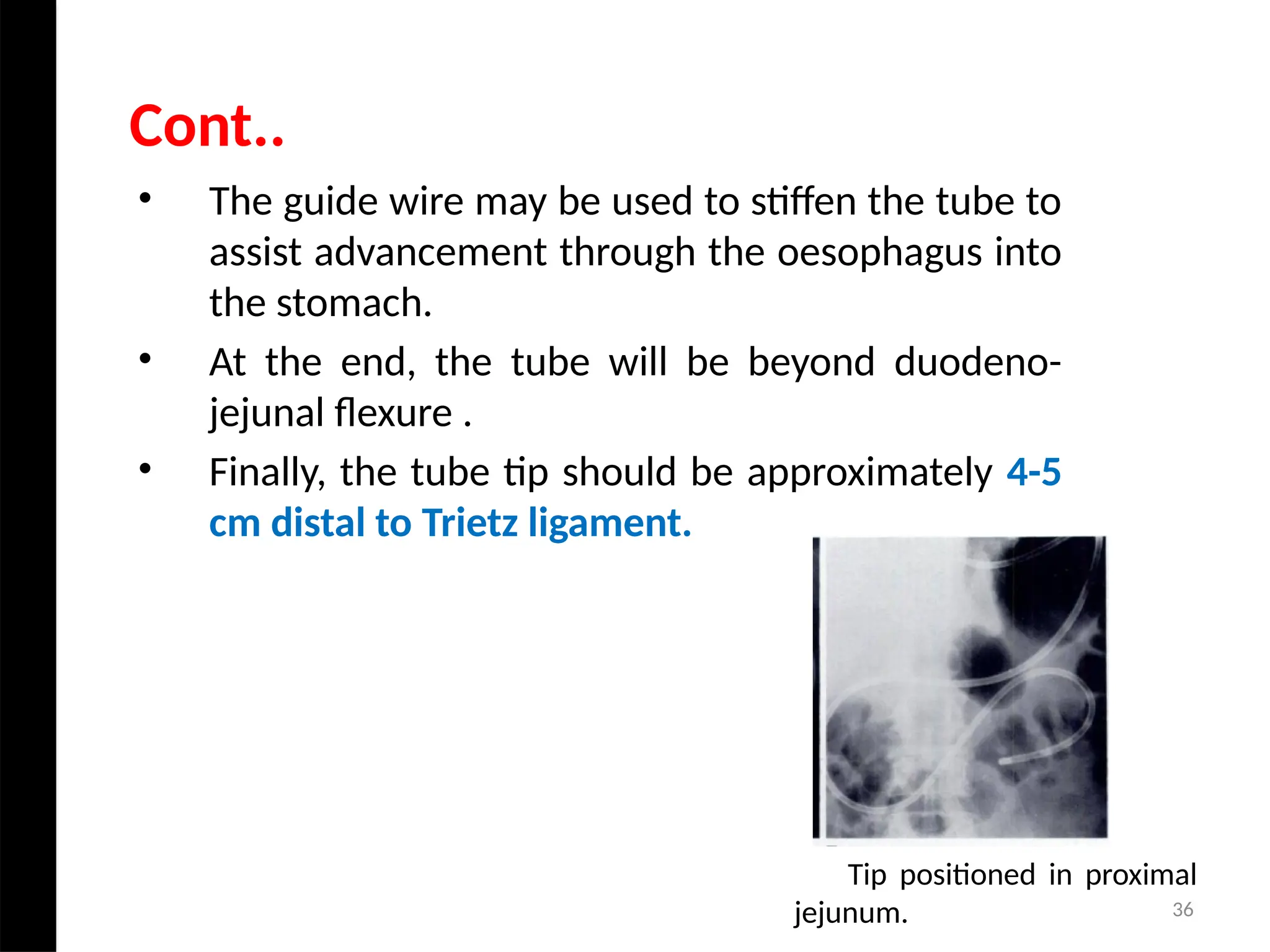
1
Barium Studies of Lower GI Tract
JOGINDER SINGH
RADIOLOGICAL TECHNOLOGIST
DEPARTMENT OF RADIODIAGNOSIS AND IMAGING
PGIMER, CHANDIGARH
2
• Anatomy
• BMFT
• Enteroclysis
• Peroral Pneumocolon
• Barium Enema
• Distal Cologram
• Loopogram
• Reduction of an Intussusception
• Newer imaging modalities
Contents
3
• Barium studies of lower gastrointestinal tract uses a
form of real-time x-ray called fluoroscopy and a
barium-based contrast material to help detect
disease and abnormalities and diagnose symptoms
such as pain, constipation or blood in the stool.
• It can often provide enough information to avoid more
invasive procedures such as colonoscopy.
Introduction
4
• The first radiographic examination of the colon is
credited to Williams.
• In 1901, he used air as a contrast medium­to outline
the colon during fluoroscopy.
• Schiile later performed the first single-contrast
enema, using an oily suspension of bismuth
subnitrate.
• The first modern double-contrast barium enema
(DCBE) with air insufflation was performed by Fischer
in 1923.
History
5
• The small intestine is continuous with
the stomach at the pyloric sphincter
and leads into the large intestine at the
ileocaecal valve.
• It is a little over 5 metres long and lies
in the abdominal cavity surrounded by
the large intestine.
• In the small intestine the chemical
digestion of food is completed and
most of the absorption of nutrients
takes place.
Anatomy of Small Intestine
6
• The small intestine comprises three main sections
continuous with each other.
• The duodenum is about 25 cm long.
• The jejunum is the middle section of the small
intestine and is about 2 metres long.
• The ileum, is about 3 metres long and ends at the
ileocaecal valve, which controls the flow of material
from the ileum to the caecum.
Conti..
7
8
• This is about 1.5 metres long, beginning
at the caecum in the right iliac fossa and
terminating at the rectum and anal
canal deep in the pelvis.
• Its lumen is larger than that of the
small intestine.
• The colon is divided into the caecum,
ascending colon, transverse colon,
descending colon, sigmoid colon,
rectum and anal canal.
Anatomy of Large Intestine
9
• The caecum- This is the first part
of the colon. Just below the
junction of the two the ileocaecal
valve opens from the ileum. It is
usually about 13 cm long.
• The ascending colon- This
passes upwards from the caecum to
the level of the liver where it curves
acutely to the left at the hepatic
flexure to become the transverse
colon.
10
• The transverse colon - This is a loop of colon which
extends across the abdominal cavity in front of the
duodenum and the stomach to the area of the spleen
where it forms the splenic flexure and curves acutely
downwards to become the descending colon.
• The descending colon - This passes down the left side of
the abdominal cavity then curves towards the midline.
After it enters the true pelvis it is known as the sigmoid
colon.
• The sigmoid colon - This part describes an S-shaped
curve in the pelvis then continues downwards to
become the rectum.
11
• The rectum - This is a slightly
dilated section of the colon about 13
cm long. It leads from the sigmoid
colon and terminates in the anal
canal.
• The anal canal - This is a short
passage about 3.8 cm long in the
adult and leads from the rectum to
the exterior. Two sphincter muscles
control the anus; the internal
sphincter, consisting of smooth
muscle fibres, and the external
sphincter, formed by skeletal muscle,
is under voluntary control .
12
13
• It is the radiographic examination of the GIT-
oesophagus, stomach, duodenum, small bowel and
ileocaecal junction by oral administration of contrast
media.
• It is so called because it is performed following a
barium meal examination of the oesophagus,
stomach and duodenum.
Barium Meal Follow Through
14
• Abdominal pain
• Diarrhea.
• Partial Obstruction.
• Suspected jejunal diverticulosis
• Malabsorption.
• Crohn's disease.
Indication
15
• Colonic obstruction
• Suspected perforation
• Paralytic ileus
Contraindications
16
• Medium density barium
suspension (50-60% w/v) is used
in dedicated Barium meal follow
through study.
Contrast medium
17
• Metaclopramide is most
frequently used to promotes bowel
motility and can decrease the
transit time of barium.
• Metaclopromide hydrochloride is
available as:
• Injection Perinorm or Injection
Maxeron.
• Dose: 2ml Injection given I.M
Drugs
18
• The colon should be cleaned by the administration of
a suitable purgative.
• A low roughage diet and a high fluid intake is also
maintained for 48 hours prior to the investigation
• No food or fluid should be taken for 12 hours before
the investigation.
• If the patient is taking tranquilizers, antispasmodics
and codeine, they should be stopped for 24-48 hrs
before the examination.
Preparation
19
• Barium(600-900 ml) 50-60% is allowed to drink
orally
• Patient is asked to drink this as rapidly as
possible. He is then put in the right side
dependent position to aid rapid gastric
emptying.
Right Lateral
Single contrast technique
20
• After 15 to 20 min, a film is taken with patient prone
to separate the bowel loops. Subsequent films are
taken at 15-30 min interval till ileocecal junction is
opacified.
Prone
Conti..
Prone abdominal radiograph
21
• To demonstrate I-C junction,
supine right side up is the best
position since ileum enters
caecum in the posteromedial part.
LPO
I-C Junction
Spot compression view of the
terminal ileum
22
• Better separation & less overlap of bowel
loops.
• In this position the center of the abdomen is
compressed making entire abdomen more
uniform and thus more uniform x-ray
penetration can be achieved.
• In this position loops of ileum tends to migrate
cephalad and becomes less compacted in the
pelvis which is often a common problem during
procedure.
Advantages of Prone Position
23
• The transit time through the small bowel can
vary greatly ranging between 15 minutes and 5
hours
24
• Same as single contrast study.
• Gas producing agent is given
when Barium reaches the
caecum.
• Patient is placed on the left
side slightly head down to
allow the gas to leave the
stomach and enter the small
bowel.
Double Contrast Technique
25
• Easily performed.
• No discomfort/ intubation to the patient unlike in
enteroclysis.
• It is a physiological process.
• Transit time can be assessed.
Advantages of BMFT
26
• Overlapping of Barium Filled bowel loops in the
pelvis.
• Poor distension of bowel loops.
• Inappropriate timing for visualization of partial/
intermittent small bowel obstruction.
• Operator dependence.
• Time consuming.
Disadvantages of BMFT
27
• Leakage of Barium from an unsuspected
perforation.
• Aspiration
• Conversion of partial large bowel obstruction into
a complete obstruction by the impaction of
Barium.
• Barium appendicitis, if Barium impacts in the
appendix.
• Side effects of pharmacological agents used.
Complications
28
• This is the radiological study of
small bowel from jejunum to the
ileocecal junction by intubation of
the jejunum & instillation of
contrast through the tube.
• Sellink refined the enteroclysis
technique, using a modification of
the Bilbao-Dotter tube in 1971.
Enteroclysis
29
• Partial small bowel obstruction
• Crohn’s disease
• Suspected diverticulum
• Malabsorption syndrome
• Tumors
• polyps
Indications
30
• Complete colonic obstruction
• Suspected perforation
• Massive dilatation of the small bowel
• Duodenal obstruction
• Paralytic ileus
Contraindications
31
• This is a 22F polyethylene
tube which is 150 cm long.
• The tube is 5cm longer than
the guide wire in order to
eliminate the risk of
perforation by the wire
protruding beyond the tip.
• The guide wire is Teflon
coated to reduce friction.
Bilbao Dotter Tube
Equipment
32
• For Single contrast enteroclysis: 20% w/v
suspension of Barium sulphate is used.
• For Double contrast enteroclysis: high density
low viscosity Barium sulphate suspension is
ideal which is 50-70%w/v.
Contrast Medium
33
• The Patient is subjected to liquid diet(2-3litres) for a
full day before the examination & is called after
overnight fasting for the procedure.
• 2-4 dulcolax tablets in the evening preceding the
enteroclysis are given.
• Drugs such as tranquilizers, sedatives &
antispasmodics should be discontinued the day
before the examination.
Preparation
34
• Preliminary plain Radiographs of the
abdomen.
• They are useful to determine
whether the Patient is adequately
prepared & to exclude the presence
of Barium from previous
examinations.
• An upright film is useful to
determine whether a large amount
of fluid is present in the stomach or
small bowel loops.
Technique
35
• The Patient sits on the edge of the X-ray table.
• The pharynx is anaesthetized with lignocaine spray.
• 2-3 cc of 2% Xylocaine jelly is introduced into the nostril
through which the tube is to be placed after ensuring that
there is no nasal blockage or mass.
• Patient’s neck is hyperextended and the Bilbao Dutter Tube
without the guide wire is inserted through one of the
nostrils and advanced with the swallowing action of
patient.
Procedure
36
• The guide wire may be used to stiffen the tube to
assist advancement through the oesophagus into
the stomach.
• At the end, the tube will be beyond duodeno-
jejunal flexure .
• Finally, the tube tip should be approximately 4-5
cm distal to Trietz ligament.
Cont..
Tip positioned in proximal
jejunum.
37
• Barium suspension 20% w /v. is injected at the rate
of 75 to 120 ml/ minute.
• Care should be taken to ensure that no air goes in
during the injection.
• An average of one to one and half litres of barium
sulfate is injected without any interruption.
• The average time taken to reach the ileo caecal
junction is about 15 minutes.
• Use interrupted fluoroscopy to follow the head of
the Barium column.
Single Contrast Enteroclysis
38
Enteroclysis
39
• 150 to 500 ml of barium suspension (high density and low
viscosity) is injected at the rate of 80-lO0ml/minute, till the
proximal ileum is reached.
• The head of the barium column is followed with intermittent
fluoroscopy and films are exposed wherever necessary. After
this, 0.5% suspension of Carboxymethyl cellulose (CMC) is
injected at a rate of around 75-120 ml/ min using a mechanical
injector.
• Ileocaecal spot films should be taken initially when the barium
column reaches the ileo caecal junction and then again when
the ileo caecal junction is in double contrast.
Double Contrast Enteroclysis
40
Double Contrast Enteroclysis
Barium in colon
Barium/Air in small
intestine
Bilbao-dutter Tube
41
• Contrast material is administered at a desired
rate & not influenced by the action of pyloric
sphincter.
• The time taken for the examination is not more
than 20-30 mins.
• Enteroclysis permits better detail of the small
bowel than that achieved by BMFT.
Advantages Of Enteroclysis
42
• Placement of nasogastric tube for the
procedure causes discomfort.
• Nausea & vomiting due to inadequate tube
placement.
• Operator dependent
Disadvantages Of Enteroclysis
43
• The Patient should be warned that diarrhoea
may occur as a result of the large volume of
fluid given.
• Patient can take full diet following the
procedure.
Complications:
1) Aspiration
2) Perforation of the bowel
After Care
44
• The peroral pneumocolon examination is performed when
barium filling of the right colon by the antegrade route
has occurred.
• At this time, air is introduced via the rectum until it
reaches the ileocecal area where it results in a double
contrast examination.
• The technique can be carried out at the conclusion of any
antegrade small bowel study as an immediate supplement
to routine compression spot films of the terminal ileum.
Peroral Pneumocolon
45
Indication:
• A poorly seen terminal ilium
• Clinically suspected inflammatory bowl disease with
an apparently normal terminal ilium
• An abnormal terminal ilium with suspicious fistula
Preparation:-
• Colonic preparation is similar to barium enema.
46
• Barium is administered orally.
• When barium has reached the right and proximal
transverse colon, air is insufflated into the rectum and
refluxed into distal ileum.
• Glucagon can be used to relax the ileocaecal valve.
• It is usually employed at the end of barium meal follow
through, when the appearance of terminal ileum is
suspicious and needs clarification.
Technique
47
48
• It is the radiographic study of the large bowel by
administration of the contrast medium through the
rectum.
Technique
• Single-contrast
• Double-contrast
Barium Enema
49
• Patient Should be given a low residue(low fiber) diet
for 2 days prior to the examination. Patient should
not have any fatty fried foods.
• Patient should drink plenty of clear fluids on the day
preceding the examination. Iron containing
medication should be stopped 2 days before the
examination because they make stools adhere to
mucosa.
Preparation
50
For removal of most solid material.
• Castor Oil (30ml)
• Dulcolax
BOWEL WASH:-
• Previous night
• In the morning, 2 hours prior to the
procedure.
Laxatives
51
Barium Enema Kit
52
• Uncooperative, or immobile patient.
• Evaluation of acute obstruction
• Reduction of intussusception
• Show configuration of colon
Single Contrast Barium Enema
Indications
53
• Allergy to Barium suspension
• Risk of perforation
• Peritonitis
• Suspicion of acute ulcerative colitis
• Following a recent deep biopsy.
Contraindications
54
• Toxic Megacolon
• If rectal biopsy has been
done in the previous 5 days
• Paralytic Ileus
Absolute Contraindication for
both DCBE & SCBE
55
• Barium suspension of Low
density 15% to 20% w/v. is
used.
• Tube is placed in the rectum
with the patient in left
lateral position.
• The height of the enema
should not be more than 1
metre above the table top.
Procedure
56
• As soon as the entire rectum is
full, the tube is clamped and a
lateral view is taken.
• Then the patient is put prone
and with the infusion running,
the frontal view film of the
rectum is exposed.
Rectum
Sigmoid
Colon
Rectum
Enema tube
in anal canal
Lateral View
Prone
57
• The patient is kept prone
with right side down oblique
position. This position helps
in the opening up the curve
of rectosigmoid junction.
Spot views of rectosigmoid
junctions with barium
flowing are taken.
Rectosigmoid Junction
Spot compression view for
recto-sigmoid junction
RAO
58
• Now the patient is kept
prone oblique with left
side down. Splenic
flexure opens out and
spot view of splenic
flexure is taken.
Splenic Flexure
LAO
59
• As barium flows towards
hepatic flexure, patient is
turned right side down
oblique and spot films of
hepatic flexure.
Hepatic Flexure
RAO
60
• With continuous flow of
barium caecum fills up. As
soon as the reflux across
ileocaecal junction takes
place, the tube is clamped
and ileocaecal spot films are
exposed.
IC Junction
RPO
61
• A full film is now
exposed to show
entire colon.
Full film
62
• Polyposis and diverticulosis can be
better visualized on post-evacuation
films.
• During the entire study, the head of
the barium column should be
followed under flouroscopy.
Post evacuation
Post Evacuating film showing
Mucosal Pattern
63
• Preferred method for routine
examination
• Rectal bleeding
• polyp
• colorectal cancer
• Polyposis
• Demonstration of sinuses or fistulas
• Severe diverticulosis
• Diarrhoea
• Presence of obstruction
• Reduction of an intussusception
Double Contrast Ba Enema
Indications
64
• Barium suspension: High density, 75% to 95% w/v.
• The Patient is in prone position with left side down
oblique & high density low viscosity Barium suspension
is allowed to flow up to splenic flexure. Now air is
introduced with Patient prone.
• Air should push the barium column and never pass
beyond the column.
• The patient is taken back in prone position with right
side dependent and air is pumped into left sided colon.
• With the right side up, more air is pumped till air
outlines the ileocaecal junction.
Procedure
65
Views
Rectum Ap, Lat
Recto sigmoid LPO,RAO
Splenic Flexure RPO, Lat
Transverse Colon PA, PA with 15 degree table
head down
Hepatic Flexure LPO, RAO
Entire Colon Supine
IC junction PA with 15 degree table
head elevation
66
Double contrast barium enema
67
Advantage of DC Over SC
1) Better surface details
2) Surface lesion can be demonstrated to the best
effect.
3) Easy unraveling of the colon as it is possible to look
through loops
Disadvantage of DC Over SC
4) Difficult in uncooperative Patient
5) Fistulae/sinuses can be missed
68
• It is a radiological study of large bowel
by administering contrast media
through colostomy opening.
• It is indicated mainly after surgery for
evaluation of operation and follow-up
of the patient.
• It is also helpful in determining the
efficacy of treatment in diverticulitis or
ulcerative colitis.
• In patient with colostomy the usual
preparation is irrigation of stoma the
night before study and again on the
morning of examination.
Distal Cologram
69
• Patient should lie down on the x-ray table in supine
position.
• Cleanse the skin around stoma appropriately
• Cannulate the colostomy with an appropriate Foley
catheter. Carefully inflate the balloon.
• Dilute barium is used for distal cologram study.
• Water soluble contrast medium can also be used.
Cont..
70
71
Indication
Following bladder resection to
demonstrate anatomy of ileal
conduit, ureters and renal
pelvicalyceal systems.
Contrast
water-soluble contrast agent.
Loopogram
72
Technique
• Cannulate ileal conduit with 14–18-F Foley catheter
and gently inflate balloon.
• Inject contrast into ileal conduit. Observe
retrograde filling of the renal collecting systems.
Stop injecting when adequately distended.
Images
• Plain AP of collecting systems.
• Post filling: AP of collecting systems, two obliques of
the kidneys with an additional, often oblique, view
of the ureteric–loop anastomosis.
74
• Pouchography is performed before closure of the
loop ileostomy to assess the integrity of the ileal
pouch and anastomosis.
Pouchography
75
• Radiographic examination of the ileal J
pouch is performed before closure of the
loop ileostomy to assess the integrity of the
ileal pouch and pouch-anal anastomosis.
• This radiographic examination is performed
by fluoroscopic visualization with a contrast
agent that could be introduced in an
antegrade position through a cannula fitted
in the efferent limb of the loop ileostomy
using a 16- to 18-French Foley catheter, or
retrogradely through the anus.
• The antegrade approach is considered the
safer technique and is commonly used for
pouch examination.
Conti..
76
This procedure should only be attempted
in full consultation with the surgeon in
charge of the case & a trained
anesthetist, when proper pediatric
anesthetic equipment is available.
Intussusceptions:- it is the enfolding of
one segment of the intestine within the
another.
Reduction of an Intussusception
77
Contraindications:
• Peritonitis or perforation
• The pneumatic method should probably not be used in
children over 4 years of age as there is a higher
incidence of significant lead points which may be
missed.
Patient preparation:
• Sedation is decided by surgeon while analgesics like
morphine are usually given.
• Correction of fluid & electrolytic balance.
78
Preliminary examinations:
Plain abdomen film: to assess bowel distension.
US: to confirm the diagnosis.
Technique
A 16-22 F catheter is inserted into the rectum and the
buttocks taped tightly together to provide a seal. It may
be necessary to inflate the balloon but if this is done it
should be performed under fluoroscopic control so that
rectum is not over distended.
The intussusception can be carried out by:
I. Pneumatic reduction.
II. Barium reduction (Rare).
Technique
79
• The child is placed in the prone position
so that it is easier to maintain the
catheter in the rectum.
• Air is instilled by a hand or mechanical
pump and the intussusception is pushed
back by a sustained pressure of up to 80
mm/hg. if this fails the pressure may be
increased to 120 mm/hg.
• Pressure should be monitored all the
times & there should be a pressure
release valve in the system.
Pneumatic Reduction
80
• An intussusception if unsuccessful once is repeated
after 3 min interval & three tries are done. A still
immovable intussusception is considered irreducible
& arrangement of surgery are made.
• Reduction is successful when there is free flow of air
into the distal ileum.
Conti..
81
• More rapid reduction, because the low viscosity
of air permits rapid filling of the colon.
• More effective reduction.
• There is more accurate control of intraluminal
pressure
• Less expensive.
Advantages
82
• Patient positioning is as for the
pneumatic method.
• The bag containing barium is raised
100 cm above the table top and
barium run in under hydrostatic
pressure. Progress of the column of
barium is monitored by intermittent
fluoroscopy.
• If the intussusception doesn’t move
for 3 min after consistent pressure,
child is given a 3 min rest & then
repeat the procedure.
Barium Reduction
83
Perforation. For the pneumatic method, if a pump
is used without a pressure- monitoring valve,
perforation may result in a tension
pneumoperitoneum, resulting in respiratory
embarrassment.
Complications
After care
Observation in hospital for 24 hours
84
Mega colon
Pathology
Crohn’s disease
85
86
Hirschsprung disease
Typical 'apple core' carcinoma in
the sigmoid colon
87
88
• 10/03/2024
8
9
ADVANCED
IMAGING MODALITIES
10/03/2024 90
• CT Enterography appears to be a promising technique for
small bowel examination which would perhaps overcome
the disadvantages of all the currently available
techniques.
Preparation and administration of neutral contrast
media:
• Neutral enteric contrast was prepared by adding 2 packet
(137.15 gm each) of PEGLEC and a small sachet of flavor in
2 Lt. of clean water.
CT Enterography
10/03/2024 91
Technique
• Patient was asked to drink 2 lt. solution within 45 minutes prior
to the scanning.
• The first 1000-1500 ml was ingested in first 30 minutes and
subsequently next 500 ml. was given in next 15 minutes.
• Immediately after completion of oral contrast CT scan should be
done.
• At the start of CT acquisition non-ionic contrast i.e. Iohexol /
Iopamidol 300 (mg/ml) was injected at the rate of 3-4 ml/ sec.
intravenously.
• The delay between start of intravenous contrast material and
start of Helical scanning should be 65-70 seconds.
Active jejunal Crohn disease in a 19-year-old woman.
CT enterogram shows mural hyperenhancement (arrows).
Compare the normal enhancement of the unaffected small bowel
(arrowhead).
Parietal adhesions. flattening (black arrowheads) of the anterior wall of small bowel
and loss of fat plane between the posterior wall of rectus sheath and bowel wall.
Anterior parietal peritoneum was thickened(arrow).
Mural stratification
• Visualization of layers of the bowel
wall at contrast material–enhanced CT.
• In mural stratification, the mucosa
and serosa enhance strongly.
• Intervening bowel wall can have any
of various degrees of attenuation,
depending on what pathologic
process.
• CT enterogram shows disease in the
neoterminal ileum with bilaminar
mural stratification (arrows) and
intramural
hyperenhancement of terminal ileal mucosa
(arrowhead) and mural thickening.
10/03/2024 96
• It displays the entire thickness of bowel wall.
• It allows the examination of ileal loops in the pelvis
without superimposition.
• It permits evaluation of surrounding mesentery &
peri enteric fat.
• It also allows assessment of solid organs & provides
global overview of the abdomen.
Advantages
10/03/2024 97
MRI Enterography may help find:-
• Internal bleeding
• Abscess
• Small tears in intestine
• MR Enterography is often recommended
when have Crohn’s disease.
• Overnight fasting
• Oral contrast – 2 packets of Peglec in 2
litres of water over 45 minutes
MRI Enterography
98
99
• Detailed knowledge about procedure including
anatomy and pathology.
• Should know about contrast media, its preparation &
its contraindications.
• He/She should be capable of tackling any emergency
arose due to contrast media.
• Knowledge of handling the equipment.
• Care & maintenance of the equipment .
• Knowledge of Quality assurance program.
• Knowledge of radiation protection.
Role of Technologist
100
• Barium Studies are safe & useful initial diagnostic
procedure for evaluating GIT abnormalities.
• It uses radiolucent & radiopaque contrast media in
combination to a fluoroscopy unit to visualize the
anatomy as well as the physiology.
• However these have limitations in the assessment of
extra mucosal extent of the pathology.
• Although newer modalities are present but Barium is still
considered as a basic and essential study.
Conclusion
101
• Radiological Procedures A Guideline by Dr.
Bhusan N.Lakhkar
• Techniques in Diagnostic Imaging, Graham H.
White house
• Radiological procedure, chapman
• Merill’s atlas of radiographic positioning and
procedure.
References
102
Thanking you

Barium Studies of lower GI Tract by joginder.pptx

  • 1.
    1 Barium Studies ofLower GI Tract JOGINDER SINGH RADIOLOGICAL TECHNOLOGIST DEPARTMENT OF RADIODIAGNOSIS AND IMAGING PGIMER, CHANDIGARH
  • 2.
    2 • Anatomy • BMFT •Enteroclysis • Peroral Pneumocolon • Barium Enema • Distal Cologram • Loopogram • Reduction of an Intussusception • Newer imaging modalities Contents
  • 3.
    3 • Barium studiesof lower gastrointestinal tract uses a form of real-time x-ray called fluoroscopy and a barium-based contrast material to help detect disease and abnormalities and diagnose symptoms such as pain, constipation or blood in the stool. • It can often provide enough information to avoid more invasive procedures such as colonoscopy. Introduction
  • 4.
    4 • The firstradiographic examination of the colon is credited to Williams. • In 1901, he used air as a contrast medium­to outline the colon during fluoroscopy. • Schiile later performed the first single-contrast enema, using an oily suspension of bismuth subnitrate. • The first modern double-contrast barium enema (DCBE) with air insufflation was performed by Fischer in 1923. History
  • 5.
    5 • The smallintestine is continuous with the stomach at the pyloric sphincter and leads into the large intestine at the ileocaecal valve. • It is a little over 5 metres long and lies in the abdominal cavity surrounded by the large intestine. • In the small intestine the chemical digestion of food is completed and most of the absorption of nutrients takes place. Anatomy of Small Intestine
  • 6.
    6 • The smallintestine comprises three main sections continuous with each other. • The duodenum is about 25 cm long. • The jejunum is the middle section of the small intestine and is about 2 metres long. • The ileum, is about 3 metres long and ends at the ileocaecal valve, which controls the flow of material from the ileum to the caecum. Conti..
  • 7.
  • 8.
    8 • This isabout 1.5 metres long, beginning at the caecum in the right iliac fossa and terminating at the rectum and anal canal deep in the pelvis. • Its lumen is larger than that of the small intestine. • The colon is divided into the caecum, ascending colon, transverse colon, descending colon, sigmoid colon, rectum and anal canal. Anatomy of Large Intestine
  • 9.
    9 • The caecum-This is the first part of the colon. Just below the junction of the two the ileocaecal valve opens from the ileum. It is usually about 13 cm long. • The ascending colon- This passes upwards from the caecum to the level of the liver where it curves acutely to the left at the hepatic flexure to become the transverse colon.
  • 10.
    10 • The transversecolon - This is a loop of colon which extends across the abdominal cavity in front of the duodenum and the stomach to the area of the spleen where it forms the splenic flexure and curves acutely downwards to become the descending colon. • The descending colon - This passes down the left side of the abdominal cavity then curves towards the midline. After it enters the true pelvis it is known as the sigmoid colon. • The sigmoid colon - This part describes an S-shaped curve in the pelvis then continues downwards to become the rectum.
  • 11.
    11 • The rectum- This is a slightly dilated section of the colon about 13 cm long. It leads from the sigmoid colon and terminates in the anal canal. • The anal canal - This is a short passage about 3.8 cm long in the adult and leads from the rectum to the exterior. Two sphincter muscles control the anus; the internal sphincter, consisting of smooth muscle fibres, and the external sphincter, formed by skeletal muscle, is under voluntary control .
  • 12.
  • 13.
    13 • It isthe radiographic examination of the GIT- oesophagus, stomach, duodenum, small bowel and ileocaecal junction by oral administration of contrast media. • It is so called because it is performed following a barium meal examination of the oesophagus, stomach and duodenum. Barium Meal Follow Through
  • 14.
    14 • Abdominal pain •Diarrhea. • Partial Obstruction. • Suspected jejunal diverticulosis • Malabsorption. • Crohn's disease. Indication
  • 15.
    15 • Colonic obstruction •Suspected perforation • Paralytic ileus Contraindications
  • 16.
    16 • Medium densitybarium suspension (50-60% w/v) is used in dedicated Barium meal follow through study. Contrast medium
  • 17.
    17 • Metaclopramide ismost frequently used to promotes bowel motility and can decrease the transit time of barium. • Metaclopromide hydrochloride is available as: • Injection Perinorm or Injection Maxeron. • Dose: 2ml Injection given I.M Drugs
  • 18.
    18 • The colonshould be cleaned by the administration of a suitable purgative. • A low roughage diet and a high fluid intake is also maintained for 48 hours prior to the investigation • No food or fluid should be taken for 12 hours before the investigation. • If the patient is taking tranquilizers, antispasmodics and codeine, they should be stopped for 24-48 hrs before the examination. Preparation
  • 19.
    19 • Barium(600-900 ml)50-60% is allowed to drink orally • Patient is asked to drink this as rapidly as possible. He is then put in the right side dependent position to aid rapid gastric emptying. Right Lateral Single contrast technique
  • 20.
    20 • After 15to 20 min, a film is taken with patient prone to separate the bowel loops. Subsequent films are taken at 15-30 min interval till ileocecal junction is opacified. Prone Conti.. Prone abdominal radiograph
  • 21.
    21 • To demonstrateI-C junction, supine right side up is the best position since ileum enters caecum in the posteromedial part. LPO I-C Junction Spot compression view of the terminal ileum
  • 22.
    22 • Better separation& less overlap of bowel loops. • In this position the center of the abdomen is compressed making entire abdomen more uniform and thus more uniform x-ray penetration can be achieved. • In this position loops of ileum tends to migrate cephalad and becomes less compacted in the pelvis which is often a common problem during procedure. Advantages of Prone Position
  • 23.
    23 • The transittime through the small bowel can vary greatly ranging between 15 minutes and 5 hours
  • 24.
    24 • Same assingle contrast study. • Gas producing agent is given when Barium reaches the caecum. • Patient is placed on the left side slightly head down to allow the gas to leave the stomach and enter the small bowel. Double Contrast Technique
  • 25.
    25 • Easily performed. •No discomfort/ intubation to the patient unlike in enteroclysis. • It is a physiological process. • Transit time can be assessed. Advantages of BMFT
  • 26.
    26 • Overlapping ofBarium Filled bowel loops in the pelvis. • Poor distension of bowel loops. • Inappropriate timing for visualization of partial/ intermittent small bowel obstruction. • Operator dependence. • Time consuming. Disadvantages of BMFT
  • 27.
    27 • Leakage ofBarium from an unsuspected perforation. • Aspiration • Conversion of partial large bowel obstruction into a complete obstruction by the impaction of Barium. • Barium appendicitis, if Barium impacts in the appendix. • Side effects of pharmacological agents used. Complications
  • 28.
    28 • This isthe radiological study of small bowel from jejunum to the ileocecal junction by intubation of the jejunum & instillation of contrast through the tube. • Sellink refined the enteroclysis technique, using a modification of the Bilbao-Dotter tube in 1971. Enteroclysis
  • 29.
    29 • Partial smallbowel obstruction • Crohn’s disease • Suspected diverticulum • Malabsorption syndrome • Tumors • polyps Indications
  • 30.
    30 • Complete colonicobstruction • Suspected perforation • Massive dilatation of the small bowel • Duodenal obstruction • Paralytic ileus Contraindications
  • 31.
    31 • This isa 22F polyethylene tube which is 150 cm long. • The tube is 5cm longer than the guide wire in order to eliminate the risk of perforation by the wire protruding beyond the tip. • The guide wire is Teflon coated to reduce friction. Bilbao Dotter Tube Equipment
  • 32.
    32 • For Singlecontrast enteroclysis: 20% w/v suspension of Barium sulphate is used. • For Double contrast enteroclysis: high density low viscosity Barium sulphate suspension is ideal which is 50-70%w/v. Contrast Medium
  • 33.
    33 • The Patientis subjected to liquid diet(2-3litres) for a full day before the examination & is called after overnight fasting for the procedure. • 2-4 dulcolax tablets in the evening preceding the enteroclysis are given. • Drugs such as tranquilizers, sedatives & antispasmodics should be discontinued the day before the examination. Preparation
  • 34.
    34 • Preliminary plainRadiographs of the abdomen. • They are useful to determine whether the Patient is adequately prepared & to exclude the presence of Barium from previous examinations. • An upright film is useful to determine whether a large amount of fluid is present in the stomach or small bowel loops. Technique
  • 35.
    35 • The Patientsits on the edge of the X-ray table. • The pharynx is anaesthetized with lignocaine spray. • 2-3 cc of 2% Xylocaine jelly is introduced into the nostril through which the tube is to be placed after ensuring that there is no nasal blockage or mass. • Patient’s neck is hyperextended and the Bilbao Dutter Tube without the guide wire is inserted through one of the nostrils and advanced with the swallowing action of patient. Procedure
  • 36.
    36 • The guidewire may be used to stiffen the tube to assist advancement through the oesophagus into the stomach. • At the end, the tube will be beyond duodeno- jejunal flexure . • Finally, the tube tip should be approximately 4-5 cm distal to Trietz ligament. Cont.. Tip positioned in proximal jejunum.
  • 37.
    37 • Barium suspension20% w /v. is injected at the rate of 75 to 120 ml/ minute. • Care should be taken to ensure that no air goes in during the injection. • An average of one to one and half litres of barium sulfate is injected without any interruption. • The average time taken to reach the ileo caecal junction is about 15 minutes. • Use interrupted fluoroscopy to follow the head of the Barium column. Single Contrast Enteroclysis
  • 38.
  • 39.
    39 • 150 to500 ml of barium suspension (high density and low viscosity) is injected at the rate of 80-lO0ml/minute, till the proximal ileum is reached. • The head of the barium column is followed with intermittent fluoroscopy and films are exposed wherever necessary. After this, 0.5% suspension of Carboxymethyl cellulose (CMC) is injected at a rate of around 75-120 ml/ min using a mechanical injector. • Ileocaecal spot films should be taken initially when the barium column reaches the ileo caecal junction and then again when the ileo caecal junction is in double contrast. Double Contrast Enteroclysis
  • 40.
    40 Double Contrast Enteroclysis Bariumin colon Barium/Air in small intestine Bilbao-dutter Tube
  • 41.
    41 • Contrast materialis administered at a desired rate & not influenced by the action of pyloric sphincter. • The time taken for the examination is not more than 20-30 mins. • Enteroclysis permits better detail of the small bowel than that achieved by BMFT. Advantages Of Enteroclysis
  • 42.
    42 • Placement ofnasogastric tube for the procedure causes discomfort. • Nausea & vomiting due to inadequate tube placement. • Operator dependent Disadvantages Of Enteroclysis
  • 43.
    43 • The Patientshould be warned that diarrhoea may occur as a result of the large volume of fluid given. • Patient can take full diet following the procedure. Complications: 1) Aspiration 2) Perforation of the bowel After Care
  • 44.
    44 • The peroralpneumocolon examination is performed when barium filling of the right colon by the antegrade route has occurred. • At this time, air is introduced via the rectum until it reaches the ileocecal area where it results in a double contrast examination. • The technique can be carried out at the conclusion of any antegrade small bowel study as an immediate supplement to routine compression spot films of the terminal ileum. Peroral Pneumocolon
  • 45.
    45 Indication: • A poorlyseen terminal ilium • Clinically suspected inflammatory bowl disease with an apparently normal terminal ilium • An abnormal terminal ilium with suspicious fistula Preparation:- • Colonic preparation is similar to barium enema.
  • 46.
    46 • Barium isadministered orally. • When barium has reached the right and proximal transverse colon, air is insufflated into the rectum and refluxed into distal ileum. • Glucagon can be used to relax the ileocaecal valve. • It is usually employed at the end of barium meal follow through, when the appearance of terminal ileum is suspicious and needs clarification. Technique
  • 47.
  • 48.
    48 • It isthe radiographic study of the large bowel by administration of the contrast medium through the rectum. Technique • Single-contrast • Double-contrast Barium Enema
  • 49.
    49 • Patient Shouldbe given a low residue(low fiber) diet for 2 days prior to the examination. Patient should not have any fatty fried foods. • Patient should drink plenty of clear fluids on the day preceding the examination. Iron containing medication should be stopped 2 days before the examination because they make stools adhere to mucosa. Preparation
  • 50.
    50 For removal ofmost solid material. • Castor Oil (30ml) • Dulcolax BOWEL WASH:- • Previous night • In the morning, 2 hours prior to the procedure. Laxatives
  • 51.
  • 52.
    52 • Uncooperative, orimmobile patient. • Evaluation of acute obstruction • Reduction of intussusception • Show configuration of colon Single Contrast Barium Enema Indications
  • 53.
    53 • Allergy toBarium suspension • Risk of perforation • Peritonitis • Suspicion of acute ulcerative colitis • Following a recent deep biopsy. Contraindications
  • 54.
    54 • Toxic Megacolon •If rectal biopsy has been done in the previous 5 days • Paralytic Ileus Absolute Contraindication for both DCBE & SCBE
  • 55.
    55 • Barium suspensionof Low density 15% to 20% w/v. is used. • Tube is placed in the rectum with the patient in left lateral position. • The height of the enema should not be more than 1 metre above the table top. Procedure
  • 56.
    56 • As soonas the entire rectum is full, the tube is clamped and a lateral view is taken. • Then the patient is put prone and with the infusion running, the frontal view film of the rectum is exposed. Rectum Sigmoid Colon Rectum Enema tube in anal canal Lateral View Prone
  • 57.
    57 • The patientis kept prone with right side down oblique position. This position helps in the opening up the curve of rectosigmoid junction. Spot views of rectosigmoid junctions with barium flowing are taken. Rectosigmoid Junction Spot compression view for recto-sigmoid junction RAO
  • 58.
    58 • Now thepatient is kept prone oblique with left side down. Splenic flexure opens out and spot view of splenic flexure is taken. Splenic Flexure LAO
  • 59.
    59 • As bariumflows towards hepatic flexure, patient is turned right side down oblique and spot films of hepatic flexure. Hepatic Flexure RAO
  • 60.
    60 • With continuousflow of barium caecum fills up. As soon as the reflux across ileocaecal junction takes place, the tube is clamped and ileocaecal spot films are exposed. IC Junction RPO
  • 61.
    61 • A fullfilm is now exposed to show entire colon. Full film
  • 62.
    62 • Polyposis anddiverticulosis can be better visualized on post-evacuation films. • During the entire study, the head of the barium column should be followed under flouroscopy. Post evacuation Post Evacuating film showing Mucosal Pattern
  • 63.
    63 • Preferred methodfor routine examination • Rectal bleeding • polyp • colorectal cancer • Polyposis • Demonstration of sinuses or fistulas • Severe diverticulosis • Diarrhoea • Presence of obstruction • Reduction of an intussusception Double Contrast Ba Enema Indications
  • 64.
    64 • Barium suspension:High density, 75% to 95% w/v. • The Patient is in prone position with left side down oblique & high density low viscosity Barium suspension is allowed to flow up to splenic flexure. Now air is introduced with Patient prone. • Air should push the barium column and never pass beyond the column. • The patient is taken back in prone position with right side dependent and air is pumped into left sided colon. • With the right side up, more air is pumped till air outlines the ileocaecal junction. Procedure
  • 65.
    65 Views Rectum Ap, Lat Rectosigmoid LPO,RAO Splenic Flexure RPO, Lat Transverse Colon PA, PA with 15 degree table head down Hepatic Flexure LPO, RAO Entire Colon Supine IC junction PA with 15 degree table head elevation
  • 66.
  • 67.
    67 Advantage of DCOver SC 1) Better surface details 2) Surface lesion can be demonstrated to the best effect. 3) Easy unraveling of the colon as it is possible to look through loops Disadvantage of DC Over SC 4) Difficult in uncooperative Patient 5) Fistulae/sinuses can be missed
  • 68.
    68 • It isa radiological study of large bowel by administering contrast media through colostomy opening. • It is indicated mainly after surgery for evaluation of operation and follow-up of the patient. • It is also helpful in determining the efficacy of treatment in diverticulitis or ulcerative colitis. • In patient with colostomy the usual preparation is irrigation of stoma the night before study and again on the morning of examination. Distal Cologram
  • 69.
    69 • Patient shouldlie down on the x-ray table in supine position. • Cleanse the skin around stoma appropriately • Cannulate the colostomy with an appropriate Foley catheter. Carefully inflate the balloon. • Dilute barium is used for distal cologram study. • Water soluble contrast medium can also be used. Cont..
  • 70.
  • 71.
    71 Indication Following bladder resectionto demonstrate anatomy of ileal conduit, ureters and renal pelvicalyceal systems. Contrast water-soluble contrast agent. Loopogram
  • 72.
    72 Technique • Cannulate ilealconduit with 14–18-F Foley catheter and gently inflate balloon. • Inject contrast into ileal conduit. Observe retrograde filling of the renal collecting systems. Stop injecting when adequately distended. Images • Plain AP of collecting systems. • Post filling: AP of collecting systems, two obliques of the kidneys with an additional, often oblique, view of the ureteric–loop anastomosis.
  • 74.
    74 • Pouchography isperformed before closure of the loop ileostomy to assess the integrity of the ileal pouch and anastomosis. Pouchography
  • 75.
    75 • Radiographic examinationof the ileal J pouch is performed before closure of the loop ileostomy to assess the integrity of the ileal pouch and pouch-anal anastomosis. • This radiographic examination is performed by fluoroscopic visualization with a contrast agent that could be introduced in an antegrade position through a cannula fitted in the efferent limb of the loop ileostomy using a 16- to 18-French Foley catheter, or retrogradely through the anus. • The antegrade approach is considered the safer technique and is commonly used for pouch examination. Conti..
  • 76.
    76 This procedure shouldonly be attempted in full consultation with the surgeon in charge of the case & a trained anesthetist, when proper pediatric anesthetic equipment is available. Intussusceptions:- it is the enfolding of one segment of the intestine within the another. Reduction of an Intussusception
  • 77.
    77 Contraindications: • Peritonitis orperforation • The pneumatic method should probably not be used in children over 4 years of age as there is a higher incidence of significant lead points which may be missed. Patient preparation: • Sedation is decided by surgeon while analgesics like morphine are usually given. • Correction of fluid & electrolytic balance.
  • 78.
    78 Preliminary examinations: Plain abdomenfilm: to assess bowel distension. US: to confirm the diagnosis. Technique A 16-22 F catheter is inserted into the rectum and the buttocks taped tightly together to provide a seal. It may be necessary to inflate the balloon but if this is done it should be performed under fluoroscopic control so that rectum is not over distended. The intussusception can be carried out by: I. Pneumatic reduction. II. Barium reduction (Rare). Technique
  • 79.
    79 • The childis placed in the prone position so that it is easier to maintain the catheter in the rectum. • Air is instilled by a hand or mechanical pump and the intussusception is pushed back by a sustained pressure of up to 80 mm/hg. if this fails the pressure may be increased to 120 mm/hg. • Pressure should be monitored all the times & there should be a pressure release valve in the system. Pneumatic Reduction
  • 80.
    80 • An intussusceptionif unsuccessful once is repeated after 3 min interval & three tries are done. A still immovable intussusception is considered irreducible & arrangement of surgery are made. • Reduction is successful when there is free flow of air into the distal ileum. Conti..
  • 81.
    81 • More rapidreduction, because the low viscosity of air permits rapid filling of the colon. • More effective reduction. • There is more accurate control of intraluminal pressure • Less expensive. Advantages
  • 82.
    82 • Patient positioningis as for the pneumatic method. • The bag containing barium is raised 100 cm above the table top and barium run in under hydrostatic pressure. Progress of the column of barium is monitored by intermittent fluoroscopy. • If the intussusception doesn’t move for 3 min after consistent pressure, child is given a 3 min rest & then repeat the procedure. Barium Reduction
  • 83.
    83 Perforation. For thepneumatic method, if a pump is used without a pressure- monitoring valve, perforation may result in a tension pneumoperitoneum, resulting in respiratory embarrassment. Complications After care Observation in hospital for 24 hours
  • 84.
  • 85.
  • 86.
    86 Hirschsprung disease Typical 'applecore' carcinoma in the sigmoid colon
  • 87.
  • 88.
  • 89.
  • 90.
    10/03/2024 90 • CTEnterography appears to be a promising technique for small bowel examination which would perhaps overcome the disadvantages of all the currently available techniques. Preparation and administration of neutral contrast media: • Neutral enteric contrast was prepared by adding 2 packet (137.15 gm each) of PEGLEC and a small sachet of flavor in 2 Lt. of clean water. CT Enterography
  • 91.
    10/03/2024 91 Technique • Patientwas asked to drink 2 lt. solution within 45 minutes prior to the scanning. • The first 1000-1500 ml was ingested in first 30 minutes and subsequently next 500 ml. was given in next 15 minutes. • Immediately after completion of oral contrast CT scan should be done. • At the start of CT acquisition non-ionic contrast i.e. Iohexol / Iopamidol 300 (mg/ml) was injected at the rate of 3-4 ml/ sec. intravenously. • The delay between start of intravenous contrast material and start of Helical scanning should be 65-70 seconds.
  • 92.
    Active jejunal Crohndisease in a 19-year-old woman. CT enterogram shows mural hyperenhancement (arrows). Compare the normal enhancement of the unaffected small bowel (arrowhead).
  • 93.
    Parietal adhesions. flattening(black arrowheads) of the anterior wall of small bowel and loss of fat plane between the posterior wall of rectus sheath and bowel wall. Anterior parietal peritoneum was thickened(arrow).
  • 94.
    Mural stratification • Visualizationof layers of the bowel wall at contrast material–enhanced CT. • In mural stratification, the mucosa and serosa enhance strongly. • Intervening bowel wall can have any of various degrees of attenuation, depending on what pathologic process. • CT enterogram shows disease in the neoterminal ileum with bilaminar mural stratification (arrows) and intramural
  • 95.
    hyperenhancement of terminalileal mucosa (arrowhead) and mural thickening.
  • 96.
    10/03/2024 96 • Itdisplays the entire thickness of bowel wall. • It allows the examination of ileal loops in the pelvis without superimposition. • It permits evaluation of surrounding mesentery & peri enteric fat. • It also allows assessment of solid organs & provides global overview of the abdomen. Advantages
  • 97.
    10/03/2024 97 MRI Enterographymay help find:- • Internal bleeding • Abscess • Small tears in intestine • MR Enterography is often recommended when have Crohn’s disease. • Overnight fasting • Oral contrast – 2 packets of Peglec in 2 litres of water over 45 minutes MRI Enterography
  • 98.
  • 99.
    99 • Detailed knowledgeabout procedure including anatomy and pathology. • Should know about contrast media, its preparation & its contraindications. • He/She should be capable of tackling any emergency arose due to contrast media. • Knowledge of handling the equipment. • Care & maintenance of the equipment . • Knowledge of Quality assurance program. • Knowledge of radiation protection. Role of Technologist
  • 100.
    100 • Barium Studiesare safe & useful initial diagnostic procedure for evaluating GIT abnormalities. • It uses radiolucent & radiopaque contrast media in combination to a fluoroscopy unit to visualize the anatomy as well as the physiology. • However these have limitations in the assessment of extra mucosal extent of the pathology. • Although newer modalities are present but Barium is still considered as a basic and essential study. Conclusion
  • 101.
    101 • Radiological ProceduresA Guideline by Dr. Bhusan N.Lakhkar • Techniques in Diagnostic Imaging, Graham H. White house • Radiological procedure, chapman • Merill’s atlas of radiographic positioning and procedure. References
  • 102.

Editor's Notes

  • #5 The valvulae conniventes, also known as Kerckring folds, plicae circulares or just small bowel folds, are the mucosal folds of the small intestine
  • #6 The valvulae conniventes, also known as Kerckring folds, plicae circulares or just small bowel folds, are the mucosal folds of the small intestine
  • #14  We found the mean diameter of the duodenum to be 24.8mm , jejunum to be 24.5mm ,proximal ileum to be 19.5mm , distal ileum to be 18.9 mm and terminal ileum to be 18.7 mm .
  • #15 Paralytic ileus: Obstruction of the intestine due to paralysis of the intestinal muscles.
  • #18 Tranquilizers a medicinal drug taken to reduce tension or anxiety. a medicinal drug taken to reduce tension or anxiety. Spasm a sudden involuntary muscular contraction or convulsive movement. antispasmodics Smooth muscle spasm Codeine a sleep-inducing and analgesic drug derived from morphine.
  • #28 Crohn's disease is an inflammatory bowel disease (IBD). Malabsorption is abnormality in absorption.
  • #29 Crohn's disease is an inflammatory bowel disease (IBD). Malabsorption is abnormality in absorption.
  • #30 perforated bowel is a hole in the wall of the intestine which can occur for a variety of reasons.
  • #33 Laxative definition is - having a tendency to loosen or relax; specifically : producing bowel movements and relieving constipation.
  • #37 at the rate of 75 to 120ml/min
  • #39 @ 80-100ml/min. atony is a condition in which a muscle has lost its strength.  The addition of methyl cellulose improves diagnostic imaging in diverticulosis by expanding the lumen
  • #41 Hypotonia Decreased muscle tone .
  • #51 ET tube functions to seal the airway One-shot-inflator has a maximum air displacement volume of 100 c.c., avoiding   over inflation. 
  • #53 Fulminant colitis condition in which patients with severe ulcerative colitis who have more than 10 stools per day, continuous bleeding, abdominal pain, distension, and acute, severe toxic symptoms including fever and anorexia. Such patients are at risk of progressing to toxic megacolon and bowel perforation.
  • #54 Pseudomembranous colitis refers to swelling or inflammation of the large intestine (colon) due to an overgrowth of Clostridium difficile (C difficile) bacteria. This infection is a common cause of diarrhea after antibiotic use. Piles are collections of tissue and vein that become inflamed and swollen.
  • #68 Colostomy irrigation is a way to manage bowel movements by emptying the colon at a scheduled time. The process involves putting water into the colon through the stoma.
  • #69 perineal is tissue b/n rproductive oragan and anus
  • #70 Colostomy irrigation is a way to manage bowel movements by emptying the colon at a scheduled time. The process involves putting water into the colon through the stoma.
  • #82 HDROSTATIC PRESSURE is the pressure with in the fluid
  • #84 The most feared complication of ulcerative colitis is the development of toxic megacolon. It occurs as a result of extension of the inflammation beyond the submucosa into the muscularis, causing loss of contractility and ultimately resulting in a dilated colon. 
  • #85 The most feared complication of ulcerative colitis is the development of toxic megacolon. It occurs as a result of extension of the inflammation beyond the submucosa into the muscularis, causing loss of contractility and ultimately resulting in a dilated colon.