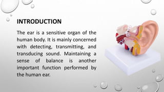
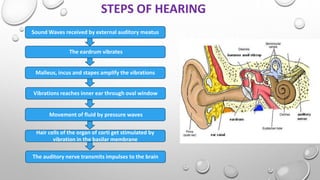
SUBMITTED TO:-
MISS SHIBANI HANSDAH
SUBMITTED BY:-
ANURADHA JENA
INTRODUCTION
The ear is a sensitive organ of the
human body. It is mainly concerned
with detecting, transmitting, and
transducing sound. Maintaining a
sense of balance is another
important function performed by
the human ear.
STRUCTURE OF HUMAN EAR
The human ear consists of three
parts:-
A. External Ear
B. Middle Ear
C. Internal Ear
External Ear
The external ear is further divided into the following parts-
I Auricle (pinna) :-
The auricle comprises a thin plate of elastic cartilage covered by a
layer of skin. It consists of funnel-like curves that collect sound waves
and transmit them to the middle ear. The lobule consists of adipose
and fibrous tissues supplied with blood capillaries.
II External auditory meatus:-
It is a slightly curved canal supported by bone in its interior part
and cartilage in the exterior part. The meatus or the canal is lined with
stratified epithelium and wax glands.
III Tympanic membrane :-
This membrane separates the middle ear and the external ear. This
part receives and amplifies the sound waves. It’s central part is known
as umbo. Its
Middle Ear
The middle ear comprises the following parts –
I. Tympanic Cavity:-
It is a narrow air-filled cavity separated from the external ear by the tympanic membrane and
from the inner ear by the bony wall. The tympanic cavity has an auditory tube known as the eustachian
tube in its anterior wall.
II. Eustachian Tube:-
The Eustachian tube is a 4-centimeter-long tube that equalizes air pressure on either side of the
tympanic membrane. It connects the tympanic cavity with the nasopharynx.
III. Ear Ossicles:-
These transmit sound waves from the ear drum to the middle ear. There are three ear ossicles in the
human ear:-
1. Malleus:
A hammer-shaped part is attached to the tympanic membrane through the handle and incus
through the head. It is the largest ear ossicle.
2. Incus:
An anvil-shaped ear ossicle connected with the stapes.
3. Stapes:-
It is the smallest ossicle and bone in the human body.
Inner Ear
It comprises two parts -
I. Bony labyrinth:-
The bony labyrinth comprises a vestibule, three
semi-circular cannels, and spirally coiled cochlea.
It is filled with perilymph.
II. Membranous labyrinth:-
The bony labyrinth surrounds the
membranous labyrinth. It comprises sensory receptors responsible for
balance and hearing. The membranous labyrinth is filled with
endolymph and comprises three semi-circular ducts, cochlear duct,
saccule, and utricle. The sensory receptors include cristae, an organ of
Corti, and ampullary maculae.
STEPS OF HEARING
Sound Waves received by external auditory meatus
The eardrum vibrates
Malleus, incus and stapes amplify the vibrations
Vibrations reaches inner ear through oval window
Movement of fluid by pressure waves
Hair cells of the organ of corti get stimulated by
vibration in the basilar membrane
The auditory nerve transmits impulses to the brain
MECHANISM OF HEARING
1. The pinna receives the sound waves and it reaches the tympanic membrane
through the meatus.
2. The eardrum vibrates the these vibrations get transmitted to the three
ossicles present in the middle ear.
3. Malleus, incus, and stapes amplify the sound waves.
4. These vibrations then reach the perilymph ( Scala vestibuli) through the oval
window.
5. Then the pressure waves get transferred to the endolymph of scala media and
reach the basilar membrane and then to the perilymph of scala tympani. This
movement of fluid is facilitated by the round window present at the end of
the scala tympani.
6. The basilar membrane movement causes the rubbing of stereocilia against the
tectorial membrane.
7. Stereocilia are bent resulting in the opening of ion channels in the plasma
membrane of hair cells. Glutamate, a neurotransmitter, is released due to Ca++
ion movement inside the cell.
8. These neurotransmitters bind to the receptors of afferent neurons, which
synapse with hair cells causing depolarization of neurons. A nerve impulse is
generated and transmitted to the auditory cortex of the brain through the
auditory nerve ( Cranial nerve viii).
9. The brain analyses the impulses and we hear the sound. The brain not only
recognizes the sound but also judges the direction, loudness, and pitch of the
sound .
BALANCE
The eustachian tube and vestibular complex are the important parts of the ear responsible for
balance.
. The eustachian tube equalizes the air pressure in the middle ear and maintains the balance.
. The vestibular complex contains receptors that maintain body balance.
Human_Ear_PPT research about human ear [1].pptx

Human_Ear_PPT research about human ear [1].pptx

  • 1.
    SUBMITTED TO:- MISS SHIBANIHANSDAH SUBMITTED BY:- ANURADHA JENA
  • 2.
    INTRODUCTION The ear isa sensitive organ of the human body. It is mainly concerned with detecting, transmitting, and transducing sound. Maintaining a sense of balance is another important function performed by the human ear.
  • 3.
    STRUCTURE OF HUMANEAR The human ear consists of three parts:- A. External Ear B. Middle Ear C. Internal Ear
  • 4.
    External Ear The externalear is further divided into the following parts- I Auricle (pinna) :- The auricle comprises a thin plate of elastic cartilage covered by a layer of skin. It consists of funnel-like curves that collect sound waves and transmit them to the middle ear. The lobule consists of adipose and fibrous tissues supplied with blood capillaries. II External auditory meatus:- It is a slightly curved canal supported by bone in its interior part and cartilage in the exterior part. The meatus or the canal is lined with stratified epithelium and wax glands. III Tympanic membrane :- This membrane separates the middle ear and the external ear. This part receives and amplifies the sound waves. It’s central part is known as umbo. Its
  • 5.
    Middle Ear The middleear comprises the following parts – I. Tympanic Cavity:- It is a narrow air-filled cavity separated from the external ear by the tympanic membrane and from the inner ear by the bony wall. The tympanic cavity has an auditory tube known as the eustachian tube in its anterior wall. II. Eustachian Tube:- The Eustachian tube is a 4-centimeter-long tube that equalizes air pressure on either side of the tympanic membrane. It connects the tympanic cavity with the nasopharynx. III. Ear Ossicles:- These transmit sound waves from the ear drum to the middle ear. There are three ear ossicles in the human ear:- 1. Malleus: A hammer-shaped part is attached to the tympanic membrane through the handle and incus through the head. It is the largest ear ossicle. 2. Incus: An anvil-shaped ear ossicle connected with the stapes. 3. Stapes:- It is the smallest ossicle and bone in the human body.
  • 6.
    Inner Ear It comprisestwo parts - I. Bony labyrinth:- The bony labyrinth comprises a vestibule, three semi-circular cannels, and spirally coiled cochlea. It is filled with perilymph. II. Membranous labyrinth:- The bony labyrinth surrounds the membranous labyrinth. It comprises sensory receptors responsible for balance and hearing. The membranous labyrinth is filled with endolymph and comprises three semi-circular ducts, cochlear duct, saccule, and utricle. The sensory receptors include cristae, an organ of Corti, and ampullary maculae.
  • 7.
    STEPS OF HEARING SoundWaves received by external auditory meatus The eardrum vibrates Malleus, incus and stapes amplify the vibrations Vibrations reaches inner ear through oval window Movement of fluid by pressure waves Hair cells of the organ of corti get stimulated by vibration in the basilar membrane The auditory nerve transmits impulses to the brain
  • 8.
    MECHANISM OF HEARING 1.The pinna receives the sound waves and it reaches the tympanic membrane through the meatus. 2. The eardrum vibrates the these vibrations get transmitted to the three ossicles present in the middle ear. 3. Malleus, incus, and stapes amplify the sound waves. 4. These vibrations then reach the perilymph ( Scala vestibuli) through the oval window. 5. Then the pressure waves get transferred to the endolymph of scala media and reach the basilar membrane and then to the perilymph of scala tympani. This movement of fluid is facilitated by the round window present at the end of the scala tympani. 6. The basilar membrane movement causes the rubbing of stereocilia against the tectorial membrane. 7. Stereocilia are bent resulting in the opening of ion channels in the plasma membrane of hair cells. Glutamate, a neurotransmitter, is released due to Ca++ ion movement inside the cell. 8. These neurotransmitters bind to the receptors of afferent neurons, which synapse with hair cells causing depolarization of neurons. A nerve impulse is generated and transmitted to the auditory cortex of the brain through the auditory nerve ( Cranial nerve viii). 9. The brain analyses the impulses and we hear the sound. The brain not only recognizes the sound but also judges the direction, loudness, and pitch of the sound .
  • 9.
    BALANCE The eustachian tubeand vestibular complex are the important parts of the ear responsible for balance. . The eustachian tube equalizes the air pressure in the middle ear and maintains the balance. . The vestibular complex contains receptors that maintain body balance.