Cornea 2
•Download as PPTX, PDF•
5 likes•297 views

Undergrduate ophthalmology lecture

Recommended
More Related Content
What's hot
What's hot (20)
Similar to Cornea 2
Similar to Cornea 2 (20)
More from faculty of medicine -benha university
More from faculty of medicine -benha university (16)
Recently uploaded
Recently uploaded (20)
Cornea 2
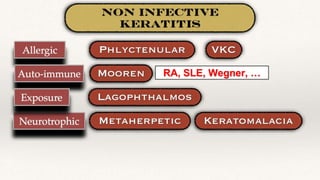
- 1. RA, SLE, Wegner, …
- 2. • Site: Limbus or corneal periphery • Size: small, 1-3 mm • Shape: round • Colour: Greyish red • Number: Solitary or multiple • Surrounded by hyperemia
- 3. Treatment • Tonsillitis • Intestinal parasites • TB • Topical steroids • Topical antibiotics to guard against 2ry bacterial infection • Topical cycloplegics A. Chemical cauterisation of the ulcer with Carbolic acid B. Diathermy & Peritomy of the feeding vessels
- 4. Aetiology • Coalescence of punctate erosions in VKC leading to large epithelial defect • If not treated properly, a plaque containing fibrin and mucus deposits (Shields) on this epithelial defect which hampers the re-epithelialization of shield ulcer
- 5. Treatment • Steroids • Mast cell stabilisers • Anti histamine • Antibiotics • Tear substitutes • Debridement of the fibrin & mucus deposits • Amniotic membrane transplantation (AMT)
- 6. Definition Rapidly progressive painful ulcerative keratitis initially affecting corneal periphery then spread circumferentially & centrally Aetiology Unknown, may be: • Auto-immune disease Limbal vasculitis Ischemic necrosis
- 7. C/P • Crescent shaped ulcer • Starts at corneal periphery (Limbal vasculitis) • Creeps Circumferentially & Centrally • Has two edges: 1. Advancing edge: undermined, creeps over the cornea 2. Peripheral Edge: Healed, Vascularized
- 8. Treatment • Steroids • Cyclosporin A • Antibiotics, Cycloplegics, Tear substitutes • Immune-suppressors: Steroids, non steroids • Conjunctival resection (Peritomy) • Keratoplasty (lamellar or penetrating) Poor Prognosis !!
- 9. Caused by loss of corneal sensation (V) C/P • Painless • Loss of corneal sensation • Resistant corneal ulcer Treatment • Preservative free tear substitutes • Soft bandage contact lens • Topical neurotrophic substances • Vit A supplement • Amniotic membrane transplantation • Conjunctival flap
- 10. Incomplete closure of the palpebral fissure when lids are gently closed A. Lid coloboma B. Ectropion C. Post ptosis surgery D. Proptosis Severe illness
- 12. Filamentary Keratitis Filamentary keratitis is a condition in which strands (“filaments”) composed of degenerated epithelial cells and mucus develop on and adhere to the corneal surface causing pain and foreign body sensation. Aetiology 1.Dry Eye 2.Neurotrophic Keratopathy 3.Long term ocular patching 4.Superior limbic kerato- conjunctivitis
- 13. Filaments stained with rose Bengal Strands of mucus admixed with epithelium, attached at one end to the corneal surface C/P
- 14. Treatment 1) Topical therapy with lubricant drops and ointment. 2)Bandage soft contact lenses. 3)Prophylactic topical antibiotic 4)Mucolytic agent such as 10% N- Acetylcysteine can be used topically to decrease the viscosity of the mucinous component of the tear film. 5)Topical Hypertonic sodium chloride 5% drops may also help by deturgescing and compacting the corneal epithelium. Medical Treatment Surgical Treatment 1) Manual removal of the corneal filaments using forceps to temporarily alleviate pain. 2)Punctal plug
- 15. Inflammation of the corneal stroma without involvement of epithelium or endothelium A. Bacteria: Syphilis, TB B. Virus: HSV, HZV C. Idiopathic: Cogan’s syndrome A. HSV B. HZV Cogan’s syndrome: • Cochlear Deafness • Optical (interstitial keratitis) • Glucocorticoids for ttt • Aortic involvement • Nerve deafnes
- 16. Aetiology Treponema pallidum (Spirochaete) • Congenital: Common - Bilateral - Young age • Acquired: Rare - Unilateral - 10 years after 1ry infection - Sexually transmitted Pathogenesis Hypersensitivity to Treponema pallidum antigen
- 17. C/P 1. Progressive stage (2 weeks): • Severe corneal symptoms (Pain, …) & signs (Ciliary injection, …) • Salmon Pink corneal patch ( dull pink hazy cornea with deep corneal vascularisation)
- 18. 2. Florid stage (2 months): • Severe corneal symptoms & signs with diminution of vision • Increased vascularisation & infiltration
- 19. 3. Regressive stage (2 years): • Corneal interstitial opacities & ghost vessels • Uveitis
- 20. Investigations A. Wasseman reaction B. Venereal Disease Research Lab (VLDL) C. Treponema Pallidum Hemagglutination Test (TPHA) D. Fluorescent Treponema Antigen (FTA) Treatment 1. Treat Keratitis: Topical steroids, antibiotics, cycloplegics 2. Treat Syphilis: Penicillins 3. Treat complications: Keratoplasty
- 21. Inflammation of the deep layers of corneal stroma & endothelium A. Bacteria: Posterior abscess B. Virus: HSV, HZV C. Fungi
- 22. • An opaque ring, grey to white in colour, that surrounds the periphery of the cornea and separated from the limbus by an area of clear cornea • Occurs primarily in elder people • Due to a lipid infiltration of the corneal stroma • Innocuous, need no treatment other than investigate for poor lipid profile
- 23. • A greyish white band-shaped corneal opacity in the inter palpebral zone • Calcium deposits at the level of Bowman membrane • Causes: A. Ocular: ❖ chronic inflammation (Keratitis, Uveitis) ❖ Phthisis Bulbi B. Systemic: metastatic calcification (hyper- parathyroidism, renal failure, sarcoisosis, malignancies, …) • Symptoms: pain, diminution of vision • Traetment: 1. Chemical chelation: di sodium edta 2. Mechanical scraping of the calcium deposits 3. PTK (Excimer laser)
- 24. • Greenish brown ring in the corneal periphery • Wilson Disease (Hepato-lenticular degeneration) (Not pathognomonic) • Due to deficiency in ceruloplasmin (Copper carrier) • Copper deposits in Cornea, lens, liver,...
- 25. • Sympyoms: Stitching pain, lacrimation, photophobia, .. • Treatment: FB removal (Search for other FBs)
- 26. • Trachoma • Floppy eyelid syndrome • Superior limbic keratoconjunctivitis • Lagophthalmos • Chronic blepharitis, Entropion • Toxicity to topical medications • Dry eye • Neurotrophic keratitis Punctate Epithelial Erosions
- 27. • Acute, Rapidly progressive • Central, usually single • Hypopyon • Start limbal, spread central & circumferential • Chronic course • Limbal • Single or multiple
- 29. Definition Progressive conical distortion of the cornea with: • irregular astigmatism • axial stromal thinning • apical protrusion • increasing myopia.
- 30. Aetiology & Pathogenesis ❖ Unknown, may be: • Repeated corneal trauma (eye rubbing) • Corneal stromal abnormalities (Collagen disorders e.g. Marfan, Down) • Hereditary (AD)
- 32. Symptoms ❖ Age: Early adulthood ❖ Gradual, progressive, painless diminution of vision: • Myopia • Astigmatism • Corneal opacities ❖ Frequent change of glasses ❖ Sudden Painful diminution of vision (Acute Hydrops)
- 33. • No Evident Clinical signs • Typical Topographic changes • Discovered accidentally e.g. patient seeking for LASIK Signs • No Evident Clinical signs • Typical Topographic changes • Scissoring on retinoscopy
- 34. Scissoring reflex in retinoscopy
- 35. • Boat shape angulation of the lower lid in down gaze
- 36. • Sharply focused beam of light near the nasal limbus ,produced by lateral illumination of the cornea in advanced keratonous
- 37. • The apex of the cone • Usually inferior to the pupillary axis
- 38. • Fine vertical lines in the deep stroma • Parallel to the axis of the cone • Disappears on gentle digital pressure Vogt ==== Vertical
- 39. • At the base of the cone • Hemosiderin pigment deposited in corneal epithelium
- 40. • Superficial linear scars • At corneal apex • Caused by ruptures in Bowman layer
- 43. Complications • Acute rupture of Descemet’s membrane, leading to corneal edema • Sudden, painful diminution of vision DDx: Disciform keratitis
- 44. Investigations • A series of concentric rings is projected onto the cornea and their reflection viewed by the examiner through a small hole in the centre of the disk. • A regular-shaped cornea should show equally spaced symmetric reflections. • If the patient is suffering from astigmatism, the rings will be distorted.
- 45. • To measure the curvature of the anterior surface of the cornea, particularly for assessing the extent and axis of astigmatism.
- 46. • A rotating camera that evaluate: 1. Corneal thickness (Pachymetry) 2. Corneal curvature & Power (Keratometry)
- 48. Grading of KCN
- 49. Treatment • Early cases with mild astigmatism
- 51. • Using Riboflavin Eye Drops • Ultraviolet A increases the cross links between the corneal stromal collagen, thus strengthens the cornea and prevents KCN progression
- 52. • Thin plastic, semi-circular rings inserted into the mid layer of the cornea. • They flatten the cornea, changing the shape and location of the cone.
- 53. • Corneal transplantation to replace the diseased cornea by a healthy donor cornea • Penetrating (Whole thickness) (PKP) • Anterior Lamellar keratoplasty • Endothelial Keratoplasty
- 54. • Therapeutic; to restore the damaged cornea, e.g. corneal scar, vascularisation • Optical; to restore the transparency & improve VA • Cosmetic; to improve or correct the distortions and any defects subjected to the cornea
- 55. • Donor cornea: ❖ Free from transmissible diseases e.g. HIV, Hepatitis ❖ Transparent with good endothelial count & morphology (Specular microscopy)
- 56. • Diseased Recipient Cornea is trephined & removed Femto laser might be used instead of the corneal trephine
- 57. • Graft is sutured
- 58. • Early: Astigmatism, Lost Anterior Chamber (Seidel) • Recurrence of the disease; e.g. HSV • Corneal graft rejection • Infection; e.g. endophthalmitis • Inflammations (Uveitis), IOP elevation
- 59. Definition Bilateral corneal disease characterised by: • thinning and protrusion of the entire corneal surface apical protrusion • irregular astigmatism
- 60. Aetiology & Pathogenesis ❖ Unknown, may be: • Congenital • Associated diseases; e.g. blue sclera, hyoer-extensibilty of joints
- 61. DDx Keratoconus Keratoglobus Conical Protrusion Protrusion of Entire Cornea Keratectasia Keratoglobus Unilateral Bilateral Corneal scar No scar Inflammatory Congenital
- 62. Treatment