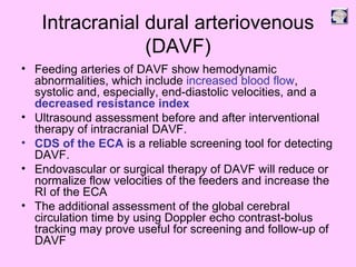
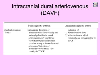
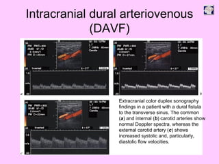
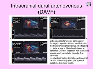
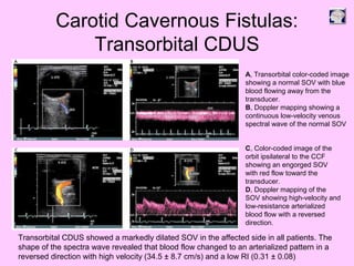
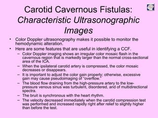
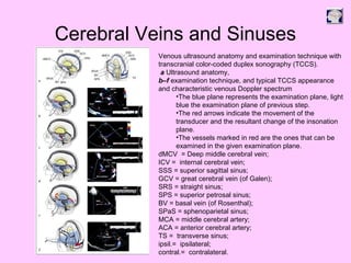
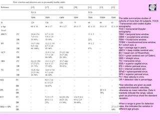
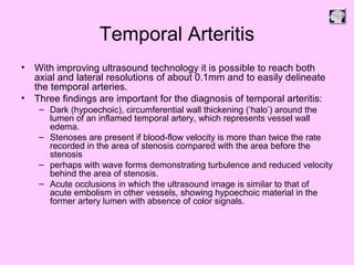
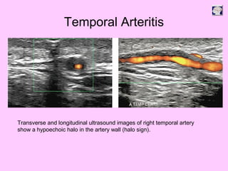
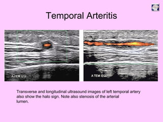
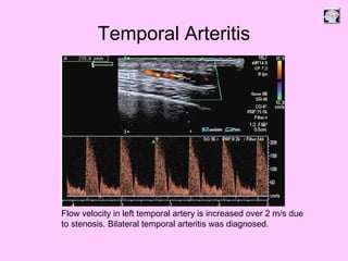
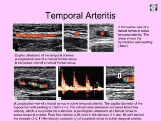
Ultrasound can be used to assess intracranial dural arteriovenous fistulas (DAVF) and carotid cavernous fistulas (CCF). For DAVF, ultrasound shows increased velocities in feeding arteries and decreased resistance indices, allowing assessment before and after treatment. For CCF, ultrasound reveals abnormal mosaic flow in the cavernous sinus and engorged veins with reversed flow. It can monitor hemodynamic changes and treatment response in a noninvasive manner. Ultrasound is also useful for assessing cerebral veins and sinuses, and can diagnose temporal arteritis by identifying hypoechoic wall thickening and stenoses in temporal arteries.
Bedside Ultrasound in Neurosurgery Part 2/3Liew Boon Seng
This document provides information from an ultrasound training course on various techniques for imaging intracranial vessels through different acoustic windows of the skull. It discusses techniques such as transorbital, temporal, and foramen magnum windows. It also covers topics like diagnosing intracranial occlusions and stenoses, grading carotid artery stenosis, diagnosing carotid dissections, assessing collateral flow, and evaluating bypass graft patency. The document discusses the use of transcranial Doppler in patients after decompressive craniectomy. It also reviews perfusion imaging with ultrasound contrast and experimental evidence for sonothrombolysis.
This document discusses hydrocephalus, including:
- Hydrocephalus is an abnormal buildup of cerebrospinal fluid in the brain, which can increase pressure and cause neurological symptoms.
- There are two main types - obstructive, caused by blockages in the flow of cerebrospinal fluid, and communicating, caused by issues with absorption of fluid.
- Common causes of obstructive hydrocephalus include aqueductal stenosis, tumors, cysts, and hemorrhages. Communicating hydrocephalus can result from subarachnoid hemorrhages.
- Normal pressure hydrocephalus is a type of communicating hydrocephalus where patients experience gait, memory, and
Bedside Ultrasound in Neurosurgery Part 1/3Liew Boon Seng
This document provides an overview of ultrasound training in neurosurgery. It introduces various applications of ultrasound in neurosurgery such as fetal neurosonograms, cranial ultrasonography of newborns, assessing ventricular shunt patency, spinal ultrasound in infants, and transcranial insonation of blood vessels. It also summarizes techniques for different types of Doppler imaging and discusses pathologies that can be detected using ultrasound like intraventricular hemorrhage.
The document discusses various vascular emergencies that can be diagnosed using ultrasound. It outlines tools like ultrasound, CT, MR and DSA that can be used and highlights advantages of ultrasound like being readily available, portable, and having good temporal resolution. It describes acute conditions like ruptured aortic aneurysm, acute carotid thrombosis, carotid and vertebral dissections, pseudoaneurysms, acute limb ischemia and graft failure. For each condition, it provides ultrasound findings, diagnostic criteria and treatment options. It emphasizes the importance of prompt diagnosis, accurate assessment and timely intervention in managing vascular emergencies.
This document provides an overview of various vascular lesions of the brain. It discusses arteriovenous malformations (AVMs), dural arteriovenous fistulas, carotid-cavernous fistulas, cavernomas, capillary telangiectasias, venous angiomas, aneurysms, and other conditions. For each type of lesion, it describes characteristics, imaging appearance, clinical presentation, and treatment options. Magnetic resonance imaging and cerebral angiography are important diagnostic tools. Treatment may involve surgery, endovascular procedures, or radiosurgery depending on the specific lesion.
Ultrasonography provides several advantages in clinical neurology. It can be used to assess neurovascular structures like arteries and veins, detect abnormalities associated with movement disorders like increased substantia nigra hyperechogenicity in Parkinson's disease, and evaluate peripheral nerves for entrapment neuropathies. Ultrasonography techniques like duplex ultrasonography and transcranial Doppler allow visualization of vessel structures, plaque composition, and blood flow velocities to diagnose vascular diseases, monitor treatment, and detect vasospasm. Transcranial Doppler is also used to evaluate movement disorders, cerebral circulation in stroke and brain injury, and support a diagnosis of brain death. Peripheral nerve ultrasonography examines cross-sectional area, echogenicity,
Collateral blood flow is important for sustaining brain tissue after an occlusion of major arteries to the brain. There are three principal pathways for collateral circulation: extracranial-intracranial communications, the circle of Willis, and leptomeningeal anastomoses. Several imaging techniques can provide insight into collateral flow, such as digital subtraction angiography, CT angiography, magnetic resonance angiography, and trans-cranial Doppler. Therapies aimed at augmenting cerebral blood flow in acute stroke have included plasma expanders, vasodilators, and induced hypertension to potentially increase collateral flow.
Neonatal sonography of the brain is an essential part of newborn care, particularly for preterm and unstable infants. It allows for portable, low-cost, and radiation-free evaluation of the brain for hemorrhages, abnormalities, and other issues like hydrocephalus. Standard imaging planes include coronal and sagittal views of the brain and ventricles. Key indications for neurosonography in newborns include detection of intraventricular hemorrhage in preterm infants and evaluation of periventricular leukomalacia, a common ischemic injury. Neurosonography is also used to identify other issues like cystic lesions, tumors, and hydrocephalus.
Details of Cerebrospinal Fluid special reference to cell count and alteration of CSF Hydrodynamics explained in brief and Different Diagnostic parameters to Hydrocephalus
This document outlines guidelines for the insertion and management of external ventricular drains (EVDs). EVDs are used to temporarily drain cerebrospinal fluid from the brain ventricles to relieve pressure. The document describes the indications for EVD placement, types of EVD systems, insertion procedure, and guidelines for drain positioning, monitoring drainage amounts and characteristics, obtaining samples, administering medications, and removing the drain. Key steps include accurately positioning the drain system based on the patient's ventricle height, monitoring hourly drainage amounts and qualities, and following aseptic technique for procedures involving the drain port.
- Transcranial Doppler (TCD) ultrasonography uses 2MHz probes to visualize intracranial vasculature non-invasively and measure blood flow velocities. It was introduced in 1982 and can monitor the middle cerebral artery, anterior cerebral artery, internal carotid artery, and other vessels.
- TCD is useful for evaluating stroke, cerebral vasospasm, intracranial stenosis, emboli, aneurysms, and raised intracranial pressure. It has good correlation with angiography and can detect stenosis with high sensitivity and specificity. Hemodynamic changes and collateral flow patterns can also be assessed.
- Monitoring emboli with TCD has helped identify the
This document discusses Doppler ultrasound of the carotid arteries. It begins with an introduction describing how carotid artery disease can cause strokes and how ultrasound is used to diagnose stenosis to determine surgical candidates. It then describes the anatomy of the carotid arteries and outlines the normal ultrasound appearance. Key points of a carotid ultrasound exam are described including using grayscale, color Doppler, power Doppler and spectral analysis. Different types of carotid plaques are defined as well as how they appear ultrasonographically. Methods for evaluating stenosis and differentiating true from pseudo-spectral broadening are also covered.
This document summarizes a seminar presentation on ultrasonography for diagnosing hydrocephalus. It begins with an introduction that defines hydrocephalus and reviews ventricular anatomy. It then presents a case study of a pregnant patient with severe hydrocephalus and spinal bifida. The procedure, images, and report of the ultrasound exam are described. The summary discusses diagnostic features of hydrocephalus on ultrasound and differential diagnoses. Management options are also reviewed.
The document discusses pulmonary angiography and CT pulmonary angiography (CTPA) for diagnosing pulmonary embolism. It describes CTPA as the standard of care and illustrates how it has a high sensitivity and specificity compared to other tests. The techniques for performing CTPA are outlined, including the use of iodinated contrast, timing of the scan, and parameters. Common findings of acute and chronic pulmonary emboli on CTPA are illustrated. The document also discusses artifacts, pitfalls and how to improve image quality.
Non traumatic Subarachnoid hemorrhage (SAH)Milan Silwal
The document discusses imaging of non-traumatic subarachnoid hemorrhage (SAH). The most common cause of SAH is the rupture of a saccular aneurysm, usually located in the circle of Willis. CT and MRI are used to diagnose and locate SAH, while CT angiography, MR angiography, and conventional angiography can identify aneurysms. Complications of SAH like hydrocephalus and cerebral ischemia are also discussed. Perimesencephalic and convexal SAH represent patterns of non-aneurysmal SAH with different etiologies and prognoses.
It's helpful in understanding various aspects of revascularization procedures, with good illustrations, easy to learn, no complexity, easy language, conclusion added, short descriptions
This document discusses shunts for treating hydrocephalus. It begins by defining hydrocephalus and explaining its causes. It then discusses epidemiology, showing hydrocephalus requiring shunts is most common in young pediatric patients. It also reviews the costs associated with hydrocephalus treatment in the US. The document goes on to describe CSF dynamics and circulation in detail. It compares pediatric and adult hydrocephalus, noting causes and treatment outcomes differ between age groups. In closing, it emphasizes the challenge in shunt treatment is to recreate natural CSF physiology.
CT pulmonary angiography (CTPA) is a medical diagnostic test that uses computed tomography to obtain images of the pulmonary arteries. It was introduced in the 1990s as a less invasive alternative to radionuclide imaging for evaluating suspected pulmonary embolism. CTPA involves acquiring breath-hold images during intravenous contrast enhancement to identify potential pulmonary embolisms as filling defects. It has high sensitivity and specificity, making it the primary imaging study for evaluation of suspected pulmonary embolism.
The document provides information on CSF nuclear imaging, including:
1. The physiology and anatomy of CSF circulation and production in the brain ventricles.
2. Radiopharmaceuticals used for CSF imaging like radiolabeled serum albumin and DTPA, and their properties.
3. Clinical applications of CSF imaging like evaluating CSF circulation in cisternography for hydrocephalus and detecting CSF leaks.
Cardiovascular CT is a valuable tool for evaluating congenital heart disease in children. It provides high spatial and temporal resolution to depict complex anatomy. Key applications include assessing pulmonary blood flow in pulmonary atresia, vascular rings prior to surgery, coronary artery anomalies, and postoperative complications. Careful patient preparation and protocols are needed given pediatric concerns. CT enables simultaneous evaluation of vascular structures, airways, and cardiac function to comprehensively evaluate complex congenital heart disease.
The caroticocavernous fistula is a specific type of dural arteriovenousfistula characterized by abnormal arteriovenous shunting within the cavernous sinus.
A caroticocavernous fistula results in high-pressure arterial blood entering the low-pressure venous cavernous sinus.
This interferes with normal venous drainage patterns and compromises blood flow within the cavernous sinus and the orbit.
Digital subtraction angiography (DSA) is the gold standard for evaluating the cerebral vasculature. It involves injecting iodinated contrast material into arteries and using subtraction techniques to visualize vessels. The normal anatomy includes the circle of Willis and branches of major arteries. Variants are common. DSA is used to diagnose conditions like aneurysms and arteriovenous malformations. Newer digital systems provide 3D reconstruction and less radiation exposure compared to older techniques. DSA remains an important tool for interventional procedures and treatment planning of complex vascular lesions of the brain.
This document discusses different techniques for angiography of the head and neck, including digital subtraction angiography (DSA), CT angiography (CTA), and MR angiography (MRA). DSA uses image subtraction to visualize contrast-filled blood vessels. CTA uses a CT scan after injection of contrast media. MRA is non-invasive and does not require contrast, using magnetic fields to visualize blood flow. The document provides details on the protocols, advantages, and images produced for each angiography technique of the head and neck vasculature.
This document discusses cerebral vasospasm (CVS), which is an abnormal constriction of cerebral arteries following subarachnoid hemorrhage. It can lead to delayed cerebral ischemia and infarction. The document covers risk factors, pathophysiology involving oxyhemoglobin and inflammation, diagnosis using tools like transcranial Doppler and angiography, and management including prevention with calcium channel blockers, treatment of symptomatic vasospasm with balloon angioplasty, and protecting the brain from ischemia.
This document discusses various surgical techniques for treating coarctation of the aorta, including:
1. Resection with end-to-end anastomosis, the original technique, but it has a high recoarctation rate.
2. Prosthetic patch aortoplasty which uses an elliptical patch to enlarge the aorta and has lower recoarctation rates.
3. Subclavian flap aortoplasty which uses the left subclavian artery as a flap to enlarge the aorta, but can cause left arm issues.
4. Balloon angioplasty and stenting are newer options that can avoid surgery for some patients, but have risks of restenosis and complications.
This document discusses contrast echocardiography, including the mechanism by which microbubble contrast agents improve echocardiographic imaging. Ideal contrast agents are described as being safe, metabolically inert, long-lasting, strong sound reflectors that are small enough to pass through capillaries. Several FDA-approved second generation contrast agents are mentioned along with their shell materials and gases. Optimal echocardiographic settings for contrast imaging are outlined. Clinical applications of contrast echocardiography include assessing shunts, venous anomalies, and leaks. Examples of its use in specific cases are provided.
1) The document discusses imaging protocols for acute ischemic stroke, including non-contrast CT (NCCT), CT angiography (CTA), MRI, and thrombectomy procedures.
2) NCCT can help identify hemorrhage as a contraindication to thrombolysis and detect early ischemia. CTA can demonstrate thrombi to guide thrombolysis or thrombectomy and assess vessel occlusion, core infarction, and collaterals.
3) MRI is more sensitive than CT for acute stroke, detecting most infarcts by 24 hours. Diffusion-weighted imaging (DWI) is most sensitive in the first few hours. Perfusion imaging assesses penumbra.
4) For severe strokes,
Posterior fossa is a shallow space accommodating brainstem and cerebellum. Bleed in the cerebellum can cost life as it leads to rapid deterioration by hydrocephalus and upward herniation.
The document provides an overview of brain anatomy and physiology for nurses. It describes the three main parts of the brain - the hindbrain, midbrain, and forebrain. It details the lobes of the cerebrum including the frontal, parietal, occipital, and temporal lobes. It discusses the composition of the brain including neurons, glial cells, cerebrospinal fluid, and blood vessels. Key structures like the ventricles, basal ganglia, and brainstem are also outlined. The document aims to educate nurses on the basic structure and function of the human brain.
Three key points about space occupying lesions of the brain:
1. Space occupying lesions include neoplasms like meningiomas and gliomas, infections like abscesses, and vascular lesions like aneurysms and hemorrhages.
2. Signs and symptoms vary depending on the location and size of the lesion but can include headaches, seizures, nausea/vomiting, and neurological deficits.
3. Neuroimaging with CT or MRI is important for diagnosis and helps characterize lesions, while lumbar puncture and blood tests help evaluate for other potential causes like infections.
This document discusses classifications and surgical methods for treating spinal injuries. It covers injuries of the cervical, thoracolumbar, and lumbar spine. For the cervical spine, it describes techniques for fractures of C1-C2 including traction, fusion, and screw fixation. For the thoracolumbar spine, it recommends a posterior approach using pedicle screws and plates or rods, and only using anterior fusion for large defects. It stresses the importance of careful technique and handling to avoid neurological complications.
Neurological Conditions and Diseases (During Development)Liew Boon Seng
No associated symptoms
- Not aggravated by routine physical activity
- Not associated with nausea or vomiting
- Bilateral and non-pulsating
- Family history of tension headaches
Current concepts in management of metastatic brain tumourLiew Boon Seng
1) Brain metastases occur in 25% of cancer patients and are most commonly diagnosed from lung cancer, breast cancer, and melanoma. Surgery is the preferred treatment for single, accessible brain metastases while radiosurgery and whole brain radiation therapy are options for multiple metastases.
2) Surgical resection provides the best chance of survival and neurological function for carefully selected patients with single brain metastases. Factors such as the patient's overall health, tumor size and location, and control of the primary cancer help determine treatment.
3) While whole brain radiation can effectively treat multiple brain metastases, it carries risks of neurocognitive side effects. Combined treatment with surgery or radiosurgery followed by whole brain radiation may improve outcomes
Applied Surgical Anatomy of the Brain and Spinal CordLiew Boon Seng
The document summarizes key anatomy related to the spinal cord and scalp. It describes the layers of the scalp from superficial to deep. It then discusses the skull vault and base, identifying several surface landmarks. It details the anatomy of the spinal cord, including its length, weight, segments, internal configuration of grey and white matter, and surrounding meninges. Finally, it lists the objectives which are to identify brain and spinal cord anatomy and relate it to surgical procedures and surface markings.
Neurological Conditions and Diseases (At birth)Liew Boon Seng
This document discusses various neurological conditions and diseases that can cause macrocephaly in infants and children. It describes conditions present at birth such as caput succedaneum, subgaleal hemorrhage, cephalohematoma, osteopetrosis, subdural hematomas, benign enlargement of the subarachnoid space, megalencephaly, vein of Galen aneurysm, and hydrocephalus. Hydrocephalus and its causes, clinical presentation, assessment, treatments including shunts, and complications are discussed in detail. Posthemorrhagic hydrocephalus as a consequence of intraventricular hemorrhage is also outlined.
CSF Shunt Infection: Diagnosis and TreatmentLiew Boon Seng
Ventricular shunt infection is a common complication of CSF shunting with an incidence rate of 8.5-15%. Risk factors include young age, prior revisions, and prolonged surgery time. Early infections are usually caused by skin flora like Staphylococcus epidermidis. Diagnosis involves CSF analysis showing pleocytosis and low glucose, along with culture of infected hardware. Treatment consists of removing the infected shunt and replacing it with a new shunt after the CSF is sterile, while administering intravenous antibiotics for 10-14 days. Prognosis depends on the organism, underlying pathology, and adequacy of treatment to prevent complications like cerebritis, abscesses, or recurrent infections.
This document discusses various medical and surgical management strategies for different types of hydrocephalus and associated conditions. It covers:
1) Medical management of hydrocephalus using diuretics and steroids to decrease CSF production.
2) The history of surgical drainage methods for hydrocephalus dating back to Hippocrates. Modern methods include ventriculostomies, shunt placements in various cavities, and endoscopic procedures.
3) Complications associated with different surgical procedures and how newer endoscopic techniques are improving outcomes compared to traditional shunting.
4) Specific guidelines for treating different causes of hydrocephalus like TB meningitis, hematocephalus, and congenital cases
Cranial surgery involves procedures to access and treat conditions within the skull and brain. The main types discussed are burr holes, craniotomies, and craniectomies. Craniotomies provide larger access than burr holes and are used for procedures like tumor removal, hemorrhage evacuation, and repairing vascular structures. Craniectomies involve removing a piece of skull that is later reconstructed. Additional topics covered include cranial procedures for vascular conditions like aneurysms, skull base surgery, and treating tumors, infections, hydrocephalus and more. Precise techniques and equipment are needed to perform surgeries near vital structures in the brain.
Neuropsychiatric manifestations of head injurySantanu Ghosh
This document summarizes a presentation on neuropsychiatric aspects of head injury. It begins with an introduction discussing the prevalence of head injuries. It then covers the history of understanding head injuries, comparative diagnostic classifications, epidemiology, types and pathophysiology of head injuries including acute and chronic behavioral consequences. The presentation also discusses clinical features such as cognitive impairment, personality changes, mood disorders, anxiety, aggression and psychosis. It concludes with discussing prognosis and predictors of outcome following head injury.
This document provides guidance on using ultrasound to assess the carotid arteries for atherosclerotic disease. It outlines the technical requirements, examination techniques, diagnostic criteria, and normal and abnormal findings. Doppler ultrasound is an accurate noninvasive method to diagnose high-grade carotid stenosis by measuring peak systolic velocities. Key findings include plaques that are hypoechoic or irregular in shape posing higher risk. Proper technique and accounting for vascular anomalies are important to avoid overestimating stenosis.
This document provides information on carotid Doppler ultrasound studies, including:
- Anatomy of the carotid arteries and branches
- Technique for performing carotid Doppler ultrasound exams, including patient positioning, transducer use, and Doppler settings
- Analysis of waveforms in normal carotid arteries versus arteries with disease
- Causes of carotid artery disease and common sites of extracranial arterial disease
- Characterization of carotid plaques based on echogenicity, morphology, and other properties.
This document provides an overview of Doppler ultrasound applications for evaluating various vascular structures and conditions. It discusses Doppler evaluation of the carotid arteries, renal arteries, portal vein, hepatic veins, and arteriovenous fistulas. Key points include diagnostic criteria for stenosis of the carotid, renal and hepatic arteries as well as portal hypertension. Imaging findings and waveforms related to conditions such as subclavian steal, Budd-Chiari syndrome, and arteriovenous fistula maturation/complications are also reviewed. The document emphasizes a thorough ultrasound technique and understanding of normal versus abnormal vascular Doppler evaluations.
This document provides an overview of carotid Doppler ultrasound. It begins with the anatomy of the carotid arteries and their branches. It then discusses the technique of carotid Doppler ultrasound, including instrumentation, examination protocol, and interpretation of ultrasound findings. It provides details on evaluating the internal carotid, external carotid, and vertebral arteries for stenosis or occlusion. The document also covers characterizing carotid plaques and differentiating true findings from artifacts.
Normal doppler spectral pattern of abdominal and limb vessels finalNipun Gupta
This document provides information on normal Doppler patterns of abdominal vessels. It begins by covering Doppler physics principles. It then discusses normal Doppler flow patterns seen in the abdominal aorta, ductus venosus, celiac artery, superior mesenteric artery, and mesenteric arteries. For each vessel, it describes anatomy, imaging recommendations, and typical Doppler waveform patterns. The document serves as an educational guide for residents to learn how to properly evaluate and interpret Doppler ultrasound of the abdominal vasculature.
This document discusses carotid Doppler ultrasound. It begins by outlining the anatomy of the carotid arteries and normal Doppler findings. It then discusses causes of carotid artery disease including atherosclerosis. Extra-carotid diseases that can affect the carotid arteries are also mentioned. The document provides detailed guidance on performing and interpreting carotid Doppler ultrasound scans including evaluating the intima-media thickness, flow patterns, spectral analysis, and grading stenosis. Imaging features of normal and diseased arteries are presented along with tips for accurate evaluation.
Carotid artery Doppler uses ultrasound to examine the carotid arteries in the neck. It can detect plaques, stenosis, dissections, and other abnormalities. A normal study shows the carotid bifurcation into the internal and external carotid arteries, with the internal carotid having low resistance flow and the external carotid having reduced diastolic flow. Doppler waveform analysis examines flow patterns to identify abnormalities. The test is used to evaluate risks of stroke and transient ischemic attacks.
Gray scale ultrasound has been used in evaluation of the eye since late 20 century.
More recently, color doppler imaging of retrobulbar vessel has been introduced as a useful adjunct to clinical examination and cross-sectional imaging for evaluating various pathologic condition in the orbit.
Although gray-scale ultrasound display the anatomy of the orbit well, color doppler imaging provides additional information about the vasculature of the orbit and information regarding direction and velocity of blood flow.
The color doppler changes in diabetes retinopathy has been a topic of discussion for decades and numerous studies have been conducted to study these changes.
Not enough standard textbook literature is available stating these changes, but meta- analysis of studies stated a common finding of increased vascular resistance in intra orbital vessels.
However, no consensus on the cut off limits of various doppler parameters for diabetic retinopathy have been reached.
This document provides an overview of carotid artery ultrasound evaluation. It describes the normal anatomy of the carotid arteries and their branches. The protocol for a carotid ultrasound examination is outlined, including patient positioning, transducer selection, scanning sequences, and evaluation of stenosis. Key anatomical structures are defined, such as the intima-media complex. Non-atherosclerotic diseases that can involve the carotid or vertebral arteries, such as fibromuscular dysplasia, dissection, vasospasm, and aneurysms are also reviewed. The limitations of carotid ultrasound are noted.
This document discusses arterial spin labeling (ASL), a noninvasive MRI technique used to measure cerebral blood flow. It describes the basic concepts and techniques of ASL, including pulsed ASL and pseudo-continuous ASL. Clinical applications include evaluating conditions like stroke, dementia, and tumors. The document outlines how ASL can detect perfusion deficits in acute ischemic stroke and discusses limitations such as lower signal-to-noise ratio compared to other perfusion techniques.
Carotico-cavernous fistula Presentation.Anas Ahmed
Caroticocavernous fistulas are abnormal connections between the carotid artery and cavernous sinus that result in high pressure arterial blood flowing into the low pressure venous sinus. They can be direct, with a connection to the internal carotid artery, or indirect, involving other meningeal or dural branches. Clinical symptoms include pulsatile exophthalmos, chemosis, and vision issues. Treatment depends on the type of fistula but commonly involves endovascular embolization to occlude the connection using coils or liquid embolic agents via a transarterial or transvenous approach. The goal is to eliminate shunting while preserving arterial patency.
A 60-year-old male presented with giddiness, vertigo, and a history of hypertension, chest pain, and prior CABG surgery. Examination found carotid bruits bilaterally. Carotid Doppler was ordered to evaluate for possible carotid artery disease or posterior circulation abnormality.
Carotid Doppler involves B-mode, color Doppler, and spectral Doppler analyses of the carotid arteries. Spectral Doppler provides information on flow patterns, velocities, and pulsatility which can identify stenosis or occlusion. Plaque characterization evaluates composition and risk of embolism. Carotid Doppler is useful for evaluating TIAs, bruits, known disease, and pre-operative screening.
The Doppler examination as
This document discusses the anatomy, embryology, and imaging of the superior vena cava (SVC). It begins with an introduction to the SVC's role as the largest central vein in the mediastinum. Imaging plays an important role in identifying congenital variants and pathologies. The document then covers the embryological development of the SVC, its normal anatomy and tributaries, and techniques for imaging it with various modalities like CT, MR, and venography. It discusses congenital variants like persistent left SVC and aneurysms. It also reviews acquired conditions like strictures, thrombus, and various tumors that can affect the SVC.
1) Carotid Doppler ultrasound is used to evaluate the carotid arteries for stenosis or occlusion. It involves using grayscale, color Doppler, and spectral Doppler to examine the anatomy and flow of the carotid arteries.
2) A normal carotid Doppler ultrasound will show triphasic waveforms within the carotid arteries with velocities under 125 cm/sec. The intima-media thickness should be less than 0.8-0.9mm.
3) Carotid artery disease is most commonly caused by atherosclerosis which can be evaluated using Doppler ultrasound parameters like peak systolic velocity, end diastolic velocity, and ICA/CCA velocity ratios to grade the severity of stenosis.
This document discusses the echocardiographic evaluation of coronary arteries. It is technically challenging to visualize the coronary arteries due to their small size and motion. Advances in ultrasound technology now allow direct visualization and Doppler analysis of multiple segments of the left main, left anterior descending, and right coronary arteries. Careful scanning techniques and Doppler measurements can provide useful anatomical and physiological information about coronary artery patency, stenosis, and blood flow velocities.
Echocardiography is a versatile imaging technique used to evaluate cardiac anatomy and function. It utilizes ultrasound to obtain standard 2D views of the heart from different windows including parasternal, apical, subcostal, suprasternal, and right parasternal. Doppler echocardiography, including color Doppler, assesses cardiac valves, chambers, and blood flow. Echocardiography is useful for diagnosing conditions such as valvular disease, heart failure, and congenital heart defects.
This document discusses the essential role of Doppler ultrasound in evaluating the kidneys and renal vasculature. It highlights how Doppler can be used to assess renal transplants, plan for dialysis access procedures, and monitor access complications. Specific applications covered include evaluating for renal artery stenosis, aneurysms, masses, fistulas and grafts. Assessment criteria and normal versus abnormal Doppler findings are presented for many common renal and vascular conditions.
This document discusses neurosonology and transcranial Doppler ultrasound (TCD). It defines neurosonology as ultrasonic imaging of the brain and neural structures. TCD provides noninvasive, real-time measures of blood flow in the brain's basal arteries. The document outlines the clinical applications of TCD, including monitoring cerebral vasospasm after subarachnoid hemorrhage, detecting intracranial stenosis, monitoring acute ischemic stroke, and screening for stroke risk in children with sickle cell disease. TCD is a useful tool for diagnosing and managing various cerebrovascular disorders.
This document discusses neurosonology and transcranial Doppler ultrasound (TCD). It defines neurosonology as ultrasonic imaging of the brain and neural structures. TCD provides noninvasive, real-time measures of blood flow in the brain's basal arteries. The document outlines the clinical applications of TCD, including monitoring cerebral vasospasm after subarachnoid hemorrhage, detecting intracranial stenosis, assessing acute ischemic stroke, and screening for stroke risk in children with sickle cell disease. TCD is a useful tool for diagnosing and monitoring various cerebrovascular disorders.
Similar to Bedside Ultrasound in Neurosurgery Part 3/3 (20)
Integrating Ayurveda into Parkinson’s Management: A Holistic ApproachAyurveda ForAll
Explore the benefits of combining Ayurveda with conventional Parkinson's treatments. Learn how a holistic approach can manage symptoms, enhance well-being, and balance body energies. Discover the steps to safely integrate Ayurvedic practices into your Parkinson’s care plan, including expert guidance on diet, herbal remedies, and lifestyle modifications.
These lecture slides, by Dr Sidra Arshad, offer a quick overview of the physiological basis of a normal electrocardiogram.
Learning objectives:
1. Define an electrocardiogram (ECG) and electrocardiography
2. Describe how dipoles generated by the heart produce the waveforms of the ECG
3. Describe the components of a normal electrocardiogram of a typical bipolar lead (limb II)
4. Differentiate between intervals and segments
5. Enlist some common indications for obtaining an ECG
6. Describe the flow of current around the heart during the cardiac cycle
7. Discuss the placement and polarity of the leads of electrocardiograph
8. Describe the normal electrocardiograms recorded from the limb leads and explain the physiological basis of the different records that are obtained
9. Define mean electrical vector (axis) of the heart and give the normal range
10. Define the mean QRS vector
11. Describe the axes of leads (hexagonal reference system)
12. Comprehend the vectorial analysis of the normal ECG
13. Determine the mean electrical axis of the ventricular QRS and appreciate the mean axis deviation
14. Explain the concepts of current of injury, J point, and their significance
Study Resources:
1. Chapter 11, Guyton and Hall Textbook of Medical Physiology, 14th edition
2. Chapter 9, Human Physiology - From Cells to Systems, Lauralee Sherwood, 9th edition
3. Chapter 29, Ganong’s Review of Medical Physiology, 26th edition
4. Electrocardiogram, StatPearls - https://www.ncbi.nlm.nih.gov/books/NBK549803/
5. ECG in Medical Practice by ABM Abdullah, 4th edition
6. Chapter 3, Cardiology Explained, https://www.ncbi.nlm.nih.gov/books/NBK2214/
7. ECG Basics, http://www.nataliescasebook.com/tag/e-c-g-basics
Rasamanikya is a excellent preparation in the field of Rasashastra, it is used in various Kushtha Roga, Shwasa, Vicharchika, Bhagandara, Vatarakta, and Phiranga Roga. In this article Preparation& Comparative analytical profile for both Formulationon i.e Rasamanikya prepared by Kushmanda swarasa & Churnodhaka Shodita Haratala. The study aims to provide insights into the comparative efficacy and analytical aspects of these formulations for enhanced therapeutic outcomes.
Promoting Wellbeing - Applied Social Psychology - Psychology SuperNotesPsychoTech Services
A proprietary approach developed by bringing together the best of learning theories from Psychology, design principles from the world of visualization, and pedagogical methods from over a decade of training experience, that enables you to: Learn better, faster!
Does Over-Masturbation Contribute to Chronic Prostatitis.pptxwalterHu5
In some case, your chronic prostatitis may be related to over-masturbation. Generally, natural medicine Diuretic and Anti-inflammatory Pill can help mee get a cure.
Travel vaccination in Manchester offers comprehensive immunization services for individuals planning international trips. Expert healthcare providers administer vaccines tailored to your destination, ensuring you stay protected against various diseases. Conveniently located clinics and flexible appointment options make it easy to get the necessary shots before your journey. Stay healthy and travel with confidence by getting vaccinated in Manchester. Visit us: www.nxhealthcare.co.uk
Adhd Medication Shortage Uk - trinexpharmacy.comreignlana06
The UK is currently facing a Adhd Medication Shortage Uk, which has left many patients and their families grappling with uncertainty and frustration. ADHD, or Attention Deficit Hyperactivity Disorder, is a chronic condition that requires consistent medication to manage effectively. This shortage has highlighted the critical role these medications play in the daily lives of those affected by ADHD. Contact : +1 (747) 209 – 3649 E-mail : sales@trinexpharmacy.com
- Video recording of this lecture in English language: https://youtu.be/kqbnxVAZs-0
- Video recording of this lecture in Arabic language: https://youtu.be/SINlygW1Mpc
- Link to download the book free: https://nephrotube.blogspot.com/p/nephrotube-nephrology-books.html
- Link to NephroTube website: www.NephroTube.com
- Link to NephroTube social media accounts: https://nephrotube.blogspot.com/p/join-nephrotube-on-social-media.html
2. Intracranial dural arteriovenous
(DAVF)
• Feeding arteries of DAVF show hemodynamic
abnormalities, which include increased blood flow,
systolic and, especially, end-diastolic velocities, and a
decreased resistance index
• Ultrasound assessment before and after interventional
therapy of intracranial DAVF.
• CDS of the ECA is a reliable screening tool for detecting
DAVF.
• Endovascular or surgical therapy of DAVF will reduce or
normalize flow velocities of the feeders and increase the
RI of the ECA
• The additional assessment of the global cerebral
circulation time by using Doppler echo contrast-bolus
tracking may prove useful for screening and follow-up of
DAVF
3. Intracranial dural arteriovenous
(DAVF)
• Measurement of flow velocities in draining veins and
sinuses using contrast-enhanced transcranial Doppler
sonography is another technique, which allows the
assessment of hemodynamic changes occurring after
the occlusion of DAVF
• Contrast-enhanced transtemporal CDS may be useful for
screening patients with clinical suspicion of DAVF of
transverse/sigmoid sinus and assessing the results of
interventional therapy.
5. Intracranial dural arteriovenous
(DAVF)
Extracranial color duplex sonography
findings in a patient with a dural fistula
to the transverse sinus. The common
(a) and internal (b) carotid arteries show
normal Doppler spectra, whereas the
external carotid artery (c) shows
increased systolic and, particularly,
diastolic flow velocities.
6. Intracranial dural arteriovenous
(DAVF)
Extracranial color duplex sonography
findings in a patient with a dural fistula to
the transverse/sigmoid sinus. The feeding
occipital artery is dilated and shows an
abnormal Doppler spectrum with increased
systolic and, especially, diastolic flow
velocities
(a), divides into two branches with normal
(b) and abnormal (c) Doppler spectra
supplying the dural fistula.
7. Carotid Cavernous Fistulas
• The confirmation of a CCF depends on cerebral vascular
angiography.
• However, the application of angiography is often limited
because it is invasive and expensive.
• Sometimes, it is not necessary to undergo invasive
diagnostic measures and treatments in low-flow CCFs
because the patients with such kinds of CCFs have more
chance of spontaneous resolution.
• Color Doppler ultrasonography (CDUS), which is much
less expensive and noninvasive, makes monitoring the
blood flow in real time possible.
8. Carotid Cavernous Fistulas
• Transcranial studies including transtemporal and
transoccipital acoustic windows were performed with
ultrasonography systems equipped with a 2- to 2.5-MHz
sector scan transducer
• The orbital window and carotid vessels were examined
with a 5- to 13-MHz linear array transducer.
• With the patient in a supine position, the internal carotid
artery (ICA), anterior cerebral artery (ACA), middle
cerebral artery (MCA), and posterior cerebral artery
(PCA) and the tip of the basilar artery (BA) were
examined through the temporal window.
• Scanning depths were regularly set between 140 and
160 mm to sufficiently visualize the contralateral skull
9. Carotid Cavernous Fistulas
• The hypoechoic mesencephalic brain stem in a “heart” or “butterfly”
shape was visualized in axial sections. This section was regarded
as a “standard section” to orientate the circle of Willis by using the
color mode.
• In the normal situation, a symmetric little round cluster of blood flow
was visualized beneath both sides of the strong echo of the anterior
clinoid.
• It is the cross section of the siphonic segment of the ICA
• The maximal cross-sectional areas of the ICA were measured from
the frozen ultrasonographic image by tracing around the round
cluster.
• From the study of healthy subjects, the area ranged from 0.2 to 0.5
cm2.
• The asymmetric cluster of blood flow in this section and the area
larger than 1 cm2 were regarded as abnormal.
10. Carotid Cavernous Fistulas
• With the patient in a prone position, the vertebral artery
(VA) and lower parts of the BA were insonated through
the occipital foramen and shown as a Y shape.
• Through the orbital window, the ophthalmic artery (OA)
and superior ophthalmic vein (SOV) were observed.
• Special emphasis was placed on maximal systolic
velocity (Vmax) and resistive index (RI).
11. Carotid Cavernous Fistulas:
Temporal window
A, Axial cross section showing a
symmetric little round cluster of
blood flow of the normal intracranial
ICA.
B, Orientation: 1 indicates ipsilateral
ICA; 2, contralateral ICA; 3,
contralateral skull bone; and 4,
ipsilateral anterior clinoid.
C, Color-coded image showing an
abnormal mosaic flow
flash of the CCF in the left
cavernous sinus area (arrow) and a
normal round cluster of the
contralateral ICA (arrow). D,
Orientation: 1 indicates CCF;
2, contralateral ICA; 3, contralateral
skull bone; and 4, anterior clinoid.
The asymmetric cluster of blood flow beneath both sides of the anterior clinoid was
visualized in axial sections. A cross section of the ipsilateral ICA was presented as
irregular mosaic color shade. Its area was much larger than that of the contralateral
ICA (range, 1.7–5.2 cm2; Figure 1, C and D).
12. Carotid Cavernous Fistulas
Spectrum of a CCF showing
turbulent blood flow. The peak
velocity ranged from 65 to 190
cm/s. Peak velocity decreased
immediately with compression of
the ipsilateral carotid artery and
increased rapidly right after relief
of the compression and ascended
slightly higher than that before
compression.
13. The RI of the ipsilateral ICA was lower than that of the contralateral ICA
and that in the control group. The Vmax of the ipsilateral MCA, ACA, and
OA was significantly lower than that of the contralateral arteries and that
in the control group. The RI of the ipsilateral ACA and bilateral VA was
lower than that in the control group. The RI of the ipsilateral PCA was
lower than that of the contralateral artery.
Carotid Cavernous Fistulas
14. Carotid Cavernous Fistulas:
Transorbital CDUS
A, Transorbital color-coded image
showing a normal SOV with blue
blood flowing away from the
transducer.
B, Doppler mapping showing a
continuous low-velocity venous
spectral wave of the normal SOV
C, Color-coded image of the
orbit ipsilateral to the CCF
showing an engorged SOV
with red flow toward the
transducer.
D, Doppler mapping of the
SOV showing high-velocity and
low-resistance arterialized
blood flow with a reversed
direction.
Transorbital CDUS showed a markedly dilated SOV in the affected side in all patients. The
shape of the spectra wave revealed that blood flow changed to an arterialized pattern in a
reversed direction with high velocity (34.5 ± 8.7 cm/s) and a low RI (0.31 ± 0.08)
15. Carotid Cavernous Fistulas:
Characteristic Ultrasonographic
Images
• Color Doppler ultrasonography makes it possible to monitor the
hemodynamic alteration.
• Here are some features that are useful in identifying a CCF.
– Color Doppler imaging shows an irregular color mosaic flash in the
cavernous region that is markedly larger than the normal cross-sectional
area of the ICA.
– When the ipsilateral carotid artery is compressed, the color mosaic
decreases or disappears.
– It is important to adjust the color gain properly; otherwise, excessive
gain may cause pseudoimaging of “overflow.”
– The blood flow draining from the high-pressure artery to the low-
pressure venous sinus was turbulent, disordered, and of multidirectional
spectra.
– The bruit is synchronous with the heart rhythm.
– The velocity decreased immediately when the carotid compression test
was performed and increased rapidly right after relief to slightly higher
than before the test.
16. • The arteriovenous direct shunting causes intracranial “steal
phenomena.”
• To compensate the ischemic area, the distribution of blood flow in
the brain has to be adjusted functionally through the circle of Willis.
• The RI of blood flow in the ICA proximal to the CCF decreased.
• The velocity of blood flow in the ICA may increase apparently if the
fistula is large.
• Blood velocity of the vascular branches distal to the fistula, such as
the MCA, ACA, and OA, decreased but increased in those of the
healthy side as compensation.
• The RI of blood flow in the ACA in the healthy side reduced through
the function of the anterior communication artery
• As the posterior vascular circle of the brain, the vertebrobasilar
artery system showed a reduced RI attributable to the adjustment of
blood flow.
• In a large fistula, the velocity of blood flow in the PCA and BA in the
affected side may obviously increase.
Carotid Cavernous Fistulas:
Ultrasonographic for Follow-up
Study
17. • Changes in the SOV were the most characteristic,
appearing as a dilated lumen, reversed blood flow, and
arterialized spectra with high velocity and a low RI.
However, sometimes these characteristics may not be
as obvious.
• Because different types of CCFs have different
hemodynamic alteration and different draining routes,
clinical features such as pulsating exophthalmos may not
come out.
• In this situation, careless detection is prone to miss the
diagnosis
Carotid Cavernous Fistulas:
Ultrasonographic for Follow-up
Study
18. Carotid Cavernous Fistulas:
Ultrasonographic for Follow-up
Study
• Color Doppler ultrasonography is more convenient than
digital subtraction angiography for the long-term follow-
up study of a CCF.
• A complete embolization means that no abnormal color
flash exits the affected site; blood flow parameters of
relevant vessels return to normal; and abnormal changes
in the SOV disappear.
• Part embolization means that the abnormal color flash
shrinks, and the velocity of blood flow decreases while
disordered spectra can be observed.
19. Carotid Cavernous Fistulas:
Ultrasonographic for Follow-up
Study
• CDUS possesses the following features in diagnosis of
CCFs.
– It is inexpensive, noninvasive, and easily repeated.
– The size and extent of the CCF focus can be
observed directly.
– Combined with the traumatic history, clinical
characteristics, hemodynamic alteration of relevant
vessels, and changes in the SOV specific to the CCF,
CDUS can confirm the diagnosis of a CCF accurately.
– In addition, CDUS is of great value in accessing the
therapeutic effect of CCFs.
20. Cerebral Veins and Sinuses
• The superficial cerebral veins cannot be imaged with ultrasound
methods.
• However, usually, three of the surface veins of the brain appear
dominant and may serve as important collaterals.
• Their recruitment can be assumed by indirect ultrasound findings.
• The superficial middle cerebral vein connects to the sphenoparietal
(SPaS) or cavernous sinus (60%), or to the pterygoid plexus (14%).
• The superior anastomotic (Trolard’s) vein drains towards the
superior sagittal sinus (SSS) and the inferior anastomotic (Labbé’s)
vein joins the transverse sinus (TS).
• Only major tributaries of paired cavernous sinuses, the SPaS and
the superior petrosal sinus (SPS), can be depicted by ultrasound.
21. Cerebral Veins and Sinuses
• The deep middle cerebral vein (dMCV) is formed by insular veins,
follows a medial course in close proximity to the middle cerebral
artery (MCA) within the lateral sulcus, and empties into the basal
vein (BV) (of Rosenthal).
• Together with the internal cerebral veins (ICV) the BV usually joins
the great cerebral vein (GCV) (vein of Galen).
• The GCV originates above the pineal gland, describes a convex
arch around the splenium of the corpus callosum, and joins the
straight sinus (SRS).
• In order to examine intracranial veins and sinuses a low-flow
sensitive color program with a low wall filter setting has to be used
and the pulse repetition frequency needs to be reduced.
22. Cerebral Veins and Sinuses
Venous ultrasound anatomy and examination technique with
transcranial color-coded duplex sonography (TCCS).
a Ultrasound anatomy,
b–f examination technique, and typical TCCS appearance
and characteristic venous Doppler spectrum
•The blue plane represents the examination plane, light
blue the examination plane of previous step.
•The red arrows indicate the movement of the
transducer and the resultant change of the insonation
plane.
•The vessels marked in red are the ones that can be
examined in the given examination plane.
dMCV = Deep middle cerebral vein;
ICV = internal cerebral vein;
SSS = superior sagittal sinus;
GCV = great cerebral vein (of Galen);
SRS = straight sinus;
SPS = superior petrosal sinus;
BV = basal vein (of Rosenthal);
SPaS = sphenoparietal sinus;
MCA = middle cerebral artery;
ACA = anterior cerebral artery;
TS = transverse sinus;
ipsil.= ipsilateral;
contral.= contralateral.
23. The table summarizes studies of
cohorts of more than 40 subjects. TCCS
Transcranial color-coded duplex
sonography;
TCD = transcranial Doppler
sonography;
TBW = temporal bone window;
OBW = occipital bone window;
FBW = frontal bone window;
TFBW = transforaminal bone window;
N = cohort size; ø
Age = average age in years;
dMCV = deep middle cerebral vein;
BV = basal vein (of Rosenthal);
GCV = great cerebral vein (of Galen);
SRS = straight sinus;
TS = transverse sinus;
SSS = superior sagittal sinus;
IPS = inferior petrosal sinus;
ICV = internal cerebral vein;
SPaS = sphenoparietal sinus;
SPS = superior petrosal sinus;
FV = flow velocity in cm/s;
DR = detection rate in percentage.
Flow velocities are given as peak
systolic/end-diastolic velocities,
otherwise as mean velocities. Data in
brackets indicate the range, values
given as plus/minus show the standard
deviation.
When a range is given for detection
rates, this indicates the variation in
different age groups.
24. Temporal Arteritis
• With improving ultrasound technology it is possible to reach both
axial and lateral resolutions of about 0.1mm and to easily delineate
the temporal arteries.
• Three findings are important for the diagnosis of temporal arteritis:
– Dark (hypoechoic), circumferential wall thickening (‘halo’) around the
lumen of an inflamed temporal artery, which represents vessel wall
edema.
– Stenoses are present if blood-flow velocity is more than twice the rate
recorded in the area of stenosis compared with the area before the
stenosis
– perhaps with wave forms demonstrating turbulence and reduced velocity
behind the area of stenosis.
– Acute occlusions in which the ultrasound image is similar to that of
acute embolism in other vessels, showing hypoechoic material in the
former artery lumen with absence of color signals.
25. Temporal Arteritis
Transverse and longitudinal ultrasound images of right temporal artery
show a hypoechoic halo in the artery wall (halo sign).
26. Temporal Arteritis
Transverse and longitudinal ultrasound images of left temporal artery
also show the halo sign. Note also stenosis of the arterial
lumen.
27. Temporal Arteritis
Flow velocity in left temporal artery is increased over 2 m/s due
to stenosis. Bilateral temporal arteritis was diagnosed.
28. Temporal Arteritis
Duplex ultrasound of the temporal arteries:
a longitudinal view of a normal frontal ramus;
b transverse view of a normal frontal ramus;
c transverse view of a
frontal ramus in active
temporal arteritis. The
arrow shows the
hypoechoic wall swelling
(‘halo’).
d Longitudinal view of a frontal ramus in active temporal arteritis. The sagittal diameter of the
hypoechoic wall swelling is 0.9mm (++). The colored area delineates increased blood flow
velocity, which is suspicious for a stenosis. e pw-Doppler ultrasound of a frontal ramus in
active temporal arteritis. Peak flow velocity is 86 cm/s in the stenosis (1+) and 19 cm/s behind
the stenosis (2+). f Inflammatory occlusion (↓) of a parietal ramus in active temporal arteritis.
29. Temporal Arteritis
• High-resolution B-mode ultrasound and Doppler
ultrasound have been used as diagnostic tools to
substitute the biopsy of the temporal artery.
• This halo sign has above 80–90 % specificity, but the
sensitivity is below 70 %, not very far from those of the
temporal biopsy.
• The presence of a halo sign is practically diagnostic in
the presence of compatible symptoms, but its absence
does not rule out vasculitis.
• Ultrasound findings disappear in about 2 weeks after the
onset of treatment with corticosteroids.
30. Deep Venous Thrombosis
• Compression ultrasonography is the diagnostic
procedure of choice for the assessment of patients with
suspected DVT.
• It has been shown to be highly sensitive and specific for
the diagnosis of DVT, particularly in the lower extremities
in symptomatic patients.
• Sensitivity and specificity are near 100 % in the
femoropopliteal segment.
• Sensitivity decreases in isolated calf thrombi and in
asymptomatic patients.
31. Deep Venous Thrombosis
A venous US scan of the right leg was performed. Transverse
ultrasound uncompressed (a) and compressed (b) images show a
non-compressible superficial femoral vein (arrowhead ).
32. Deep Venous Thrombosis
Longitudinal ultrasound image
shows the distended vein
(posterior to the artery) with
intraluminal clot ( arrows ).
Color Doppler longitudinal image shows normal flow in
the superficial femoral artery and no flow in the
superficial femoral vein.
33. Functional Transcranial Doppler
Sonography
• Since the first demonstration of noninvasive Doppler ultrasound
pericranial recordings of the cerebral blood flow velocities (CBFV) in
the basal cerebral arteries, functional transcranial Doppler
sonography (fTCD) has been established as a complementary
perfusion-sensitive neuroimaging tool.
• Like functional magnetic resonance imaging (fMRI) and positron
emission tomography, fTCD is based on the close linkage between
local neural activity and regional cerebral blood flow (rCBF) changes
(neurovascular coupling)
• Recent developments in fTCD have shown that the technique can
assess language function reliably in adults and children.
• Functional TCD thus has the potential to assess hemispheric
differences in activation for a wide range of brain functions in normal
subjects and patients.
34. Functional Transcranial Doppler
Sonography
Measurement of the CBFV in the
basal arteries through the
transtemporal cranial window:
In most fTCD studies the M1-
segment of the MCA is insonated at
a depth of 40–60 mm.
The two probe positions illustrate
the dependency of the recorded
flow velocity as a cosine function of
the insonation angle.
In case of the lower probe position,
this results in a negative deviation
of 15% of the absolute velocity in
the insonated vessel segment.
ACA, MCA, and PCA = Anterior,
middle, and posterior cerebral
arteries.
35. Functional Transcranial Doppler
Sonography
Prototype fTCD setup for language lateralization: blood flow velocities in the
basal arteries (here the middle cerebral arteries; MCAs) are bilaterally monitored by a
commercially available TCD device. Stimulus presentation is accomplished by computer
display. CBFV envelope curve are stored with the simultaneously recorded marker signal for
segmentation of event-related signal intervals. Offline analysis is performed by AVERAGE, a
program for automated blood flow data analysis
36. Functional Transcranial Doppler
Sonography
Averaged event-related CBFV modulations resulting from 30 repetitions of a language lateralization
paradigm (PDLT; picture description language task) in an 8-year-old child.
The left y-axis indicates the relative perfusion increases between the left and right middle cerebral arteries
(MCAs) during resting period (baseline) and task performance (PDLT) as the difference between the
averaged relative perfusion changes in the left and right MCA (right y-axis in blue).
Positive values by calculation indicate left hemispheric dominance and vice versa.
An fTCD language lateralization index (LI; red column; 7.8%) with confidence bands (indicated in green) is
calculated by the maximum increase during a defined period of interest
37. Pain: Non-invasive functional
neurosurgery using ultrasound
• Researchers in Switzerland (2009) have shown that chronic pain
can be alleviated through thermal ablation of thalamic tissue by
high-intensity focused ultrasound.
• Mri-guided focused ultrasound (mrgFus), is used to treating brain
tissues with ultrasound.
• Martin and colleagues used transcranial mri-guided highintensity
focused ultrasound (tcmrgHiFu) to perform lesions of the central
lateral thalamic nuclei in nine patients with chronic neuropathic pain
• These interventions relied on the capacity of tcmrgHiFu to heat
brain tissues to temperatures >53 °C to achieve thermal ablations
through multiple, repeated sonication events.
• All patients receiving the outpatient tcmrgHiFu procedure remained
awake and responsive during the entire neurosurgical intervention,
which resulted in thalamotomies ≈4 mm in diameter.
38. Non-invasive functional
neurosurgery using ultrasound
Potential applications of ultrasonic neuromodulation for the noninvasive treatment of
brain disorders.
Through both thermal and non-thermal (mechanical) mechanisms, ultrasound has been
shown to exert numerous bioeffects on brain tissues that could provide a basis for non-
invasive therapies for neurological and psychiatric disorders.
39. • The integration of external beam radiotherapy (EBRT) and
highintensity focused ultrasound (HIFU), with a case history taken
from their collection of bone metastasis treatments using magnetic
resonance-guided high-intensity focused ultrasound (MRgFUS)
• HIFU is only employed for palliative treatment of bone metastases
(pain reduction), when the disease has become systemic.
• In several cases, complete success (pain palliation and total
metastasis ablation) has been obtained.
40. Measurement of optic nerve
sheath diameter by ultrasound: a
means of detecting acute raised
intracranial pressure in hydrocephalus
• The evaluation of the optic nerve
sheath diameter is a simple non-
invasive procedure, which is a
potentially useful tool in the
assessment and monitoring of
children with hydrocephalus
suspected of having raised
intracranial pressure.
• A non-invasive method of
determining whether ICP is raised by
using ocular ultrasound to detect
dilation of the optic nerve sheath 3
mm behind the eye has recently
been described
41. Delayed reversal of ONS distension- initial
scan. ONUS performed in a 66-year-old
patient who became acutely unresponsive
two days after removal of an EVD. Scan
performed prior to placement of new EVD.
The ocular globe is the hypoechoic
structure in the upper part of the images
and the ONS the linear hypoechoic
structure behind the globe.
Caliper A identifies a point 3 mm behind
the retina while Caliper B measures the
ONSD.
ONSD is 0.55 cm, suggestive of raised
ICP. Opening pressure was >50 mmHg.
Measurement of optic nerve
sheath diameter by ultrasound: a
means of detecting acute raised
intracranial pressure in hydrocephalus
42. Delayed reversal of ONS distension- early
post-treatment scan. ONUS performed
twenty minutes after first measurement.
The ocular globe is the hypoechoic structure
in the upper part of the images and the ONS
the linear hypoechoic structure behind the
globe.
Caliper A identifies a point 3 mm behind the
retina while Caliper B measures the ONSD.
ONSD is 0.53 cm, suggestive of continued
intracranial hypertension.
Simultaneously recorded ICP from EVD was
only 10 mmHg.
Measurement of optic nerve
sheath diameter by ultrasound: a
means of detecting acute raised
intracranial pressure in hydrocephalus
43. Conclusions
The specificity and PPV of ONSD measurement for the detection of raised
ICP are substantially decreased in the presence of significant acute ICP
fluctuation.
ONSD measured on the initial brain CT scan is independently associated
with ICU mortality rate (when ≥ 7.3 mm) in severe TBI patients.
The sensitivity and specificity of bedside sonographic measurement of optic
nerve sheath diameter is inadequate to aid medical decision making in
children with suspected increased intracranial pressure.
Key messages
• Optic nerve sheath distension on bedside ultrasound imaging is less
predictive of intracranial hypertension when intracranial pressure is acutely
fluctuating between high and normal.
• Delayed reversal of optic nerve sheath distension is one possible
explanation for this decrease in positive predictive value.
Measurement of optic nerve
sheath diameter by ultrasound: a
means of detecting acute raised
intracranial pressure in hydrocephalus