
Recommended
More Related Content
What's hot
What's hot (20)
Similar to General histology || Nervous tissue
Similar to General histology || Nervous tissue (20)
Recently uploaded
Recently uploaded (20)
General histology || Nervous tissue
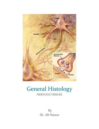
- 1. General Histology NERVOUS TISSUES By Dr. Ali Nasser
- 2. PAGE 1 FUNCTION 1. reception of information from the external and internal environment. 2. integration and analysis of the incoming information. 3. generation of new signals. 4. conduction of these neural messages to special responding tissue (effectors). DIVISIONS OF THE NERVOUS SYSTEM 1. Central nervous system. - The central nervous system (CNS) consists of the brain and spinal cord. 2. Peripheral nervous system. - The peripheral nervous system (PNS) consists of all the nervous tissue lying outside the brain and spinal cord. It consists of 12 pairs of cranial nerves and 31 pairs of spinal nerves. This PNS is further divided into the somatic nervous system, which supplies motor fibers to skeletal muscles that are under conscious control, and the autonomic nervous system (ANS), which supplies motor fibers to smooth muscle, cardiac muscle, and glands. The ANS consists of the sympathetic (thoracolumbar) nervous system and the parasympathetic (craniosacral) nervous system. CLASSIFICATION OF NEURONS On the basis of their structure, neurons can also be classified into Three main types : 1. Unipolar Neurons. - Sensory neurons have only a single process or fibre which divides close to the cell body into two main branches (axon and dendrite). Because of their structure they are often referred to as unipolar neurons. 2. Multipolar Neurons. - Motor neurons, which have numerous cell processes (an axon and many dendrites) are often referred to as multipolar neurons. Interneurons are also multipolar. 3. Bipolar Neurons. - Bipolar neurons are spindle-shaped, with a dendrite at one end and an axon at the other. An example can be found in the light-sensitive retina of the eye.
- 3. PAGE 2 PARTS OF A NEURON 1. the cell body / containing the nucleus and synthetic organelles. 2. the axon / a long cytoplasmic process associated with the cell body used to communicate with target organs. 3. the dendrites / shorter cytoplasmic processes off the cell body used to communicate between neurons. CENTRAL NERVOUS SYSTEM Spinal Cord. - The spinal cord is composed of grey and white matter (fig 1). The grey matter is composed of nerve cell bodies and in a cross-sectional cut appears as a darker stained "H"-like central area. The white matter, composed entirely of axonal projections, surrounds the grey matter and is lighter staining (fig 2). Fig (1) fig (2) Brain. - Like the spinal cord, the brain is composed of areas of grey and white matter. In some areas an addition outer gray layer is present. We will study two regions of the brain, in which this additional grey matter has a distinct morphology. Cerebral Cortex. - Starting at the surface of the cortex and moving inward, you can identify the first three layers of cell bodies (fig 3): (1) superficial molecular layer, containing only a few small cell bodies; (2) outer
- 4. PAGE 3 granular layer, containing small round cell bodies; (3) pyramidal cell layer, containing cell bodies triangular in shape (fig 4). In total, the cerebral cortex consists of six layers with the inner most three represented by an inner granular layer, internal pyramidal layer, and polymorphic cell layer, the innermost layer containing cell bodies of many shapes. Fig 3 fig 4 Cerebellar Cortex. - This portion of the brain's gray matter is arranged into three layers (fig 5): (1) the superficial molecular layer containing mostly unmyelinated axons and few cell bodies; (2) a deeper layer of large flask-shaped cells called Purkinje cells (fig 6); and (3) an inner granular layer containing many small cell bodies. The purkinje cells send long dendritic projections into the molecular layer (fig 7). Fig 5 fig 6 fig 7 The Meninges - Connective Coverings of the Brain. - The brain is enclosed in three layers of connective tissue. The outer most (dura mater) consists of dense connective tissue. Underlying the
- 5. PAGE 4 dura is the arachnoid layer, often described as a "roof with pillars" made of dense connective tissue. Spaces within the arachnoid are filled with cerebrospinal fluid. The inner most layer, the pia mater, consists of loose connective tissue on the surface of the brain and lining channels which penetrate the brain carrying the vascular system (fig 8). Fig 8 Glial Cells. - Regular connective tissue types surround the components of the central nervous system, but they are not found within the tissue. The glial cells, derived from neuroectoderm, serve roles of connective tissue within the CNS tissue. Three types are found: (1) microglia, which represent macrophages of the CNS; (2) oligodendrocytes, which myelinate the axons within the CNS; and (3) astrocytes, which are a fibroblast-like supportive cell (fig 9). These cells are characterized as fibrous astrocytes with unbranched processes and protoplasmic astrocytes with branched processes. Fig 9 Choroid plexus. - The choroid plexus produces the cerebrospinal fluid. It consists of a small tuft of capillaries surrounded by epithelium which "hangs" in the brain ventricles (fig 10). The capillaries are covered by a layer of simple
- 6. PAGE 5 cuboidal epithelium, the ependymal cells, which also surround the ventricular space (fig 11). Fig 10 fig 11 PERIPHERAL NERVOUS SYSTEM Nerve Fibers. -Axonal projections travel in bundles through the body. These bundles are encapsulated in fibroconnective tissue in a manner similar to that seen in muscle tissue. Entire nerve bundles are surrounded by the epineurium (fig 12). Branching from the epineurium and dividing the nerve bundle into fascicles is the perineurium (fig 13). Finally each individual axon is surropunded by the endoneurium (fig 14). Observed in cross section, in a routine H & E preparation, a clear area will be seen around each axon. This is the location of the myelin sheath, which due to its high lipid content is dissolved in this type of preparation (fig 15). Only in specimens fixed in osmium tetroxide (OsO4) will the myelin be preserved and, in this case, stained black (fig 16). The myelin sheath is also visible in teased preparations. Here the gaps in the sheath, the Nodes of Ranvier, are easily observed (fig 17). Many of the nuclei seen within a nerve bundle are fibroblast nuclei. Their presence is needed to synthesize and maintain the connective tissue wrappings. Other nuclei represent the Schwann Cells, a PNS glial cell responsible for myelination of nerve processes (fig 18). These cell types can also be distinguished in nerves cut in longitudental
- 7. PAGE 6 section (fig 19). Fibroblasts contain darker stained nuclei when compared to Schwaan cells (fig 20). Fig 12 fig 13 fig 14 Fig 15 fig 16 fig 17 Fig 18 fig 19 fig 20 Ganglions. - Usually more than one nerve is needed to reach from the CNS to or from the peripheral effector organs. These chains of nerve fibers interconnect in structures called ganglions (fig 21). Within the ganglion very large cells are visible. These are the cell bodies of the neurons (fig 22). Within the cell body you should be able to see the Nissl substance, an accumulation of the basophilic stain of ribosomes (fig 23). The cell bodies are supported by small surrounding cells called capsule cells or amphicytes. Between this arrangement, you will find a number of bundles of axonal projections. Ganglions of the sympathetic system (fig 24) are easily distinguished from others by
- 8. PAGE 7 the presence of lipofuscin pigment within the cytoplasm of the cell bodies (fig 25). Fig 21 fig 22 fig 23 Fig 24 fig 25 REFERENCES "Nervous Tissue". Sidwell School. Retrieved 27 January 2015. "Cellular Components of Nervous Tissue" (PDF). RMC faculty. Randolph-Macon College. Retrieved 20 January 2015.