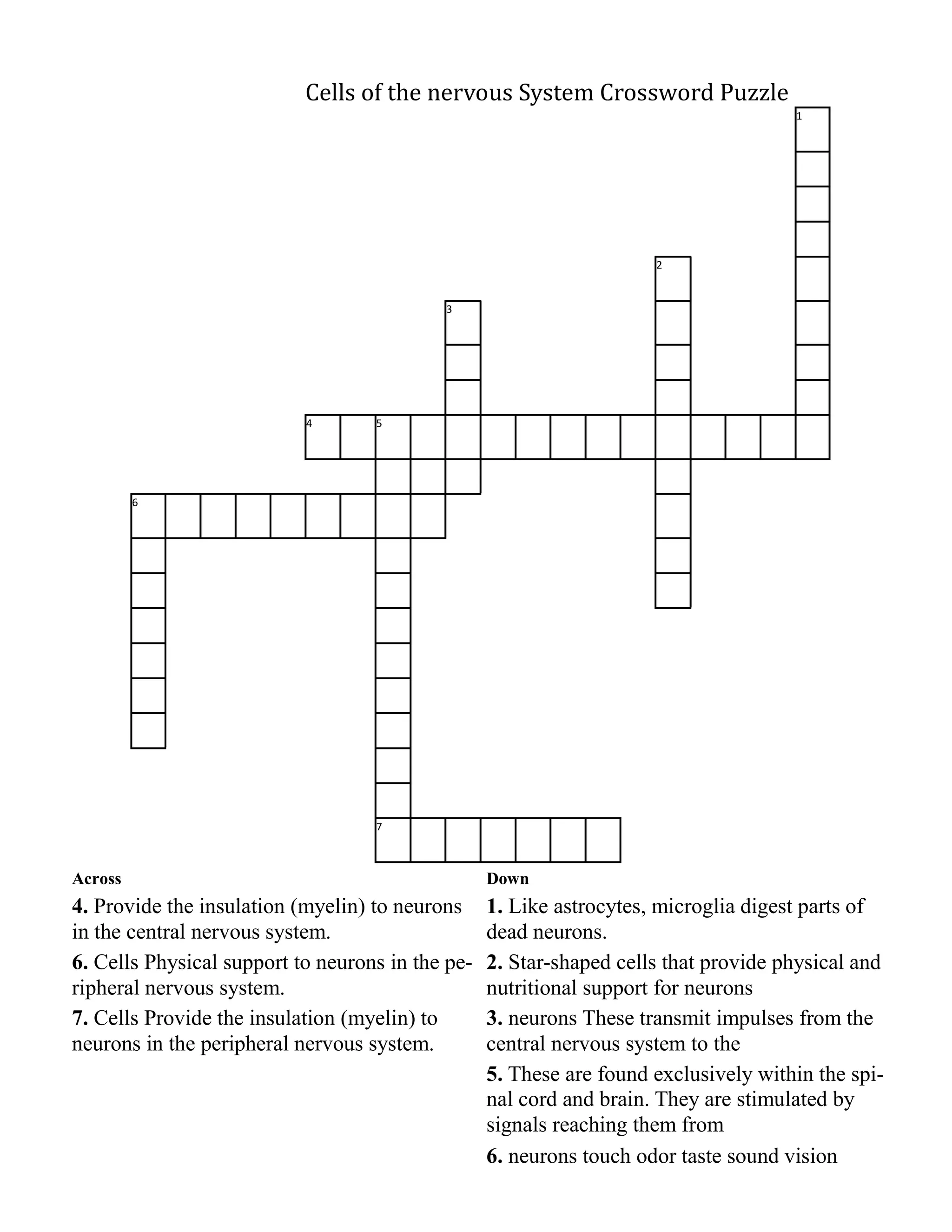
The document provides information on the nervous system, including:
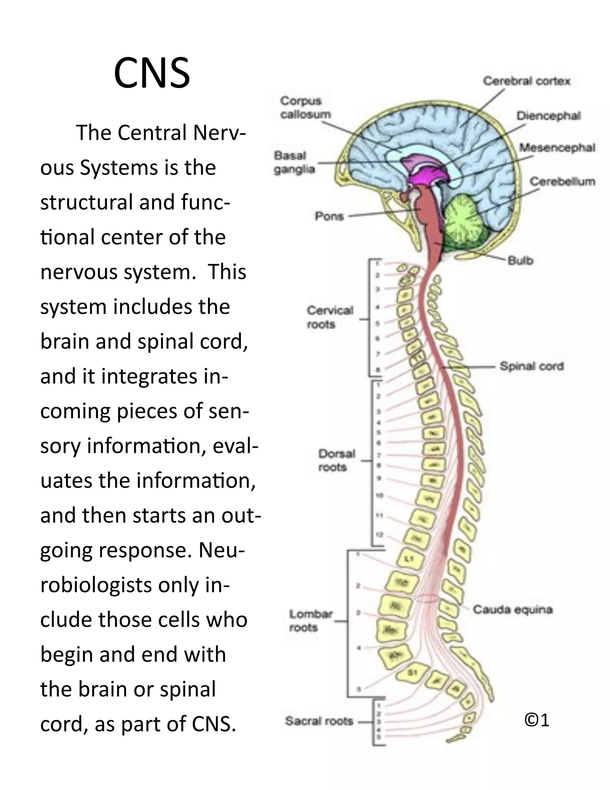
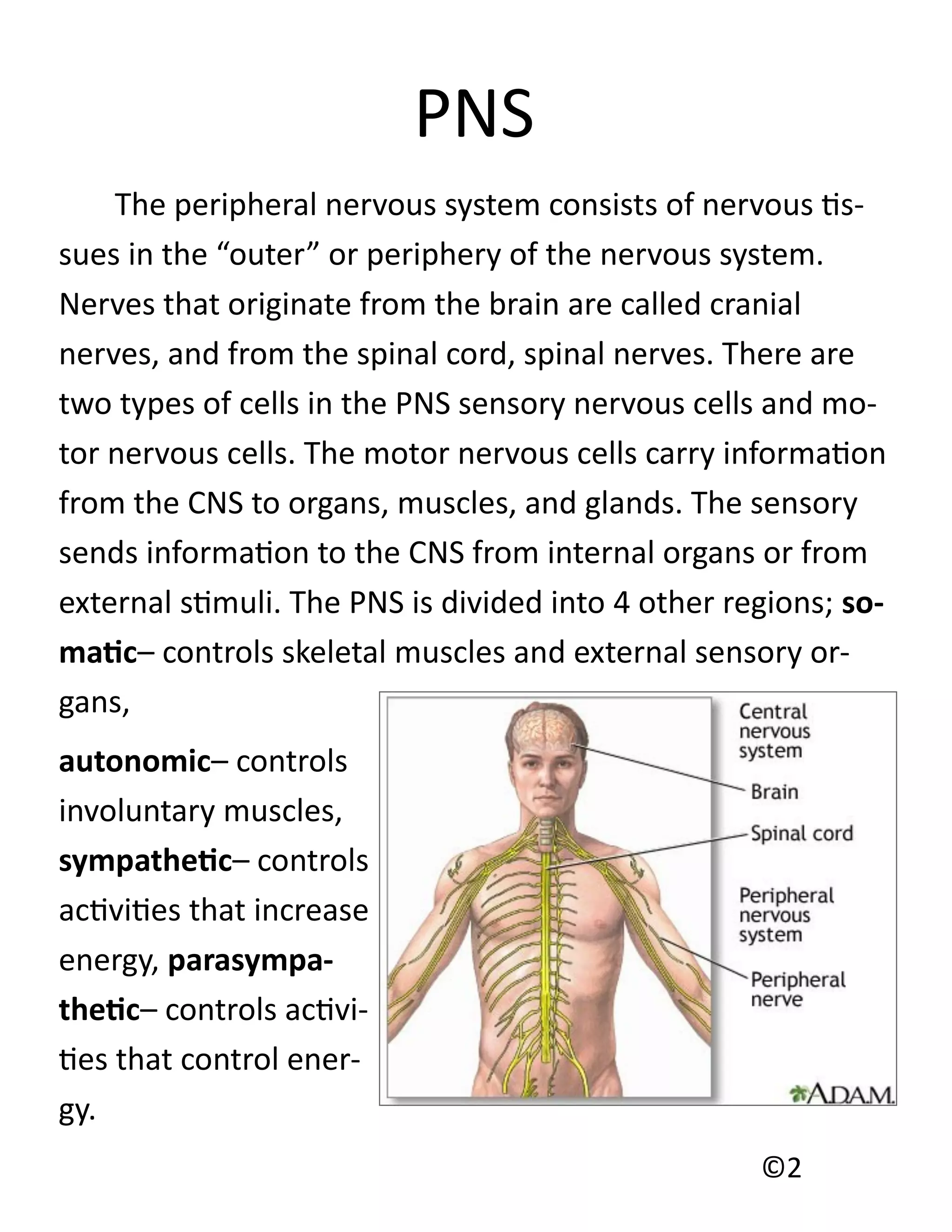
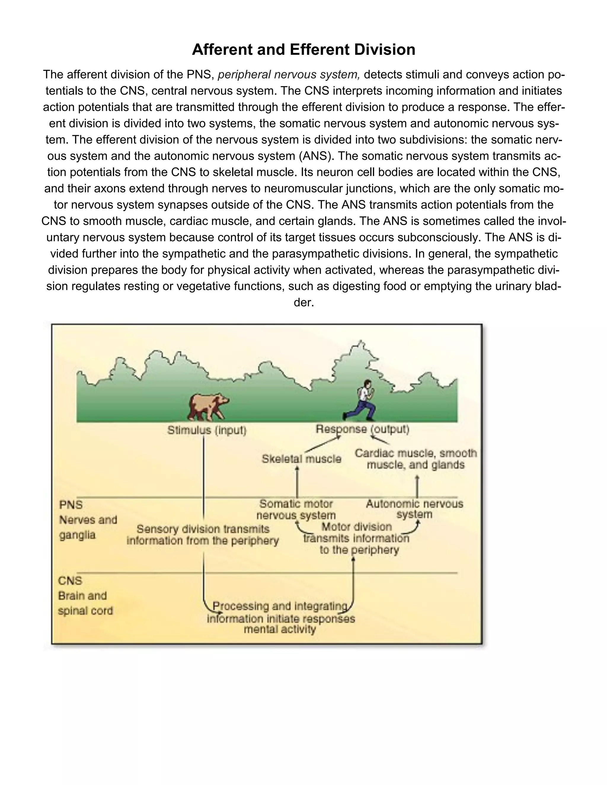
1) It describes the central nervous system (CNS), which includes the brain and spinal cord, and integrates sensory information and initiates responses. The peripheral nervous system (PNS) consists of nerves that originate from the brain and spinal cord.
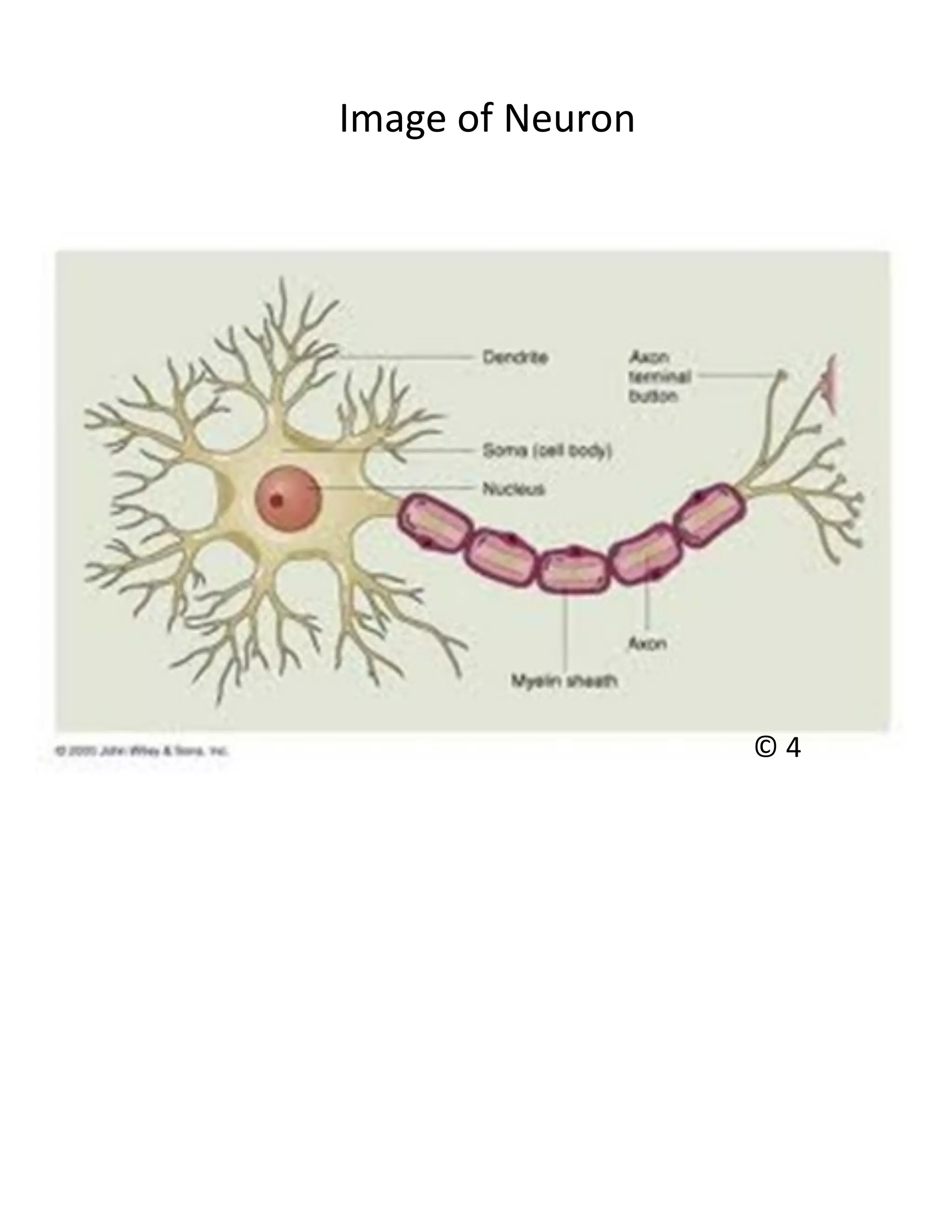
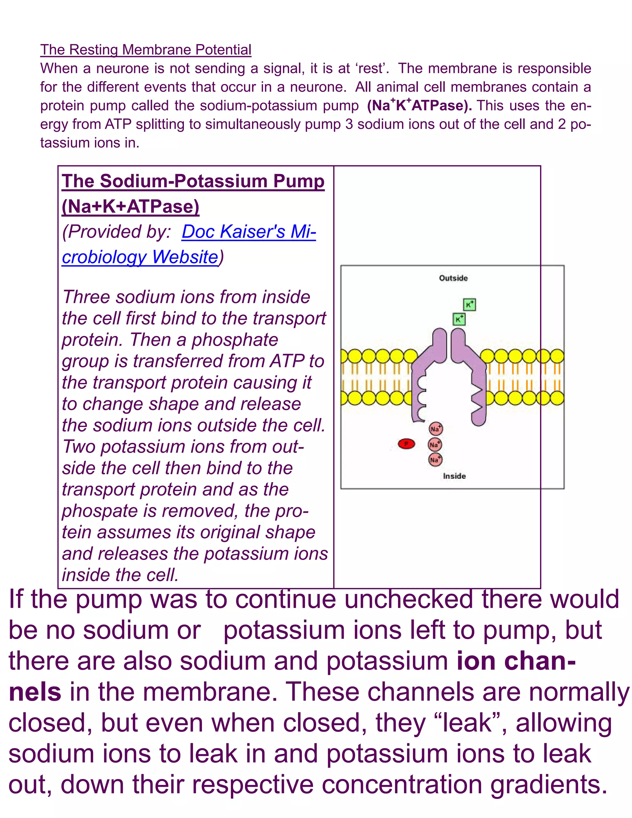
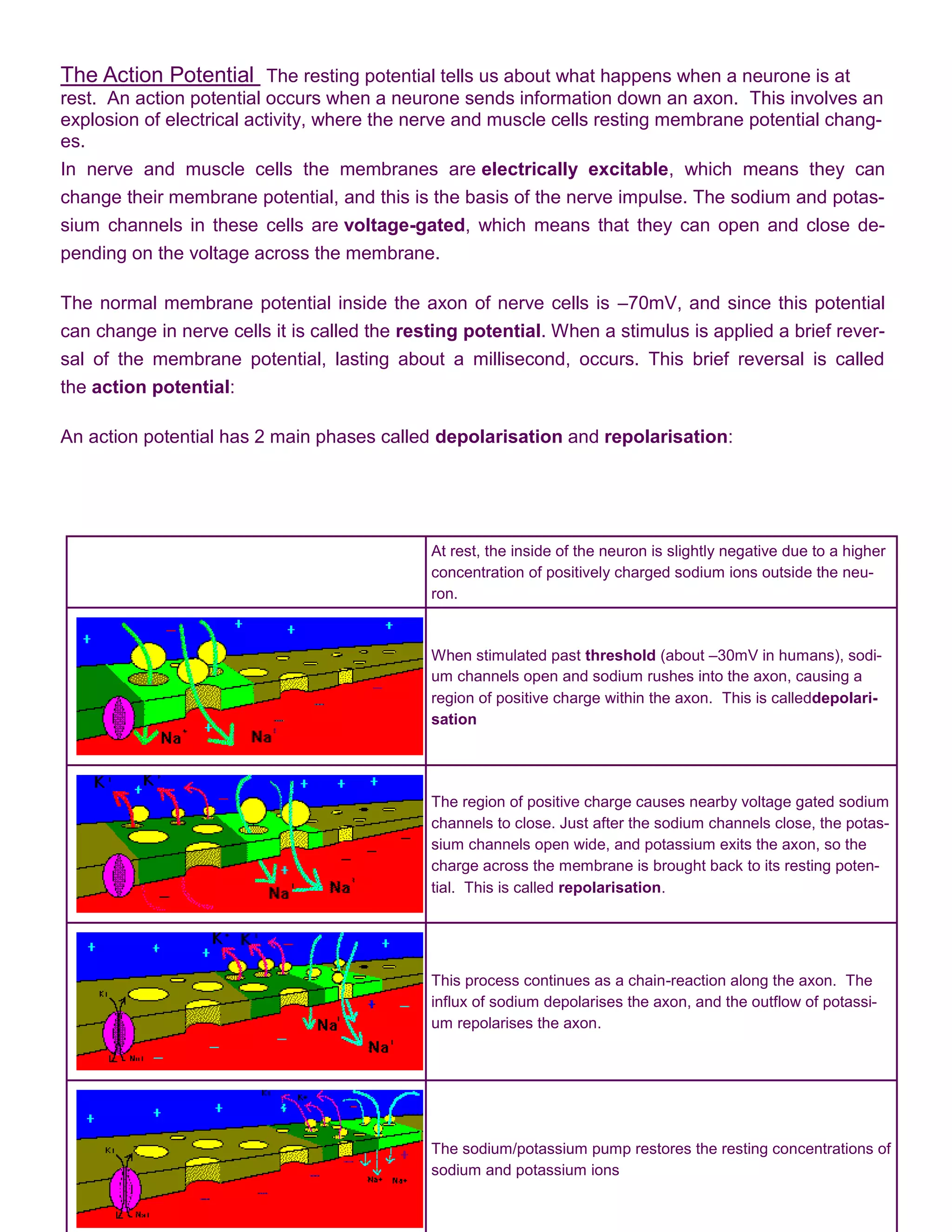
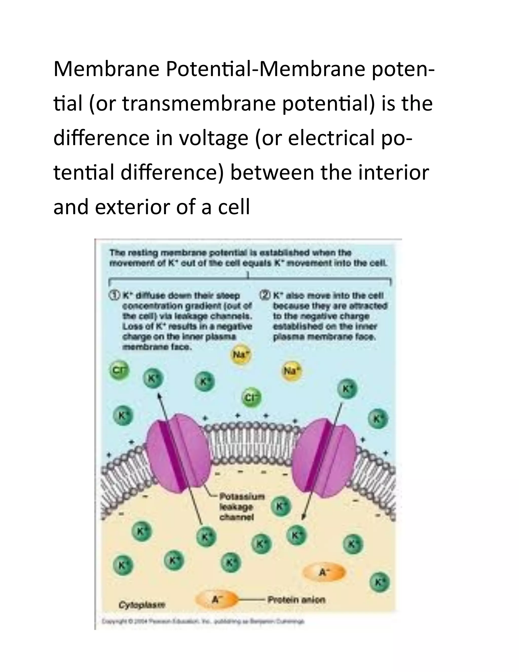
2) It explains how neurons transmit signals via nerve impulses through changes in their membrane potentials, including the processes of depolarization and repolarization during an action potential.
3) Synapses are the junctions between neurons where neurotransmitters are released to stimulate the next neuron and transmit signals throughout the nervous system.
The document contains 14 questions about skeletal muscle structure and function from a magazine editor. Responses are provided that explain:
1) How skeletal muscles provide movement, heat and posture through muscle contraction triggered by acetylcholine.
2) Excitability is shared by muscles and nerves, and contractility relates to the agonist/antagonist concept.
3) Structures unique to skeletal muscle fibers and those involved in contractility vs excitability.
The document summarizes a reflex arc lab experiment. [1] Voluntary reactions took longer than involuntary reactions due to additional processing time for the brain. [2] The speed of stimulus travel in the reflex arc was calculated to be 11.5 m/s based on distance and reaction time measurements. [3] The slower speed compared to literature values of 100 m/s is likely due to less concentrated nerve cells.
This document summarizes the key components of muscle tissue and muscle contraction. It discusses nerve impulses, motor units, neuromuscular junctions, and the sliding filament theory of muscle contraction. The main components that allow for contraction are described, including myosin, actin, tropomyosin, troponin, and the calcium release initiated by a nerve impulse that allows for crossbridge binding and sarcomere shortening. Regions of the sarcomere such as the Z-line, A-band, I-band, H-zone, and M-line are also outlined.
When walking, a series of neural processes occur to facilitate coordinated muscle contraction. Signals are sent from sensory neurons in the spinal cord to motor neurons, activating motor units and causing muscle fibers to contract. Both fast twitch and slow twitch fibers are used, with more reliance on slow twitch fibers for continuous walking. Muscle spindles and golgi tendon organs provide feedback on muscle length and force to ensure proper gait. Over time, motor unit plasticity results in neural adaptations that optimize walking performance.
Striated muscle contracts to move limbs and maintain posture. The contraction of skeletal muscles is an energy-requiring process. In order to perform the mechanical work of contraction, actin and myosin utilize the chemical energy of the molecule adenosine triphosphate (ATP).Muscle contraction results from a chain of events that begins with a nerve impulse traveling in the upper motor neuron from the cerebral cortex in the brain to the spinal cord.When the signal to contract is sent along a nerve to the muscle, the actin and myosin are activated. Myosin works as a motor, hydrolyzing adenosine triphosphate (ATP) to release energy in such a way that a myosin filament moves along an actin…
Excitation–Contraction Coupling
Excitation–contraction coupling is the link (transduction) between the action potential generated in the sarcolemma and the start of a muscle contraction.
Sliding Filament Model of Contraction
For a muscle cell to contract, the sarcomere must shorten. However, thick and thin filaments—the components of sarcomeres—do not shorten. Instead, they slide by one another, causing the sarcomere to shorten while the filaments remain the same length. The sliding filament theory of muscle contraction was developed to fit the differences observed in the named bands on the sarcomere at different degrees of muscle contraction and relaxation. The mechanism of contraction is the binding of myosin to actin, forming cross-bridges that generate filament movement
Skeletal muscles provide movement through contractions, generate heat through catabolism, and maintain posture through partial contractions. Muscles are excitable through nerve signals and use structures like myofibrils, sarcomeres, and troponin to generate force through the sliding filament theory when calcium ions bind during excitation and relax when calcium ions unbind. Different fiber types allow for specialized functions in sprinters versus marathon runners.
- Skeletal muscle contraction is controlled voluntarily through nervous signals from the brain. When a nerve impulse reaches the motor end plate, it causes acetylcholine to be released, which stimulates a muscle impulse.
- The muscle impulse travels through the muscle fiber and causes calcium ions to be released from the sarcoplasmic reticulum. Calcium ions bind to troponin and allow muscle contraction by exposing actin binding sites for myosin.
- Repeated stimuli can cause muscle fibers to contract without fully relaxing through summation, resulting in a sustained tetanic contraction. Forceful exercise causes muscle hypertrophy through recruitment of fast twitch fibers, while disuse leads to muscle atrophy.
Muscles contract through a sliding filament mechanism where actin and myosin filaments interact. Energy from ATP hydrolysis causes the myosin head to undergo conformational changes, attaching and detaching from actin and sliding the filaments past each other. This shortens the sarcomere. Tropomyosin normally blocks the actin binding sites, but calcium released during muscle stimulation displaces tropomyosin, allowing the cross-bridge cycle and contraction. When calcium is reabsorbed, tropomyosin resets and the muscle relaxes.
Mechanism Of Muscle Contraction&Neural Controlraj kumar
- Skeletal muscle is attached to bone by tendons and contains fascicles of striated muscle fibers.
- Muscle contraction occurs via the sliding filament theory where myosin cross-bridges attach to actin and pull the thin filaments towards the center of the sarcomere, shortening the muscle.
- Calcium released from the sarcoplasmic reticulum binds to troponin, allowing cross-bridge formation and muscle contraction.
Molecular basis of Skeletal Muscle ContractionArulSood2
The ppt aims to explain the molecular basis of skeletal muscle contraction and certain applied aspects of the same. Sources include Guyton and Hall's Textbook of Physiology (South-Asia edition, Vol. 2) and C.L. Ghai's Textbook for Practical Physiology.
This document discusses the neuromuscular system and muscle physiology. It begins by explaining how stimuli are transmitted through nerves to muscles via action potentials, causing muscle contraction. It then describes the structure and function of neurons, neuromuscular junctions, and muscle fibers. Specific topics covered include muscle fiber types, the sliding filament model of contraction, and factors that can cause fatigue. Diagrams illustrate key concepts such as the generation and propagation of action potentials.
The document discusses the three main types of muscle in the body - skeletal, smooth, and cardiac muscle. It describes the structure, function, and control mechanisms of skeletal muscle in detail. Key topics covered include skeletal muscle fiber structure, the sliding filament model of contraction, excitation-contraction coupling, and the properties of skeletal muscle contractions.
The document describes various aspects of muscle contraction including:
1) Excitation-contraction coupling which involves depolarization of the muscle membrane leading to calcium release and muscle contraction.
2) The roles of the sarcoplasmic reticulum, t-tubules, and troponin-tropomyosin complex in regulating calcium levels and exposing actin binding sites during contraction.
3) The sliding filament theory of how myosin heads binding to actin causes muscle shortening through an ATP-driven cycling of cross-bridge formation and breaking.
2nd and 3rd September 2011,a General Lecture Theatre, Dr Chirantan Mandal, Dr Avik Basu, Dr Dipayan Sen Dr Ushnish Adhikari,Dr Srimanti Bhattacharya, Dr Shubham Presided by Dr Arnab Sengupta (Physiology Dept Medical College Kolkata)
The document summarizes several topics related to muscular physiology:
1) Rigor mortis is the stiffening of muscles that occurs after death due to a lack of ATP preventing muscle relaxation.
2) Four factors influence muscle contraction strength: number of cross bridges, fiber length, stimulation frequency, and recruitment.
3) The document then discusses several other muscle-related topics such as phases of muscle contraction, the staircase phenomenon, muscle fiber structure, and the roles of skeletal muscle.
1) Skeletal muscle contraction occurs via the sliding filament theory where the binding of myosin heads to actin causes sarcomeres to shorten.
2) Motor neurons innervate muscle fibers to produce either contraction or relaxation in response to neural signals.
3) Calcium release from the sarcoplasmic reticulum in response to action potentials is key to initiating muscle contraction by exposing binding sites on the actin thin filaments.
The document defines key terms related to muscle contraction such as sarcomeres, myofibrils, and cross-bridges. It then summarizes the sliding filament model of muscle contraction which involves calcium binding to troponin, exposing actin binding sites, ATP binding to myosin heads, power strokes that cause actin to slide over myosin, and cross-bridge detachment. It also describes muscle twitches, summation that results from high stimulation frequencies, tetanus which is a sustained contraction, and recruitment where additional motor neurons are activated to involve more muscle fibers and produce more force.
Factors influencing force of contractonRajesh Goit
The document discusses factors that influence the force of muscle contraction, including motor unit recruitment, fatigue, and the muscle twitch. It describes how motor unit recruitment involves activating additional motor units to generate more force. Fatigue results from depletion of energy stores and accumulation of metabolites in the muscle over time. The properties of the muscle twitch, such as peak tension and duration, depend on the fiber type - fast or slow twitch. Slow twitch fibers generate low force for a long duration while fast twitch fibers contract quickly but fatigue rapidly.
1. The document discusses the structure and function of the neuromuscular junction and skeletal muscle contraction. It describes how an action potential causes the release of acetylcholine from the motor neuron, leading to depolarization of the muscle fiber membrane.
2. Calcium release within the muscle fiber initiates cross-bridge cycling between actin and myosin, producing muscle contraction. Contraction ceases as calcium is reabsorbed by the sarcoplasmic reticulum, relaxing the muscle.
3. The length-tension relationship states that skeletal muscle generates maximum force when sarcomeres are at their optimal resting length, with overlapping actin and myosin filaments. Shorter or longer lengths reduce the number of cross-
The document summarizes the structure and function of skeletal muscle myosin and actin filaments during muscle contraction. It describes how myosin filaments are composed of myosin molecules that form cross-bridges with heads that can interact with actin filaments. Actin filaments contain actin, tropomyosin and troponin. Calcium released from the sarcoplasmic reticulum activates contraction by allowing the myosin heads to bind to active sites on actin. The myosin heads then tilt and pull the actin filaments towards the center in a ratcheting motion, powered by ATP hydrolysis.
This document summarizes the physiology of muscle contraction. It describes the three types of muscle tissue: striated skeletal muscle, striated cardiac muscle, and smooth muscle. It explains the structure and function of the sarcomere, the basic unit of skeletal muscle contraction. Contraction occurs via a sliding filament mechanism when myosin cross-bridges interact with and pull actin filaments, shortening the sarcomere. Neurotransmitter acetylcholine released at the neuromuscular junction triggers an action potential in the muscle fiber membrane, allowing calcium entry and the initiation of contraction.
1. Muscle contraction occurs via a sliding filament mechanism where calcium ions released by the sarcoplasmic reticulum allow actin and myosin filaments to interact.
2. Myosin filaments contain cross-bridges that can attach to actin filaments and generate a pulling force via ATP hydrolysis.
3. Tropomyosin and troponin on the actin filaments regulate the exposure of actin's binding sites depending on calcium levels.
It includes the basic anatomy physiology of skeletal muscles, the thorough working of the muscles, at superficial level to molecular level, the energy input, smooth muscle-cardiac-skeletal muscles differences, smooth muscle anatomy physiology.
The document discusses skeletal muscle electrophysiology. It describes how skeletal muscles generate action potentials via voltage-gated sodium channels, leading to depolarization. The same voltage then opens potassium channels to cause repolarization. At the neuromuscular junction, acetylcholine is released from motor neurons and binds to receptors on muscle fibers. This excitation causes contraction via excitation-contraction coupling, where the action potential initiates mechanical contraction. There is a latent phase between excitation and contraction as events occur to trigger the sliding of actin and myosin filaments and initiation of muscle contraction.
General and molecular mechanism of Muscle contractionShiv Patel
Skeletal muscle contraction occurs through the interaction of the thin filament proteins actin and tropomyosin with the thick filament protein myosin. An action potential causes calcium release from the sarcoplasmic reticulum, allowing myosin to bind to actin and generate a power stroke that slides the thin filaments past the thick filaments, shortening the muscle. Repeated binding and detachment of the myosin heads along the actin filaments through ATP hydrolysis drives sustained muscle contraction.
Muscle movement plays an important role in day to day life where the contraction and relaxation of muscle is significant. The current slide has been developed with the focus on different phases during muscle contraction and the physiological change involved on it.
Neurons are electrically excitable cells that communicate with each other and the body. The human nervous system contains around 100 billion neurons. There are three main types of neurons - sensory neurons relay signals from sense organs to the central nervous system, motor neurons relay signals from the CNS to effector organs, and interneurons connect sensory and motor neurons. Each neuron has a cell body, dendrites that receive signals, and an axon that transmits signals. When a neuron is stimulated, it generates an action potential down its axon via changes in membrane potential. Neurotransmitters are released at synapses to transmit signals between neurons.
The document summarizes the structure and function of the human nervous system. It describes the three main types of neurons - sensory, motor, and interneurons - and how they transmit nerve impulses via electrical and chemical signals. It also explains the process of synaptic transmission between neurons and how homeostasis is maintained through negative feedback mechanisms that monitor and respond to changes in the internal environment.
These slides contain the basic information and principle of nervous transduction, It also includes the information about the type of the neurons, structure of the neuron, resting and active membrane potential, synapes and events occurring in it, and introduction to the neurotransmitters.
Nerve Impulse is defined as a wave of electrical chemical changes across the neuron that helps in the generation of the action potential in response to the stimulus. This transmission of a nerve impulse across the neuron membrane as a result of a change in membrane potential is known as Nerve impulse conduction.
Mechanism of Nerve Impulse Conduction
Nerve impulse conduction is a major process occurring in the body responsible for organized functions of the body. So, for conduction of nerve impulse there are two mechanisms:
Continuous conduction
Saltatory conduction
Coordination is achieved through the nervous and endocrine systems. The nervous system uses electrical and chemical signaling via nerve impulses and neurotransmitters to achieve rapid transmission and localized responses. The endocrine system uses slower acting chemical signaling via hormones transported through the bloodstream to regulate longer term processes like growth. At synapses, the arrival of a nerve impulse triggers the release of neurotransmitters which may excite or inhibit the next neuron, allowing information to be passed through the nervous system. This coordination allows the body's organs to function in a synchronized manner.
This document provides an overview of nerve tissue physiology. It discusses the two principal cell types in the nervous system - neurons and neuroglial cells. Neurons are specialized for signal conduction while neuroglial cells provide support and protection. The document then examines the structure and function of neurons, including their cell body, dendrites, axon, and synaptic transmission. It also explores concepts such as membrane potentials, action potentials, refractory periods, and the mechanisms of electrical and chemical synaptic transmission.
The nervous system is composed of the central nervous system (CNS), which includes the brain and spinal cord, and the peripheral nervous system (PNS). The CNS receives sensory input, integrates information, and directs motor responses. Within the CNS, the brain is responsible for higher functions like thinking and memory, while the spinal cord transmits signals between the brain and body. Neurons are the basic functional units and communicate via electrical and chemical signals across synapses. The nervous system allows animals to integrate internal and external sensory information to direct activities and maintain homeostasis.
The nervous system is divided into the central nervous system (CNS) and peripheral nervous system (PNS). The CNS contains the brain and spinal cord, and receives and processes sensory information. The PNS transmits signals between the CNS and body. Within the nervous system are neurons, which transmit signals, and glial cells, which support neurons. Neurons communicate via electrical and chemical signals to coordinate bodily functions.
types of neurons, structure and functions, types of glia cells, their structure and function, functioning of a neuron - resting potential, action potential, graded potential, absolute and relative refractory period.
This document discusses neurons and neuronal communication. It describes two main types of communication - neuronal communication via electrical signals transmitted by neurons, and hormonal communication via slower-acting chemical signals from hormones. It provides details on the structure and function of neurons, including the key parts of neurons (cell body, dendrites, axon), properties of excitability and conductivity, and types of neurons (sensory, intermotor, motor). The document explains the mechanisms of neuronal signaling, including resting membrane potential, action potentials, and synaptic transmission. It compares chemical and electrical synapses and provides illustrations and animations of these processes.
Indian Dental Academy: will be one of the most relevant and exciting training center with best faculty and flexible training programs for dental professionals who wish to advance in their dental practice,Offers certified courses in Dental implants,Orthodontics,Endodontics,Cosmetic Dentistry, Prosthetic Dentistry, Periodontics and General Dentistry.
The document summarizes key concepts about the nervous system including:
- Neurons are the basic structural and functional units that transmit electrochemical signals called nerve impulses. Nerves are bundles of axons.
- The central nervous system (CNS) contains gray matter with neuron cell bodies and unmyelinated axons, and white matter with bundles of myelinated axons.
- There are three main types of neurons - sensory, interneurons, and motor neurons. Neuroglial cells provide support and insulation for neurons in the CNS.
- The peripheral nervous system connects the CNS to other body parts and allows sensory input, integrative processing, and motor output functions.
The Resting Potential And The Action Potentialneurosciust
An action potential occurs when a neuron is stimulated enough to reach its threshold of excitation. This causes sodium channels to open, allowing sodium to rush into the neuron and depolarize it. The neuron then repolarizes as potassium leaves and the sodium-potassium pump restores the ion gradients. After an action potential, the neuron enters an absolute refractory period where it cannot fire again, followed by a relative refractory period where a stronger stimulus is needed to trigger another action potential. This process allows neurons to rapidly transmit signals down axons to synapse with other neurons.
Unit 1 Nervous System.pptx by Nutan KambleNutanKamble7
The human nervous system is a complex of interconnected systems in which larger systems are comprised of smaller subsystems each of which have specific structures with specific functions.
The nervous system is very important in helping to maintain the homeostasis (balance) of the human body.
A series of sensory receptors work with the nervous system to provide information about changes in both the internal and external environments.
The Nervous system consist of vast number of cells called as Neurones.
Consist of special type of connective tissue, Neuroglia.
Neuron consist of cell body,axon,dendrites.
The neurons are actively conducting nerve impulses (action potentials).
Irritability and Conductivity- characteristics of neuron
membrane of the axon- axolemma
Large axons and those of peripheral nerves are surrounded by myelin sheath.(series of shwann cell arranged along length of axon)
Each one is wrapped around the axon so that it is covered by a number of concentric layers of Schwann cell plasma membrane.
Between layers of plasma membrane-small amt of fatty sub.-Myelin
Axons and dendrites are extensions of cell bodies & form the white matter of the nervous system.
Axons are found deep in brain and in groups,called tracts, at the periphery of the spinal cord.
Refferd as nerves or nerve fibers outside the brain and spinal cord.
he nervous system includes the brain, spinal cord, and a complex network of nerves. This system sends messages back and forth between the brain and the body. The brain is what controls all the body's functions. The spinal cord runs from the brain down through the back.The nervous system uses tiny cells called neurons (NEW-ronz) to send messages back and forth from the brain, through the spinal cord, to the nerves throughout the body.
Billions of neurons work together to create a communication network. Different neurons have different jobs. For example, sensory neurons send information from the eyes, ears, nose, tongue, and skin to the brain. Motor neurons carry messages away from the brain to the rest of the body to allow muscles to move. These connections make up the way we think, learn, move, and feel. They control how our bodies work — regulating breathing, digestion, and the beating of our hearts.In biology, the nervous system is the highly complex part of an animal that coordinates its actions and sensory information by transmitting signals to and from different parts of its body. The nervous system detects environmental changes that impact the body, then works in tandem with the endocrine system to respond to such events.[1] Nervous tissue first arose in wormlike organisms about 550 to 600 million years ago. In vertebrates, it consists of two main parts, the central nervous system (CNS) and the peripheral nervous system (PNS)The CNS consists of the brain and spinal cord. The PNS consists mainly of nerves, which are enclosed bundles of the long fibers, or axons, that connect the CNS to every other part of the body.
- Nerve cells transmit electrical signals through long fibers called axons. The cell body contains organelles that produce proteins transported via axoplasmic flow to nerve endings. Myelination increases conduction velocity. Injury can cause temporary (neuropraxia) or permanent (neurotmesis) dysfunction. Regeneration involves Wallerian degeneration clearing debris, axon regrowth, and reinnervation.
The document discusses the nervous system and sense organs. It begins by describing the basic functions and components of the nervous system, including neurons, action potentials, and synapses. It then provides details on the types of neurons, glial cells, and how the resting membrane potential and action potentials work. The document also discusses the evolution of nervous systems in invertebrates and vertebrates. It concludes by describing the peripheral nervous system and different types of sense organs.
The document discusses how nerve cells transmit impulses through the nervous system. It explains that:
- Nerve cells have a resting potential of around -70mV due to the distribution of ions inside and outside the cell.
- When a nerve is stimulated, it causes voltage-gated sodium channels to open, allowing sodium ions to rush in and depolarize the cell. This creates an action potential.
- The cell then repolarizes as sodium channels close and potassium channels open, restoring the resting potential and allowing another impulse to be transmitted.
- This process of depolarization and repolarization via ion channel activation allows rapid and efficient transmission of nerve impulses through the nervous system.
The nervous system consists of the brain, spinal cord and nerves. It detects changes inside and outside the body and responds through electrical signals called nerve impulses. Neurons conduct these impulses while neuroglia provide support. There are two main types of synapses - electrical and chemical. At chemical synapses, a neurotransmitter is released from the presynaptic neuron and binds to receptors on the postsynaptic neuron.
The document summarizes key aspects of nerve physiology:
- The nervous system is divided into the central nervous system (brain and spinal cord) and peripheral nervous system. The peripheral nervous system is further divided into the somatic and autonomic nervous systems.
- A neuron consists of a cell body, dendrites, and an axon. Neurons transmit electrical signals called action potentials via their axons.
- An action potential occurs when a neuron is stimulated - sodium ions rush into the neuron, depolarizing the membrane. Then potassium ions exit, repolarizing the membrane back to its resting potential. This allows signals to propagate along axons.
The nervous system allows for coordination in the body through electrochemical signaling between neurons. It consists of neurons and neuroglia. Neurons receive and transmit signals via dendrites, the cell body, and the axon. There are three types of neurons - sensory, motor, and inter. A nerve impulse is generated through changes in the neuron's membrane potential and the opening and closing of ion channels, causing the signal to propagate along the axon. At a synapse, neurotransmitters transmit the signal to the next neuron. Reflexes are automatic responses to stimuli.
This document discusses lung volumes and capacities, including tidal volume, inspiratory reserve volume, expiratory reserve volume, vital capacity, residual volume, total lung capacity, and minute ventilation. It provides definitions and normal values for these measurements and describes how they are obtained through spirometry. The objectives are to obtain graphical representations of lung volumes, compare volumes between males and females, and correlate volumes with clinical conditions.
An electrocardiogram (EKG or ECG) records the electrical activity of the heart over time. The main components of a heartbeat are labeled P, Q, R, S, and T waves. The P wave represents electrical activity spreading through the atria, while the QRS complex represents ventricular activation. The T wave occurs as the ventricles recover. By examining intervals between these waves, as well as their presence, shape, and consistency, doctors can detect disorders like abnormal heart rhythms or damage to heart muscle tissue. In this experiment, students will record their own EKG, identify the wave components, and calculate their heart rate. They will also compare EKGs recorded from different electrode placements on the arms
This document analyzes blood typing results from 4 patients and determines their blood types based on agglutination reactions with different blood serum samples. It finds that patient 3, Mr. Green, has type AB blood based on agglutinating with anti-A, anti-B, and anti-Rh serum. The document also discusses how blood types are determined, similarities and differences between agglutinogens and agglutinins, causes of erythroblastosis fetalis, and the importance of proper blood lab procedures and sterilization to minimize disease transmission.
The document provides an illustrated overview of the 206 bones in the human skeletal system, including both the axial skeleton (skull, vertebral column, rib cage) and appendicular skeleton (shoulder girdle, upper limbs, pelvic girdle, lower limbs). Key bones are labeled for each anatomical region, with close-up views highlighting important features like processes, tubercles, and articulation surfaces. An accompanying text labels and describes the functions of individual bones and bone groups.
The document discusses the endocrine system and hormones, explaining how hormones are released from glands and target specific cells through receptor proteins to regulate bodily functions like growth, metabolism, and reproduction. It provides information on different types of hormones based on their chemical structure and source, and includes tables listing major hormones and their functions.
The nervous and endocrine systems work together to monitor and regulate cells in the body. The endocrine system uses hormones to initiate responses in target cells by binding to specific receptors. Hormones are classified by their structure and function, with some made from cholesterol and others from amino acids. Hormones are released into the bloodstream and bind with matching receptors on target cells to initiate various responses and maintain homeostasis. The major endocrine glands and their hormones are the pituitary, thyroid, parathyroid, adrenal, pancreas, gonads, placenta and pineal glands.
This document discusses sensory receptors, including their types, structures, and functions. It covers exteroceptors, visceroceptors, proprioceptors, mechanoreceptors, chemoreceptors, thermoreceptors, nociceptors, photoreceptors, olfactory receptors, taste receptors, hearing receptors, and balance receptors. Key points include that sensory receptors receive stimuli from both internal and external environments and relay this information to the nervous system, and that different receptor types are activated by mechanical, chemical, thermal, or light stimuli.
The document summarizes the anatomy and functions of the central nervous system and its parts. It describes the three meninges (dura mater, arachnoid mater, pia mater) that protect the CNS. It then discusses the structures and functions of the brainstem, cerebellum, diencephalon, and cerebral cortex. It also outlines the somatic sensory and motor pathways, as well as the autonomic nervous system including its sympathetic and parasympathetic divisions.
(1) The document describes various organelles and cellular structures that can be "sold", including the cell membrane, mitochondria, lysosomes, endoplasmic reticulum, Golgi apparatus, ribosomes, chloroplasts, and cilia.
(2) It then provides more detailed information about the structures and functions of the cell membrane, nucleic acids, lipids, carbohydrates, proteins, and inorganic molecules.
(3) The organelles and molecules play essential roles in processes like ATP synthesis, protein synthesis, waste breakdown, toxin removal, and genetic coding that allow cells to function properly.
The document discusses the seven levels of organization of the human body from chemical to organism, describing cells, tissues, organs, organ systems, and the two main body cavities. It also examines anatomical planes used to divide the body, directional terms, characteristics of living things, and homeostasis, giving examples of positive feedback loops that help maintain stability in the body. Diagrams are included showing body organization levels and anatomical planes.
Transcription and translation are the two main processes involved in protein synthesis. Transcription involves copying a segment of DNA into mRNA. Translation then uses the mRNA to assemble a protein, with the help of tRNA and rRNA, according to the genetic code. Both transcription and translation are essential for cells to make the proteins they need.
Active transport requires energy to move materials from areas of low concentration to high concentration inside cells. It uses protein pumps to transport particles, like endocytosis which moves items into the cell and exocytosis which moves them out. Cell growth and reproduction are essential life processes where cells grow using genetic information and DNA to make needed proteins, and reproduce through mitosis and cytokinesis to pass genetic material to new daughter cells. Catabolism breaks down complex molecules while anabolism constructs complex living tissue from simpler substances using enzymes.
Organic molecules like carbohydrates and proteins are made of carbon, hydrogen, and oxygen and are essential to living organisms. Carbohydrates are sugars and starches that provide energy, while proteins are chains of amino acids that perform vital functions. Inorganic molecules like oxygen, carbon dioxide, and salts are not as complex and are usually found in non-living things, but still play important roles like in respiration and bodily functions. The balance of acids and bases is also important and measured by the pH scale.
(1) The document discusses several types of organelles and their functions, including mitochondria which generates ATP for energy, lysosomes which break down waste, the endoplasmic reticulum which synthesizes lipids and proteins, and ribosomes which synthesize proteins.
(2) It also discusses the cell membrane, which forms the boundary of the cell and regulates what enters and exits, as well as cilia and flagella, which help move substances on the cell surface.
(3) The Golgi apparatus packages proteins and lipids into vesicles for transport within and outside the cell.
The document summarizes the key levels of organization in the human body from chemical to organism. It then provides explanations of anatomical planes, directional terms, homeostasis, and other anatomical and physiological concepts. Diagrams are included to illustrate body structures and organization.
4. Afferent and Efferent Division
The afferent division of the PNS, peripheral nervous system, detects stimuli and conveys action po-
tentials to the CNS, central nervous system. The CNS interprets incoming information and initiates
action potentials that are transmitted through the efferent division to produce a response. The effer-
ent division is divided into two systems, the somatic nervous system and autonomic nervous sys-
tem. The efferent division of the nervous system is divided into two subdivisions: the somatic nerv-
ous system and the autonomic nervous system (ANS). The somatic nervous system transmits ac-
tion potentials from the CNS to skeletal muscle. Its neuron cell bodies are located within the CNS,
and their axons extend through nerves to neuromuscular junctions, which are the only somatic mo-
tor nervous system synapses outside of the CNS. The ANS transmits action potentials from the
CNS to smooth muscle, cardiac muscle, and certain glands. The ANS is sometimes called the invol-
untary nervous system because control of its target tissues occurs subconsciously. The ANS is di-
vided further into the sympathetic and the parasympathetic divisions. In general, the sympathetic
division prepares the body for physical activity when activated, whereas the parasympathetic divi-
sion regulates resting or vegetative functions, such as digesting food or emptying the urinary blad-
der.
5. Cells of the nervous System Crossword Puzzle
1
2
3
4 5
6
7
Across Down
4. Provide the insulation (myelin) to neurons 1. Like astrocytes, microglia digest parts of
in the central nervous system. dead neurons.
6. Cells Physical support to neurons in the pe- 2. Star-shaped cells that provide physical and
ripheral nervous system. nutritional support for neurons
7. Cells Provide the insulation (myelin) to 3. neurons These transmit impulses from the
neurons in the peripheral nervous system. central nervous system to the
5. These are found exclusively within the spi-
nal cord and brain. They are stimulated by
signals reaching them from
6. neurons touch odor taste sound vision
7. The Resting Membrane Potential
When a neurone is not sending a signal, it is at ‘rest’. The membrane is responsible
for the different events that occur in a neurone. All animal cell membranes contain a
protein pump called the sodium-potassium pump (Na+K+ATPase). This uses the en-
ergy from ATP splitting to simultaneously pump 3 sodium ions out of the cell and 2 po-
tassium ions in.
The Sodium-Potassium Pump
(Na+K+ATPase)
(Provided by: Doc Kaiser's Mi-
crobiology Website)
Three sodium ions from inside
the cell first bind to the transport
protein. Then a phosphate
group is transferred from ATP to
the transport protein causing it
to change shape and release
the sodium ions outside the cell.
Two potassium ions from out-
side the cell then bind to the
transport protein and as the
phospate is removed, the pro-
tein assumes its original shape
and releases the potassium ions
inside the cell.
If the pump was to continue unchecked there would
be no sodium or potassium ions left to pump, but
there are also sodium and potassium ion chan-
nels in the membrane. These channels are normally
closed, but even when closed, they “leak”, allowing
sodium ions to leak in and potassium ions to leak
out, down their respective concentration gradients.
8. The Action Potential The resting potential tells us about what happens when a neurone is at
rest. An action potential occurs when a neurone sends information down an axon. This involves an
explosion of electrical activity, where the nerve and muscle cells resting membrane potential chang-
es.
In nerve and muscle cells the membranes are electrically excitable, which means they can
change their membrane potential, and this is the basis of the nerve impulse. The sodium and potas-
sium channels in these cells are voltage-gated, which means that they can open and close de-
pending on the voltage across the membrane.
The normal membrane potential inside the axon of nerve cells is –70mV, and since this potential
can change in nerve cells it is called the resting potential. When a stimulus is applied a brief rever-
sal of the membrane potential, lasting about a millisecond, occurs. This brief reversal is called
the action potential:
An action potential has 2 main phases called depolarisation and repolarisation:
At rest, the inside of the neuron is slightly negative due to a higher
concentration of positively charged sodium ions outside the neu-
ron.
When stimulated past threshold (about –30mV in humans), sodi-
um channels open and sodium rushes into the axon, causing a
region of positive charge within the axon. This is calleddepolari-
sation
The region of positive charge causes nearby voltage gated sodium
channels to close. Just after the sodium channels close, the potas-
sium channels open wide, and potassium exits the axon, so the
charge across the membrane is brought back to its resting poten-
tial. This is called repolarisation.
This process continues as a chain-reaction along the axon. The
influx of sodium depolarises the axon, and the outflow of potassi-
um repolarises the axon.
The sodium/potassium pump restores the resting concentrations of
sodium and potassium ions
9. Membrane Potential-Membrane poten-
tial (or transmembrane potential) is the
difference in voltage (or electrical po-
tential difference) between the interior
and exterior of a cell
10. The local potential is the depolariza-
tion of a cell below threshold. After
the cell is sufficiently depolarized (and
reaches threshold), it fires an action
potential down the axon.
11. Synapse
1. Summation- The potentials spread far enough to
reach the axon hillock, where they add together. When
they add together and reach threshold pontential, they
produce an actional potential called Spatial Summa-
tion. When in rapid succession they produce an action
potential it is called temporal summation.
2. Nuerotransmitters- some trigger the opening or
closing of ion channels directly. They will bind to re-
ceptors linked to G proteins. Small-molecule neuro-
transmitters are amino acids or are derived from ami-
no acids. Large-molecule neurotransmitters are two
chains of 2-40 amino acids.
12. Neuromuscular Reflex lab Graphs
5
4
3 Delta T (s) striking
Delta T(S) sound
2
1
0 0.2 0.4 0.6 0.8 1
5
4
Reflex with reinforcement
3
Relex without
reinforcement
2
1
0 1 2 3 4
13. DATA ANALYSIS FOR NEOMUSCULAR REFLEX LAB
1. Compare the reaction times for voluntary vs. involuntary activation of the quadriceps mus-
cle. What might account for the observed differences in reaction times?
The voluntary times are substantially higher due to the test subject being aware of the
need for his leg to move in order to collect data. Because the test subject could focus
more on voluntarily moving his leg when the table was hit, he could in fact increas the
time of stimulous. While involuntarily, his nervous system took over and was being
tested. He was given no warning for when his knee was to be hit so he did not have
time to contol any outcome of the time of stimulus.
2. Using data from Table 2, calculate speed at which a stimulus traveled from the patellar ten-
don to the spinal cord and back to the quadriceps muscle (a complete reflex arc). To do this,
you must estimate the distance traveled. Using a cloth tape measure, measure the distance
in cm from the mark on the patellar tendon to the spinal cord at waist level (straight across
from the anterior-superior iliac spine–see Figure 9). Multiply the distance by two to obtain
the total distance traveled in the reflex arc. Once this value has been obtained, divide by the
average ∆t from Table 2 and divide by 100 to obtain the speed, in m/s, at which the stimu-
lus traveled.
41.58 m/s
3. Nerve impulses have been found to travel as fast as 100 m/s. What could account for the
difference between your answer to Question 2 and this value obtained by researchers?
Possible miscalculations could have caused our answer to question two, along with the
differenciating intenisties of the subjects knee being hit.
4. Assume the speed of a nerve impulse is 100 m/s. How does this compare to the speed of
electricity in a copper wire (approx. 3.00 ´ 108 m/s)?
The speed of the electricity of a copper wire is 300,000,000,000 x faster than the speed
of a nerve impluse.
5. Compare the data you obtained in this experiment with other members of your group/class.
Can individual differences be attributed to any physical differences (body shape/size, mus-
cle mass, physical fitness level)?
Yes, because the physiology of humans are all diffirent. All of the data collected
throughout the class is all diverse as well due to each test subject being anatomical
ly diverse.
14. DATA NAME: ZACH
Table 1
Kick 1 Kick 2 Kick 3 Kick 4 Kick 5 Average
Time of muscle contraction (s) 12.27 15.39 18.23 21.11 23.86
Time of stimulus (s) 12.09 15.19 17.53 20.30 23.17
∆t (s) 00.18 00.20 00.70 00.81 00.69 .516
Table 2
Reflex 1 Reflex 2 Reflex 3 Reflex 4 Reflex 5 Average
Time of muscle contraction (s) 8.00 11.27 17.74 21.85 25.98
Time of stimulus (s) 7.91 11.25 17.71 21.83 25.95
∆t (s) 0.09 0.02 0.03 0.02 0.03 .038
Table 3
Reflex without reinforcement Reflex with reinforcement
Reflex response Max (mV) Min (mV) ∆mV Max (mV) Min (mV) ∆mV
1 2.158 .716 1.442 2.61 .633 1.988
2 3.442 .535 2.907 2.101 .726 1.375
3 1.982 .676 1.306 2.281 .687 1.594
4 2.379 .612 1.767 2.212 .729 1.483
5 2.776 .679 2.1 2.558 .575 1.983
Average values 1.904 1.684