The document summarizes key information from an anatomy and physiology weekly newsletter, including:
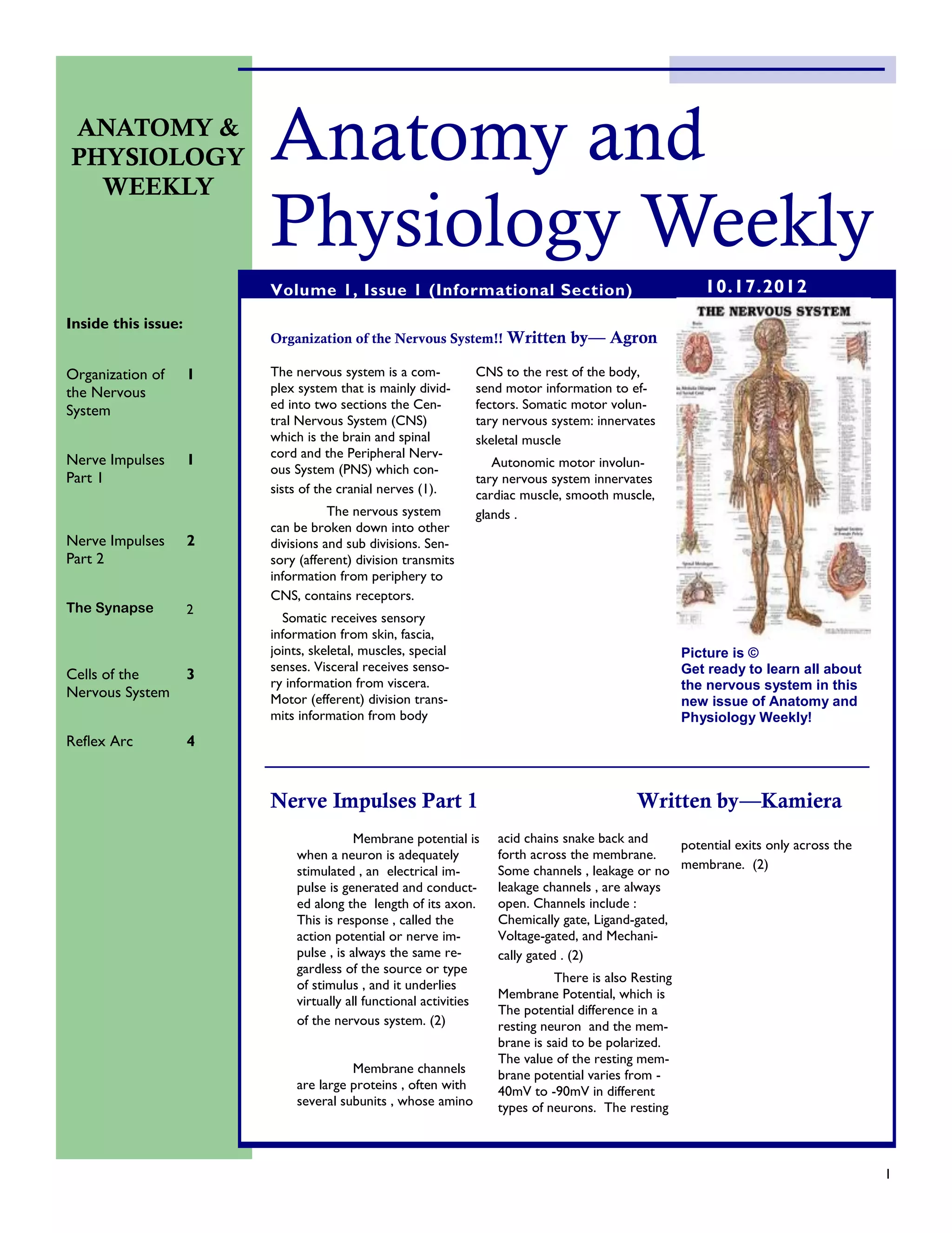
1. It describes the organization of the nervous system into the central and peripheral nervous systems.
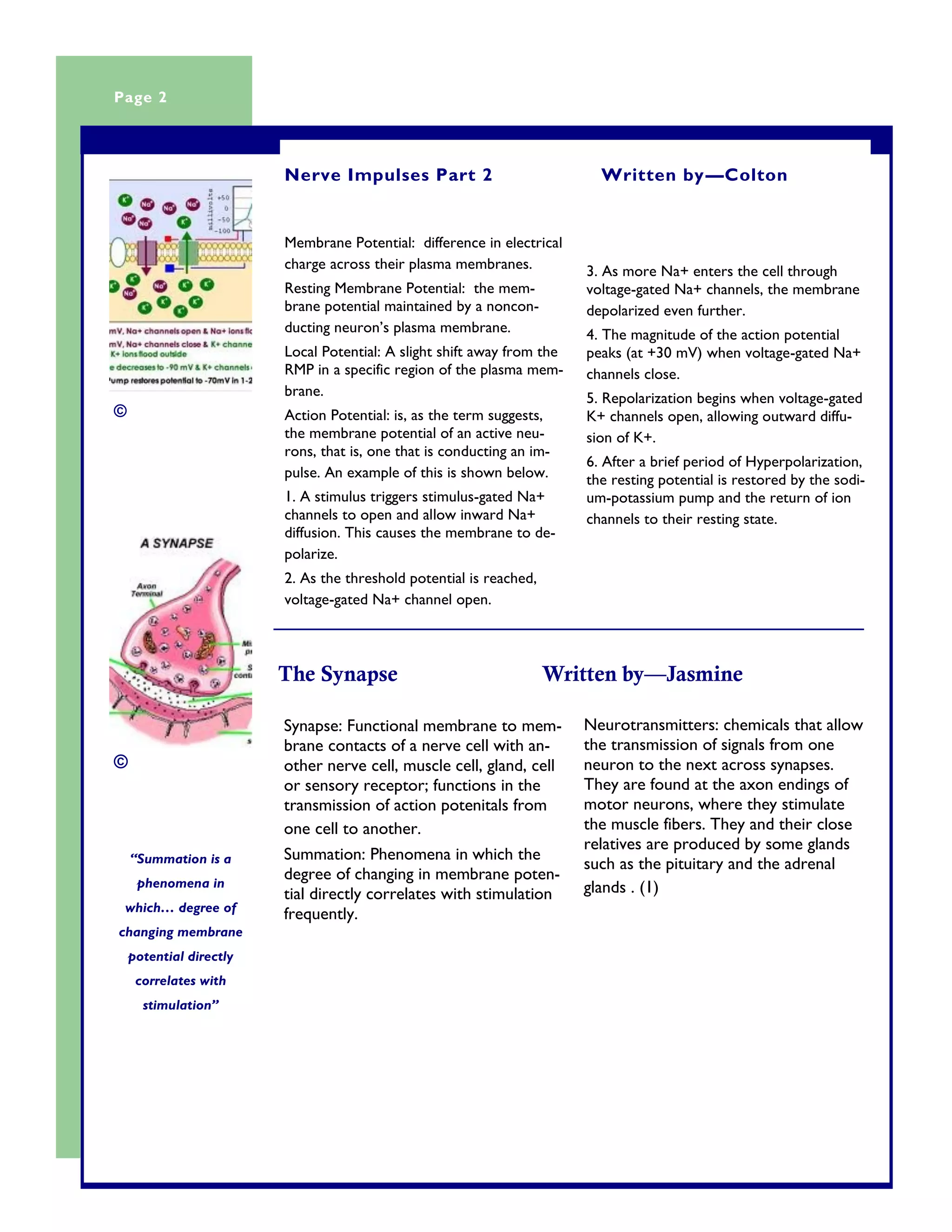
2. It provides two articles summarizing nerve impulses and the synapse.
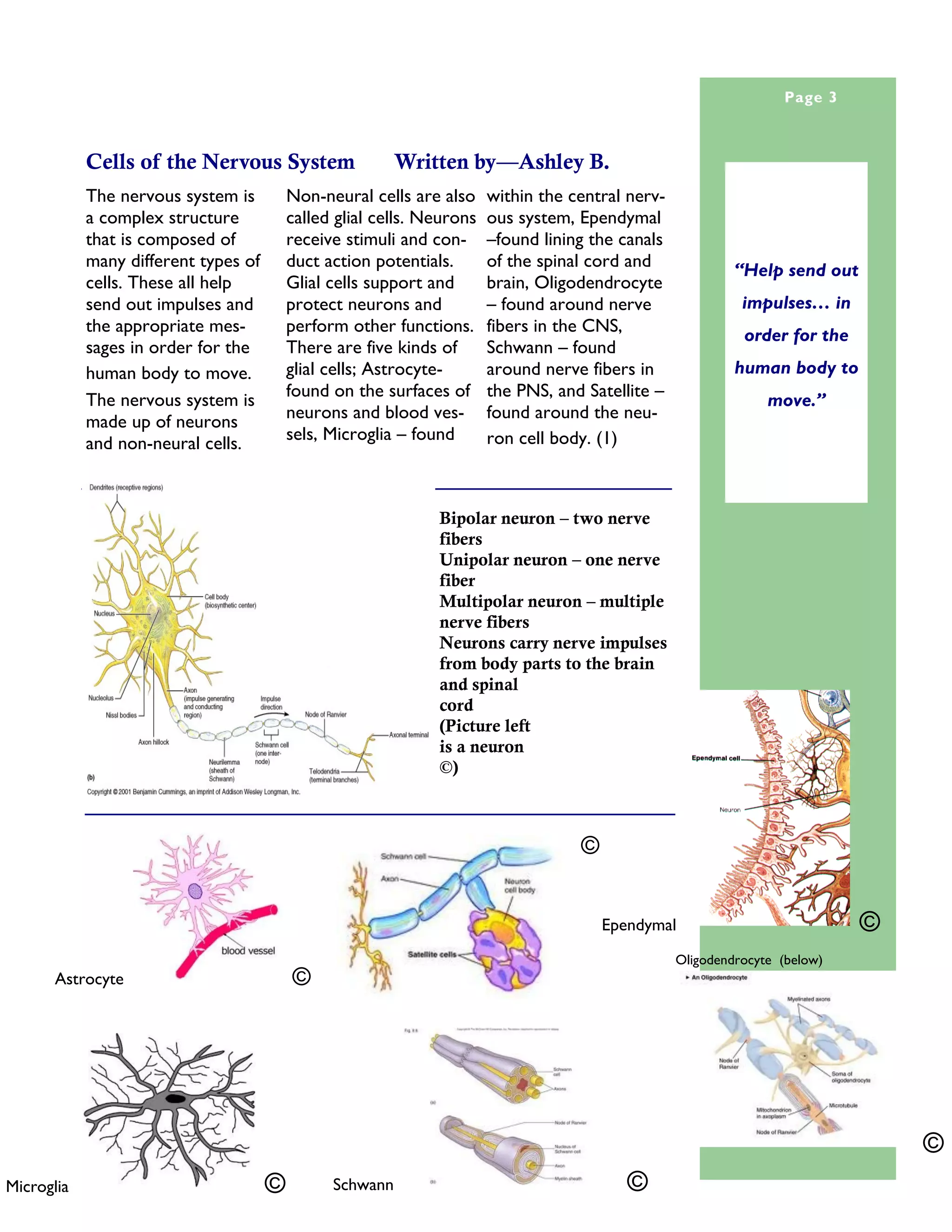
3. It lists the main cell types in the nervous system that help send out impulses to move the human body.
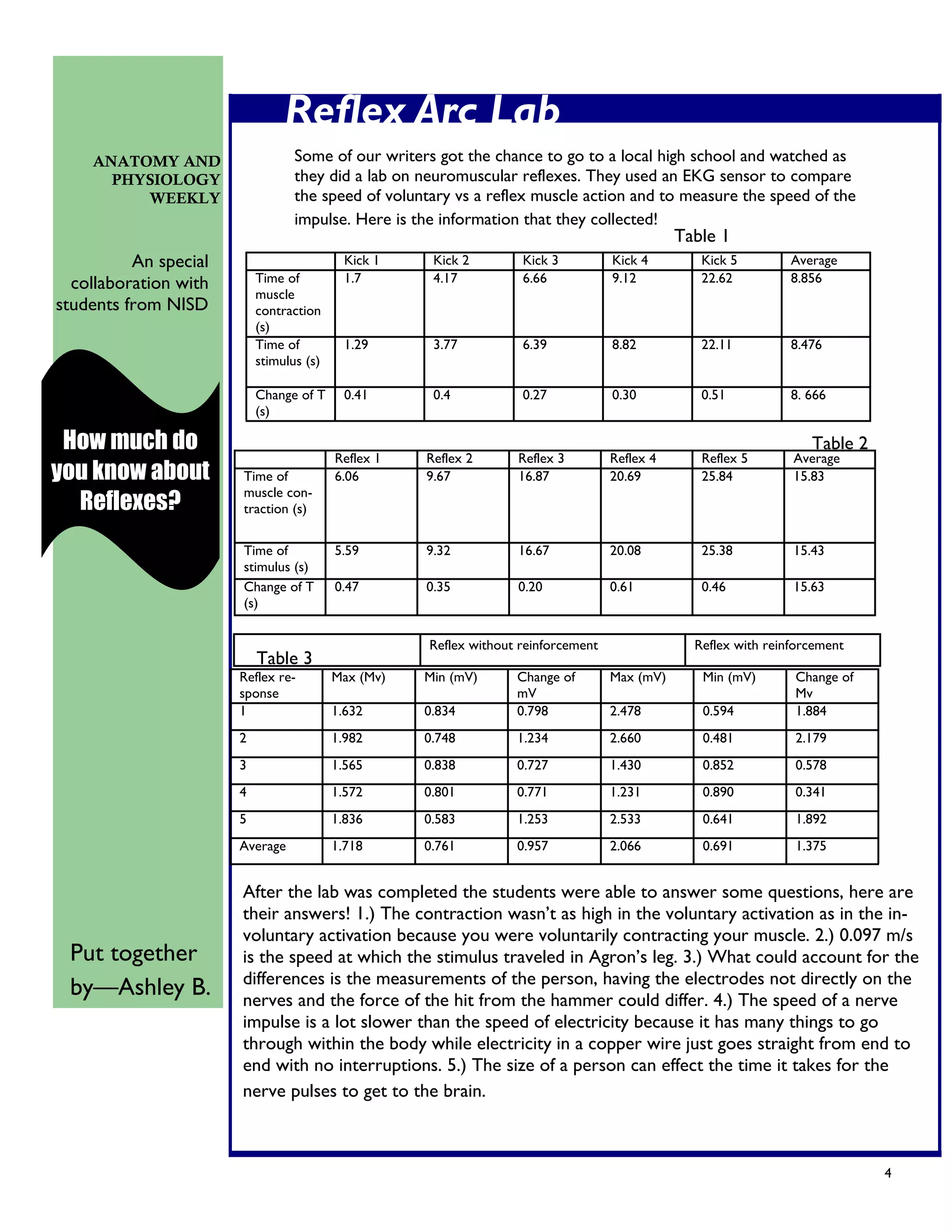
4. It describes a student lab that measured the speed of voluntary kicks versus reflexes using an EKG sensor. The students were able to answer questions about their findings.


![[biurowi 2 - en] identification of threats and occupational risk assessment](https://cdn.slidesharecdn.com/ss_thumbnails/2-identificationofthreatsandoccupationalriskassessment11-110221072313-phpapp01-thumbnail.jpg?width=640&height=640&fit=bounds)


