
Recommended
More Related Content
Similar to INTRODUCTION TO NERVOUS SYSTEM.pptx
Similar to INTRODUCTION TO NERVOUS SYSTEM.pptx (20)
Recently uploaded
Recently uploaded (20)
INTRODUCTION TO NERVOUS SYSTEM.pptx
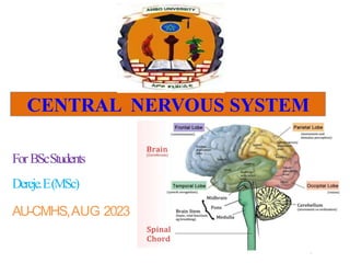
- 1. For BScStudents Dereje.E(MSc) AU-CMHS,AUG 2023 CENTRAL NERVOUS SYSTEM 1
- 2. Divisions of the Nervous System 2
- 4. Overview of the NS… The NS is an intricate, highly organized network of billions of neurons & even more neuroglia. Human brain has an estimated number of 1011 Neurons and 1014 Synapses. Principal cell types that make up the nervous system are: – Neurons & – Neuroglial cells
- 5. Overview function of the NS… Neurons Functional, signal conducting cells specialized for: – Sensory function – Generation of thought – Storage of memory – Integrates idea – Coordinates muscular activities
- 6. 6 Overview of the NS… Neuroglia Supporting cells. 20x outnumber neurons. (the guy to the right had an inordinate amount of them) Can multiply after maturation. Potential causes of glioma. (brain tumour)
- 7. 7 The Neuroglia Non neural cells found in association with neurons. Provide supporting functions to the nervous system. They are: 1. Microglial cells 2. Astrocytes 3. Oligodendrocytes 4. Ependymal cells 5. Schwann cells 6. Satelite cells
- 8. 8 The Neuroglia… 1. Microglial cells Specialized immune cells that act as the macrophages of the CNS. Why is it important for the CNS to have its own army of immune cells? 2. Astrocytes Star-shaped, abundant & versatile. Provide nourishment to the CNS & involved in the formation of the blood brain barrier (BBB).
- 9. 9 The Neuroglia 3. Oligodendrocytes Produce myelin sheath which provides electrical insulation for certain neurons in the CNS. 4. Ependymal cells Low columnar epithelial cells that line ventricles of the brain & the central canal of the spinal cord. Some are ciliated which facilitates the movement of cerebrospinal fluid (CSF).
- 10. 10 Neuroglia…cont’d 5. Schwann cells • Form myelin sheaths around the larger nerve fibers in the PNS. • Vital to neuronal regeneration. 6. Satellite cells • Small cells that line the exterior surface of the PNS. • Regulate the external chemical environment.
- 12. NEURON Defined as the structural and functional unit of NS. Similar to any other cell in the body, having nucleus and all the organelles in cytoplasm. However, it is different from other cells by two ways: 1. Has axon and dendrites 2. Neuron does not have centrosome. 3. So, it cannot undergo division.
- 13. 13 Neurons • There are different types of neurons but most have certain structural and functional characteristics in common: – Cell body (soma) – One or more specialized, slender processes (axons/dendrites) – An input region (dendrites/soma) – A conducting component (axon) – A secretory (output) region (axon terminal)
- 14. 14 Soma • Contains nucleus plus most normal organelles. • Biosynthetic center of the neuron. • Contains a very active & developed rough endoplasmic reticulum (RER) which is responsible for the synthesis of neurotransmitters (NTs). – neuronal RER is referred to as the Nissl body. • Contains many bundles of protein filaments (neurofibrils) which help to maintain the shape, structure, and integrity of the cell.
- 15. 15 Neuronal Processes Clusters of somata in the: – CNS are known as nuclei – PNS are known as ganglia Armlike extensions emanating from every neuron. – Tracts = Bundles of processes in the CNS – Nerves = Bundles of processes in the PNS 2 types of processes that differ in structure and function: – Dendrites and Axons
- 16. 16 Dendrites Thin, branched processes whose main function is to receive incoming signals. Effectively increase the surface area of a neuron to increase its ability to communicate with other neurons. Convey info towards the soma thru the use of graded potentials. Neuronal Processes…
- 17. 17 Axons (Myelinated /unmylinated) Most neurons have single axon (long up to 1m) process designed to convey info away from the cell body. – Originates from a special region of the cell body called the axon hillock. Transmit APs from the soma toward the end of the axon where they cause NT release. Often branch sparsely, forming collaterals. – Each collateral may split into telodendria which end in a synaptic knob, which contains synaptic vesicles- membranous bags of NTs. Neuronal Processes…
- 18. 18
- 19. Neuronal Processes… Axolemma = axon plasma membrane. Surrounded by a myelin sheath, a wrapping of lipid which: – Protects the axon – electrically isolates it and – Increases the rate of AP transmission • This wrapping is never complete. • There are gaps where there is no myelin – these are nodes of Ranvier. • The myelin sheath is made by ________ in the CNS and by _________in the PNS.
- 20. 20 Functional classification of neurons There are three classes of neurons: 1. Sensory (afferent) neurons - conduct impulses from periphery to the center 2. Motor (efferent) neurons - conduct impulses from CNS to the periphery 3. Interneuron (association neurons) - integrative - conduct impulses from sensory to motor area.
- 21. 21 Morphological classification of Neurons • Structurally neurons are classified into 3 classes: 1. Multipolar neurons found in the CNS motor in function 2. Bipolar neurons: found in retina & inner ear sensory in function 3. Unipolar neurons: located in the ganglia of spinal & cranial nerves. sensory in function
- 22. 22 Synapse - The region where there is a transfer of message from a neuron to the effector cell (postsynaptic neuron, muscle). - The junction between 2 cells in which 1 must be a neuron. - Two types: 1. Electrical synapse (direct) & 2. Chemical synapse (indirect)
- 23. 23 Synapse… 1. Electrical Synapse AP is transmitted through a gap junction. Very fast transmittance used for : – escape responses in invertebrates. – coordinated activity of cardiac muscle cells.
- 24. 24 Synapse… 2. Chemical Synapses Chemical messenger (neurotransmitter) is released from a neuron into the synaptic cleft. NT in the synaptic cleft binds to a receptor on the target cell. Acts slower than electrical synapses because the NT must diffuse across the synaptic cleft to bind the receptor. Advantages over electrical synapses = one-way direction of communication; presynaptic cell to postsynaptic cell.
- 25. Synapse… 25
- 26. 26 Synaptic Transmission There are 3 types of synapses 1. Neuro-neuronal junction (between 2 neurons) 2. Neuro-muscular junction (between neuron & muscle) 3. Neuro-glandualr junction (between neuron & gland) There are 3 types of neuroneuronal junctions (axo-dendritic, axo-somatic & axo-axonic junctions) Two modes of transmission (chemical and electrical) One neuron will transmit impulse to another neuron or to a muscle or gland cell by releasing chemicals called neurotransmitters.
- 27. 27 Components of Axo-Somatic synapse 1. Presynaptic terminal contains neurotransmitter (NT) 2. Synaptic cleft contains ECF and Enzymes 3. Postsynaptic neuron contains receptor for the action of NT
- 28. 28 Mechanism of Chemical Synaptic Transmission 1. AP reaches the presynaptic axon terminal of the presynaptic cell & causes V-gated Ca2+ channels to open. 2. Ca2+ rushes in, binds to regulatory proteins & initiates NT release by exocytosis. 3. NTs diffuse across the synaptic cleft and then bind to specific receptors on the postsynaptic membrane & initiate postsynaptic potentials.
- 29. 29 Mechanism of…cont’d 4. NT-Receptor interaction results in either EPSP/IPSP. • When the NT-R combination triggers the opening of ligand gated Na-channels, this leads to membrane depolarization, EPSP. e.g. Ach on Nicotinic receptor • When the NT-R combination triggers the opening of ligand gated K or Cl-channels, this leads to membrane hyperpolarization, IPSP. e.g. GABA on GABAb receptor
- 30. 30 Excitatory Vs Inhibitory Synapses 1. Excitatory - more likely to have action potential - depolarization 2. Inhibitory - less likely to have action potential - hyperpolarization - membrane stabilization
- 31. 31 1. Excitatory Synapses • Depolarizes postsynaptic cell – brings membrane potential closer to threshold by opening or closing ion channels • Opens channels that are equally permeable to Na and K – causes depolarization because of the stronger force of Na to flow into the cell. • Depolarization=EPSP (excitatory postsynaptic potential)
- 32. 32 2. Inhibitory Synapses • Neurotransmitter binds to receptor, channels for either K or Cl open hyperpolarizes the cell • If K channels open – K moves out IPSP (inhibitory postsynaptic potential) • If Cl channels open, either – Cl moves in IPSP – Cl stabilizes membrane potential
- 33. 33 Neurotransmitter Removal Why do we want to remove ACh from the neuro-muscular junction? How was ACh removed from the NMJ? NTs are removed from the synaptic cleft via: – Enzymatic degradation – Diffusion – Reuptake
- 34. 34 Properties of synaptic transmission Unidirectional conduction Synaptic delay (0.5 -1.0m/s) Fatigue - Decrease in response of postsynaptic neurons after repetitive stimulation by the presynaptic neurons Synaptic potentiation (facilitation) - Increase in postsynaptic responses caused by previous post synaptic stimulation
- 35. 35 PH - Alkalosis ↑ Synaptic transmission - Acidosis ↓ Synaptic transmission Hypoxia ↓ Synaptic transmission Drugs - Caffeine, Theophylline, Theobromine ↑Synaptic transmission - Strychnine ↑ Synaptic transmission - Hypnotics, Anesthetics, tranquilizers ↓ Synaptic transmission Factors Affecting Synaptic transmission
- 36. Nervous System Controls all activities of the body. It is quicker than other control system. Primarily, nervous system is divided into two parts: 1. Central nervous system 2. Peripheral nervous system.
- 37. „ Central Nervous System CNS includes brain and spinal cord. It is formed by neurons and supporting cells called neuroglia. The CNS contains more than 100 billion neurons. Brain and spinal cord are arranged in two layers, – Gray matter consists of somata, dendrites, and unmyelinated axons. – White matter consists primarily of myelinated axons.
- 38. Cont.…… In brain, white matter is placed in the inner part and gray matter in outer part. In spinal cord vis versa, Brain is situated in the skull. It is continued as spinal cord in the vertebral canal through the foramen magnum. surrounded by three layers of meninges – Dura mater – Arachnoid mater and – Pia mater
- 39. Cont.… The space between arachnoid mater and pia mater is known as subarachnoid space. This space is filled with a fluid called cerebrospinal fluid (CSF).
- 40. 40 Brain Regions Cerebrum Diencephalon - Thalamus - Hypothalamus Brainstem - Midbrain - Pons - Medulla ob. Cerebellum Cerebellum
- 41. Spinal cord lies loosely in the vertebral canal. It foramen magnum up to the lower border of first lumbar vertebra. Covered by meninges – Pia matter – Dura matter and – Arachnoids
- 42. Spinal Cord The spinal cord has two functions: 1. Common passageway for ascending and descending tracts. Neurons in the white matter of the spinal cord transmit sensory signals from peripheral regions to the brain motor signals from the brain to peripheral regions. 2. Centre for Spinal Cord reflexes. Neurons in the gray matter of the spinal cord integrate incoming sensory information and respond with motor impulses that control muscles (skeletal, smooth, or cardiac) or glands. 42
- 43. Spinal cord Ascending fibers of spinal cord Anterior spinothalamic tract – crude touch like itching and tickling Lateral spinothalamic tract – sensations of pain and temperature Ventral spinocerebellar – subconscious kinesthetic sensation (proprioceptive impulses from muscles, tendons and joints)
- 44. Spinal cord Dorsal spinocerebellar tract – subconscious kinesthetic sensation – This tract is uncrossed – Lesion affects on the same side Spinotectal tract is concerned with – spinovisual reflex Fasciculus gracilis and fasciculus cuneatus – are together called ascending posterior column tracts. – Fasciculus gracilis contains the fibers from lower extremities and lower parts of the body – Fasciculus cuneatus contains fibers from upper part of the body, – Both terminate in the medulla oblongata
- 45. Spinal cord Tracts of the posterior white column convey impulses of following sensations: – Fine (epicritic) tactile sensation – Tactile localization – Tactile discrimination – Sensation of vibration – Stereognosis and – to differentiate the weight of different objects
- 46. Spinal cord Descending tracts These tracts carry motor impulses from brain to spinal cord. Descending tracts of spinal cord are of two types: A. Pyramidal tracts cerebral cortex towards spinal cord For voluntary motor activity B. Extrapyramidal tracts. position of head and body during angular and linear acceleration. maintenance of muscle tone, respiration and diameter of blood vessels reflex movements
- 47. 47 Brain Regions Cerebrum Diencephalon - Thalamus - Hypothalamus Brainstem - Midbrain - Pons - Medulla ob. Cerebellum Cerebellum
- 48. Brainstem Is the part of brain formed by; – medulla oblongata – pons and – midbrain. Contains; – ascending and descending tracts between brain and spinal cord – centers for regulation of vital functions
- 49. Medulla oblongata Is the lowermost part of brain. Functions of medulla – Respiratory centers, which maintain normal rhythmic respiration. – Vasomotor center controls blood pressure and heart rate. – Deglutition center regulates the pharyngeal and esophageal stages of deglutition. – Vomiting center induces vomiting – Salivatory nuclei control the secretion of saliva – Nuclei of 12th, 11th, 10th cranial nerves are located in the medulla oblongata.
- 50. Pons A bridge between medulla and mid brain Nuclei of 8th, 7th, 6th and 5th cranial nerves are located in pons Connects cerebellum with cerebral cortex. Pneumotaxic and apneustic centers for regulation of respiration
- 51. MIDBRAIN Lies between pons and diencephalon. It consists of two parts: A. Tectum B. Cerebral peduncles. Tectum is formed by two structures: 1. Superior colliculus-light reflex 2. Inferior colliculus-auditory reflexes
- 52. 52 Diencephalon • Forms the central core of the forebrain • 3 paired structures: 1. Thalamus 2. Hypothalamus 3. Epithalamus All 3 are gray matter
- 53. 53 Thalamus… – Composes 4/5 of the diencephalon. – Forms most of the walls of the 3rd ventricle. – Acts as relay center through which all sensory information (except olfactory) passes to the cerebrum. • Lateral geniculate nuclei: –Relay visual information. • Medial geniculate nuclei: –Relay auditory information. • Intralaminar nuclei: –Activated by many sensory modalities. –Projects to many areas. »Promotes alertness and arousal from sleep.
- 54. Hypothalamus The hypothalamus (hypo- under) is the small portion of the diencephalon that lies below the thalamus and above the pituitary gland. Although its size is small, the hypothalamus controls many important body activities, most of them related to homeostasis. The chief functions of the hypothalamus are as follows:-
- 55. 55 Function of Hypothalamus • Controls the ANS • Influences HR, BP, resp. rate, GI motility, pupillary diameter. • Can you hold your breath until you die? Anterior nuclei acts as a parasympathetic center Posterior nuclei acts as a sympathetic center • Endocrine function – Controls adenohypophyseal hormones Releases hormones that influence hormonal secretion from the anterior pituitary gland. – Controls neurohypophyseal hormones Releases oxytocin and vasopressin – Controls adrenal medulla
- 56. Function of Hypothalamus • Regulation of body temperature The heat losing center (anterior HT) Heat gaining center (posterior HT • Contributes to the regulation of sleep, wakefulness, emotions, sexual arousal, anger, fear, pain, and pleasure. • Controls food intake (hunger sensation): Feeding center (lateral HT), Satiety center (ventromedial HT) 56
- 57. 57 Function of Hypothalamus Control of water-electrolyte balance Thirst center (lateral HT, OVLT) Osmoreceptors (anterior HT, SFO) Control of sexual behavior: libido, sexual activities are controlled by cerebral cortex, limbic system and HAT. Relation to sleep: Lesion to posterior HT leads to somnelence. Hypothalamic neurons project on RAS where sleep center is located. Effect of HT lesion: Diabetes inspidus, hypo-/hyperthermia, sleep disturbance, hormonal disturbance, hyperphagia, emotional diturbance
- 58. 58 Epithalamus • Located above the thalamus. • Contains the pineal gland which releases melatonin. Because the pineal gland secretes the hormone melatonin, it is part of the endocrine system. Melatonin promotes sleepiness and contributes to the setting of the body’s biological clock. • Contains a structure called the habenula – involved in food and water intake.
- 59. 59 Cerebellum Lies inferior to the cerebrum and occupies the posterior cranial fossa. 2nd largest region of the brain. 10% of the brain by volume, but it contains 50% of its neurons.
- 60. 60 Cerebellum The 2 cerebellar hrs. are separated by a shallow groove called vermis In the vermis, most motor function of cerebellum controlling movt of axial body; neck, shoulder, & lips are located. The intermediate zone of cerebellar h. controls muscle movt of upper and lower limbs. The lateral zone of cerebrall h. controls timing & planning of sequential motor movts. Anterior lobe Posterior lobe Flocculnodular lobe Vermis Primary fissure
- 61. 61 Functional Parts of Cerebellum 1. Vestibulocerebellum/archicerebellum: It is the oldest part of the cerebellum It consists of flocculonodular lobe It is mainly connected to the vestibular apparatus Function: controls equilibrium and posture 2. Spinocerebellum/paleocerebellum: It comprises vemis and paravermal (medial) parts It receives signal from muscle spindle and Golgi tendon organs Function: it is concerned mainly with control of muscle tone 3. Cerebrocerebellum/pontocerebellum/neocer. It includes the lateral cerebellar hemispheres It is the newest part, connected to cerebrum Function: Concerned w/t control of skilled voluntary movts initiated by cerebral cortex
- 62. 62 Afferent Cerebellar Connections A. From the brain 1. Tecto-cerebellar fibers: originate from the tectum (sup. & inf. Colliculus) in the MB --- SCP --- different parts of the cerebellum. Transmit visual and auditory signals to the cerebellum. 2. Cortico-ponto-cerebellar fibers: Started from motor, somatosensory & association areas---pontin relay nuclei--- MCP---contra lateral cerebro-cerebellum. They transmit signals from the cerebral cortex to the cerebellum to produce intended motor plan of movt. 3. Olivo-cerebellar pathway: Originates from the inf. Olive--- ICP---to all parts of cerebellum. Inf. Olive is stimulated by fibers from the motor cortex, BG, RF & the spinal cord about muscle tone and movts.
- 63. 63 Afferent Cerebellar connections (cont´d) 4. Vestibulo-cerebellar tract: originates from the vestibular apparatus--- vestibular nuclei---ICP---vestibulocerebellum. Transmits signals about body posture and equilibrium. 5. Reticulo-cerebellar pathway: Originates from the different parts of RF---MCP & ICP---Spinocerebellum (vermis). Transmits signals of various sensations particularly muscle tone and movts.
- 64. 64 Function of Cerebellum 1. Control of posture and equilibrium It is the function of the vermis and archicerebellum Cerebellum compares signals coming from the vestibular apparatus and proprioceptive signals from periphery to maintain posture and equilibrium. 2. Control of muscle tone Generally the neocerebellum is facilitatory to muscle tone, while the paleocerebellum is inhibitory. The former is dominant. Cerebellar output signals through reticulspinal tract, vestibulospinal tract, rubrospinal tract increase muscle tone.
- 65. Function of Cerebellum 65 3. Control of voluntary movt Cerebellum influences voluntary movt through the following functions: Planning: Cerebrocerebellum is concerned with the intention & plan of movt. Timing of movt: Cerebellum determines the start and termination of sequential movts. Damping of movt: ending of movt without osscillation. Ballistic movt: rapid & short movts such as typing.
- 66. 66 Cerebellar Syndrome Produced by lesion to the cerebellar nuclei. Cerebellar syndrome appeared on the same side of the lesion. There are three main types 1. Atonia/Atetonis/Hypotonis: marked decrease in muscle tone due to loss of the excitatory effects of dentate nucleus and interposituse nuclei on muscle tone. Manifestation: Flaccid feel of muscle and Pendular kneejerk 2 Asthenia: Lack of strength Manifestation: Muscle weakness due to difficulty in initiating and maintaining muscle contraction. 3. Ataxia: Incoordination of voluntary movts. Cerebellar ataxia is manifested by:-
- 67. 67 Cerebellar syndrome (cont´d) • The cerebellum can be permanently damaged by trauma or stroke or temporarily affected by drugs such as alcohol. • These alterations can produce ataxia – a disturbance in balance.
- 68. 68 Cerebrum • Largest portion of brain (80% mass). • Most developed in man • Responsible for higher mental functions, concerning perception of Fine sensation Learning Memory Speech Judgment and planning.
- 69. Cerebral cortex It consists of two hemispheres. Connected by Corpus callosum Surface of the cerebral cortex is characterized by sulci and gyri – Sulcus is a slight depression – groove and gyrus is a raised ridge. Cerebral cortex consists of gray matter that surrounds the deeper white matter.
- 70. Lobes of cerebral cortex 1. Frontal 2. Parietal 3. Occipital 4. Temporal lobe.
- 71. Lobes of cerebral cortex Lobes of each hemisphere are demarcated by four main fissures and sulci: 1. Central sulcus- between frontal and parietal lobes 2. Parieto-occipital sulcus- between parietal and occipital lobe 3. Sylvian fissure or lateral sulcus- between parietal and temporal lobes 4. Callosomarginal fissure -between temporal lobe and limbic area.
- 72. Cerebral hemisphere Right hemisphere is called representational hemisphere it is associated with; – Artistic and visuospatial functions like; – Judging the distance, – Determining the direction, – Recognizing the tones, etc. Lesion in representational hemisphere causes only mild effects like astereognosis.
- 73. Cerebral hemisphere Left hemisphere is the dominant hemisphere About 75% of the right-handed persons. Lesion in dominant hemisphere leads to language disorders.
- 74. Wernicke’s area When Wernicke's area in the dominant hemisphere of an adult person is destroyed:-the person normally loses almost all intellectual functions associated with language or verbal symbolism. Such as:- the ability to read, the ability to perform mathematical operations, and even the ability to think through logical problems. – Lateral prefrontal cortex is also used for language comprehension and complex word analysis 74
- 75. 75 Broca’s Area • Typically found in only one hemisphere (often the left), anterior to the inferior portion of the premotor cortex. • Directs muscles of tongue, lips, and throat that are used in speech production. • Involved in planning speech production and possibly planning other activities. • Involves articulation of speech. • In damage, comprehension of speech is impaired.
- 76. 76 Memory Memory is the ability of the brain to store information and recall it at a later time. It was calculated that 10 neurons are required to store 1 bit of information The total storage capacity of the human brain is about 3x108 bits Medial temporal lobe: Consolidates short term into long term memory. Hippocampus is critical component of memory. Acquisition of new information, facts and events requires both the medial temporal lobe and hippocampus.
- 77. 77 Types of Memory There are 4 types of memories I. Sensory memory (immediate memory) II. Primary memory (short-term memory) III. Secondary memory (long-term memory) IV. Tertiary memory (Permanent memory) Sensory memory It is the storage of sensory info for few seconds Forgetting starts immediately after the info is acquired. A gradual decline in the amount of info is called fading of info. The spontaneous disappearance of info from the memory is called extinction of info. Infos in sensory memory can be transferred into primary or secondary memory.
- 78. 78 Types of memory (cont´d) Primary memory This is a memory that lasts from a few minutes to few Hrs Info enters this memory by verbalization, ie. Describing the items in words. Primary memory is not stored in infants and animals The capacity of primary memory is small, but rate of retrieval is rapid.
- 79. Types of memory (cont´d) Secondary memory This is a memory that lasts for Hrs, days or years. Info is introduced into this memory by two means:- 1. From the sensory memory, through stimulation of reward or punishment system. 2. From the sensory and primary memories by practice or rehearsal, ie. Attentive repetition of information or experience. The capacity of secondary memory is very large Information are stored according to their significance Retrieval is time taking Forgetting of info in the secondary memory occurs through interference, by previously stored info (pro active inhibition) or subsequently stored info (retro active inhibition). 79
- 80. 80 Types of memory (cont´d) Tertiary memory This is the permanent memory. The info stored never forgotten. eg. One´s name, ability to read and write Infos in the tertiary memory comes from secondary memory by years of practice, which consolidates memory. Tertiary memory can not be erased by brain injury and diseases. Access to retrieve tertiary memory is rapid
- 81. 81 Characteristics of different types of memories Characters Sensory Primary Secondary Tertiary Capacity: Very small Small Very large Large Duration: Few seconds Several min-hrs Several hrs-yrs Permanent Entry into Automatic duringVerbalization Practice, reward/ Frequent Storgae: perception punishment practice Rate of retrieval: Very rapid Rapid Slow Very rapid Type of Info: Sensory Verbal All forms All forms Mechanis of Synaptic Long-term Structural and functional Storage: potentiation potentiation modification of memory traces Mehanism of Fading & New info Proactive or retro- No forgett- Forgetting extinction replaces the old active inhibition ing
- 82. 82 Memory Disordes Amnesia (Gr = forgetfulness) = It means that inability to remember past experiences. Types of amnesia: 1. Retrograde amnesia: 2. Antrograde Retrograde amnesia: Inability to recall events occurred shortly before the onset of brain malfunction without affecting past memories. It occurs due to brain concussion (post-traumatic amnesia), anesthesia, ECT.
- 83. Memory Disordes (cont´d) Antrograde amnesia: Inability to form new memories. Consolidated memories before the onset of amnesia are retained. Primary memory is functional, but not consolidated. Caused by bilateral lesion to hippocampus and related structures involved in memory encoding. Pschogenic or hysterical amnesia A rare condition chara/zed by sudden loss of memory of all info in the secondary and tertiary memories. It is purely functional disorder without any organic disease. 83
- 84. 84 Memory Disordes (cont´d) Alzheimer's disease and Senile dementia AD is caused by degeneration of the cholinergic nerve fibers in the limbic system (basal forebrain, amygdala, and hippocampus). The disease is chara/zed by deterioration of intellectual abilities as impairment of memories, lack of judgment and inattentiveness The disease occurs at any age. In old age, it is called senile dementia An anticholinesterase drug, physiostegmin (eserine) produces improvement but does not stop progression of the disease.
- 85. Cerebrospinal fluid Is the clear, colorless and transparent fluid. It circulates through ventricles of brain, subarachnoid space and central canal of spinal cord. It is a part of extracellular fluid (ECF). SITE OF FORMATION CSF is formed by choroid plexuses situated within the ventricles. Choroid plexuses are tuft of capillary CSF is formed by the process of secretion that involves active transport mechanism.
- 87. „Functions of cerebrospinal fluid 1. Protective Function CSF acts as fluid buffer acts like a cushion (countercoup injury). 2. Medium of Exchange 3. Diagnostic purpose 4. To remove weast products