Download as PDF, PPTX
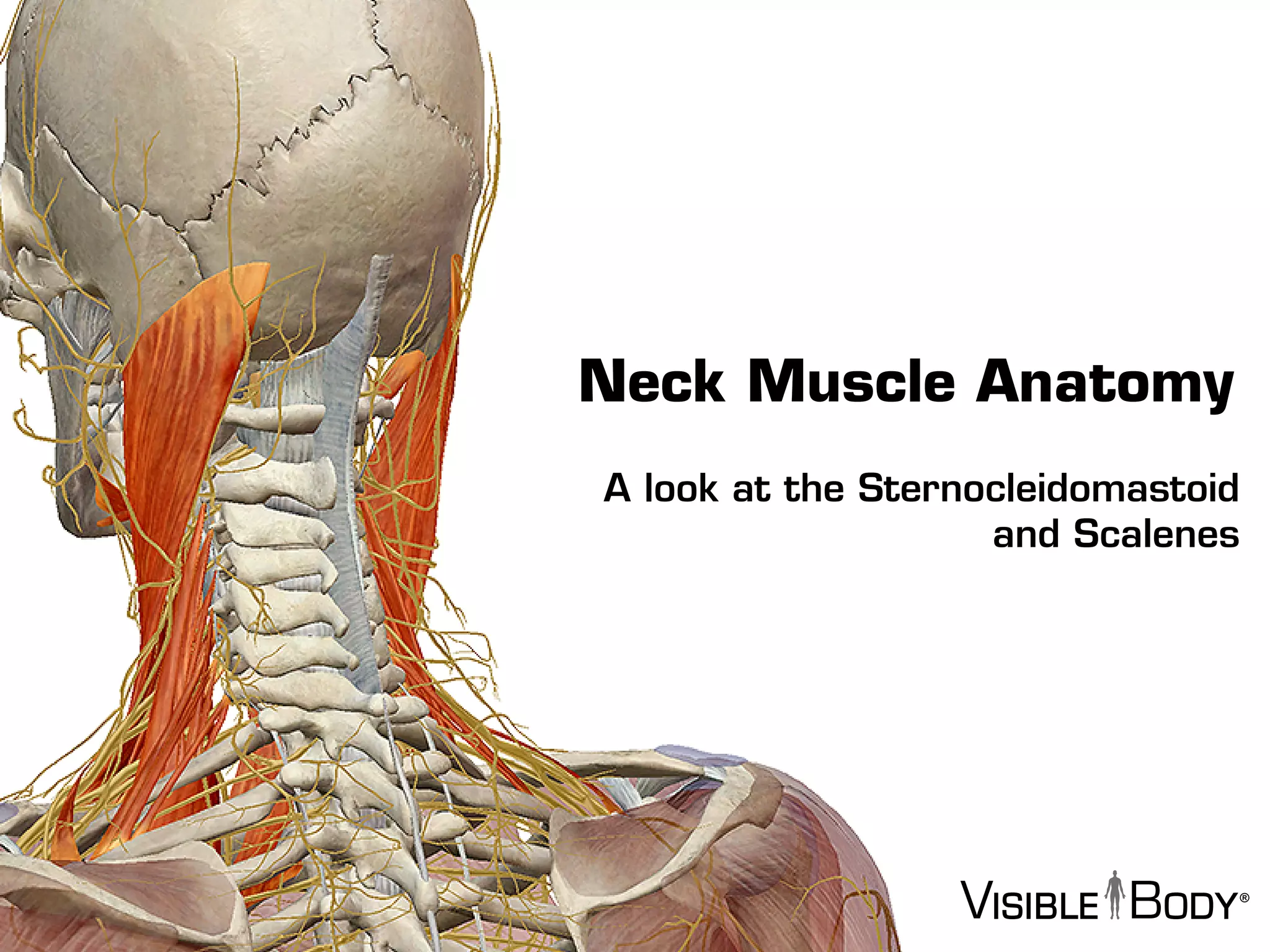


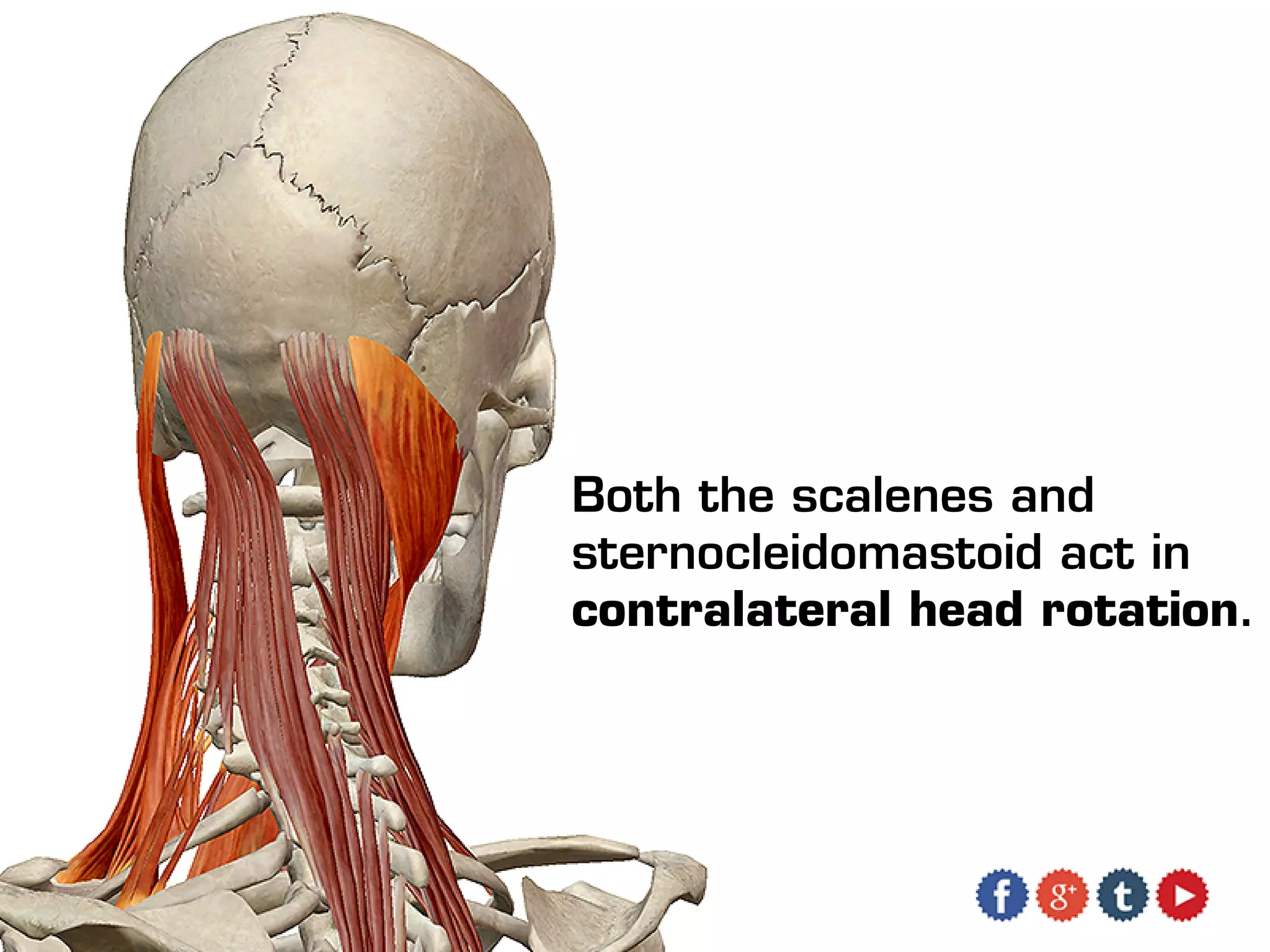


The document provides an overview of neck muscle anatomy, focusing on key muscles such as the sternocleidomastoid and scalenes, which are vital for head movement and upper body stabilization. It details the innervation of these muscles and their specific functions, including lateral neck flexion and head rotation. Additionally, it promotes the Visible Body's Muscle Premium app as a resource for further anatomical exploration.


