
Download to read offline

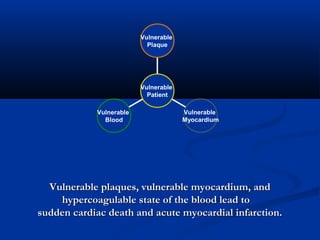
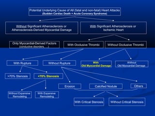
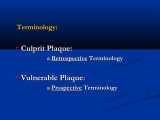
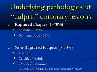
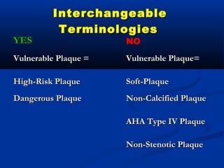
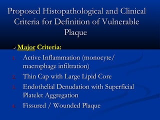
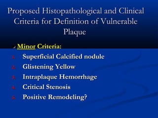
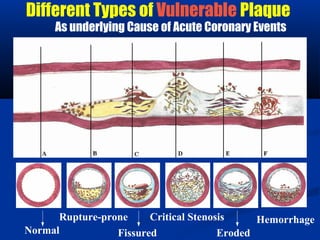
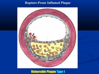
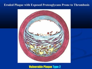
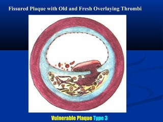
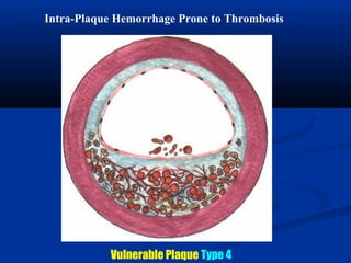
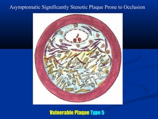
This document provides guidelines for defining vulnerable plaques and vulnerable patients from the Association for Eradication of Heart Attack. It outlines major and minor histopathological and clinical criteria for vulnerable plaques, including active inflammation, thin fibrous cap with large lipid core, endothelial denudation, and stenosis. Potential screening and diagnostic methods are discussed at the plaque, systemic, and blood levels. Various types of vulnerable plaques that can cause acute coronary events are also described.


