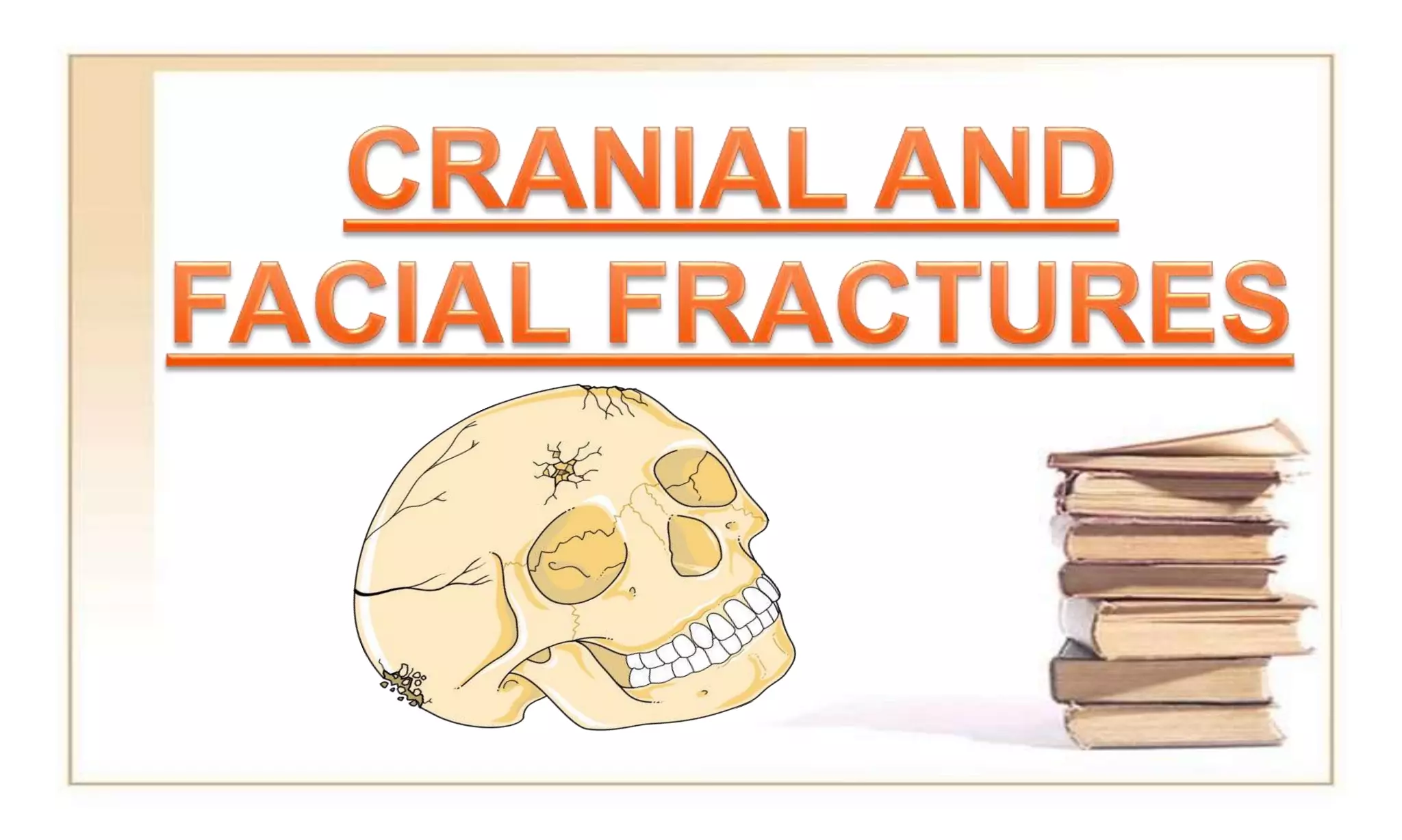
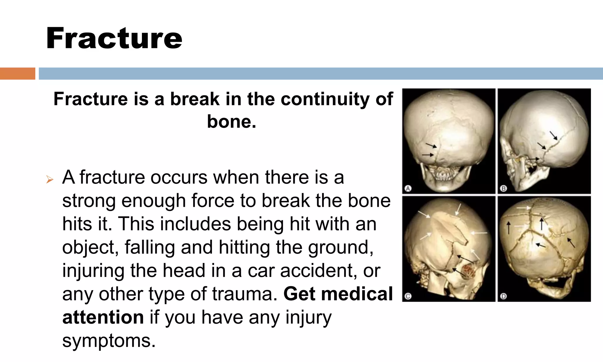
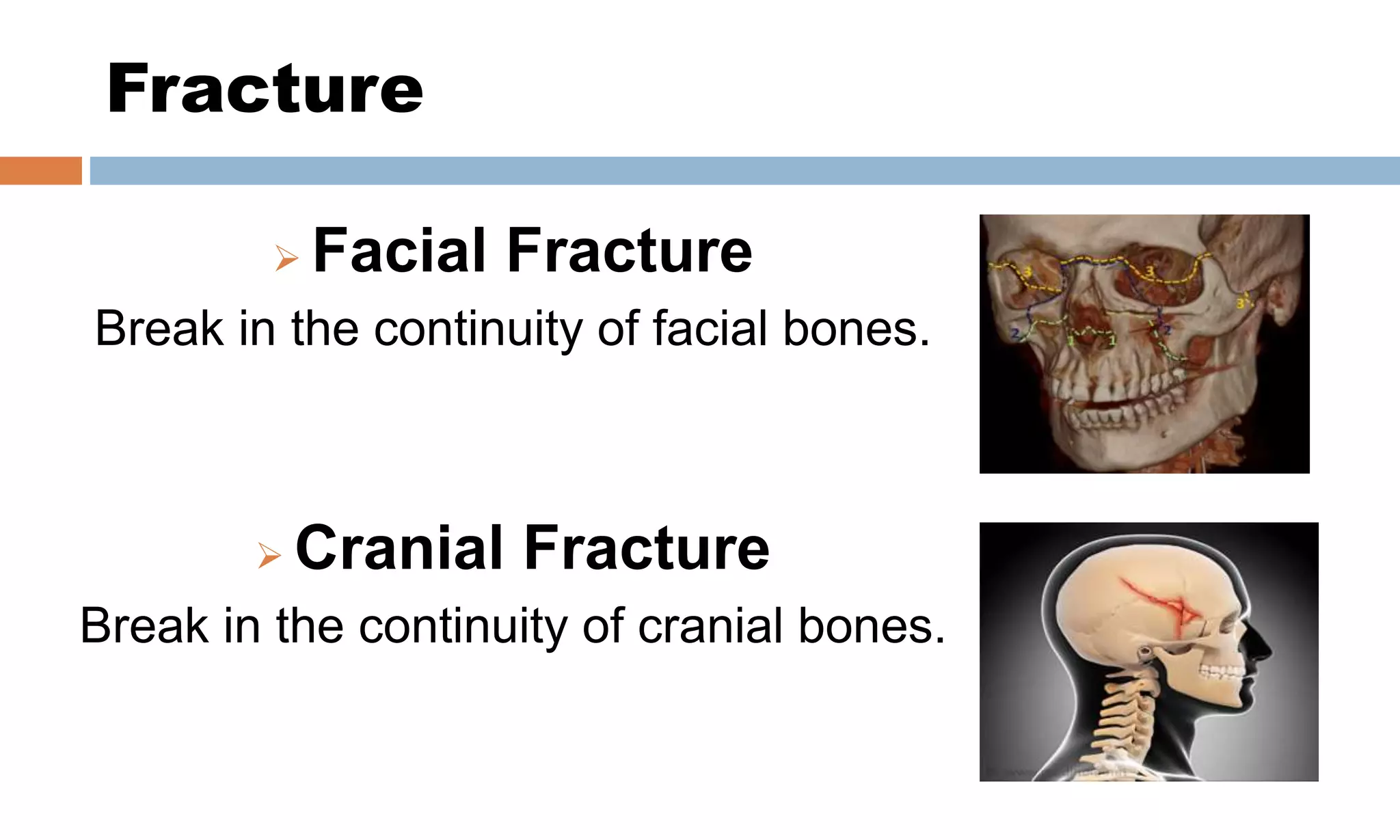
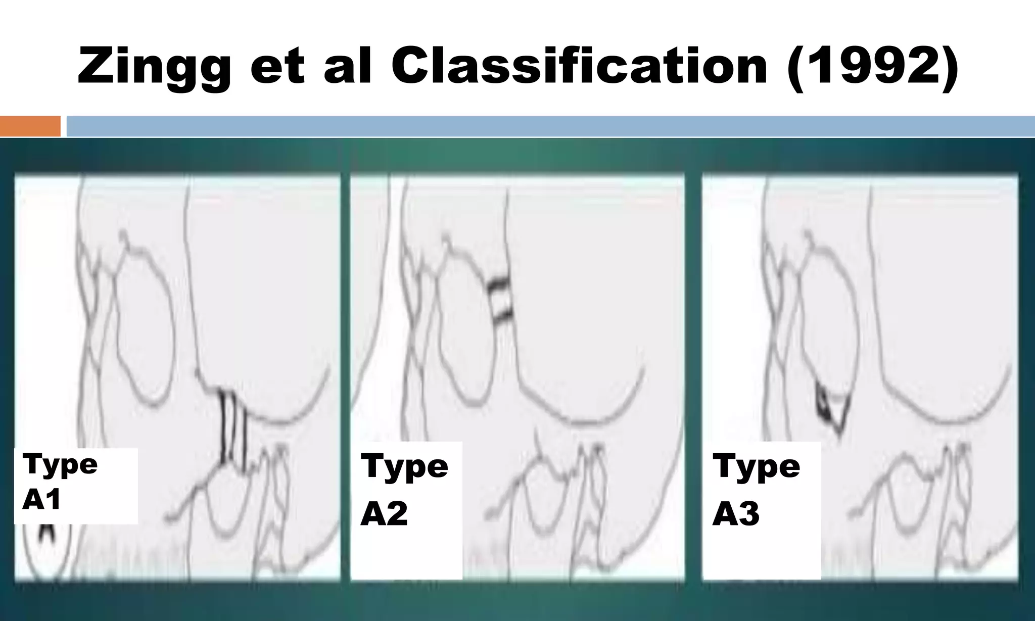
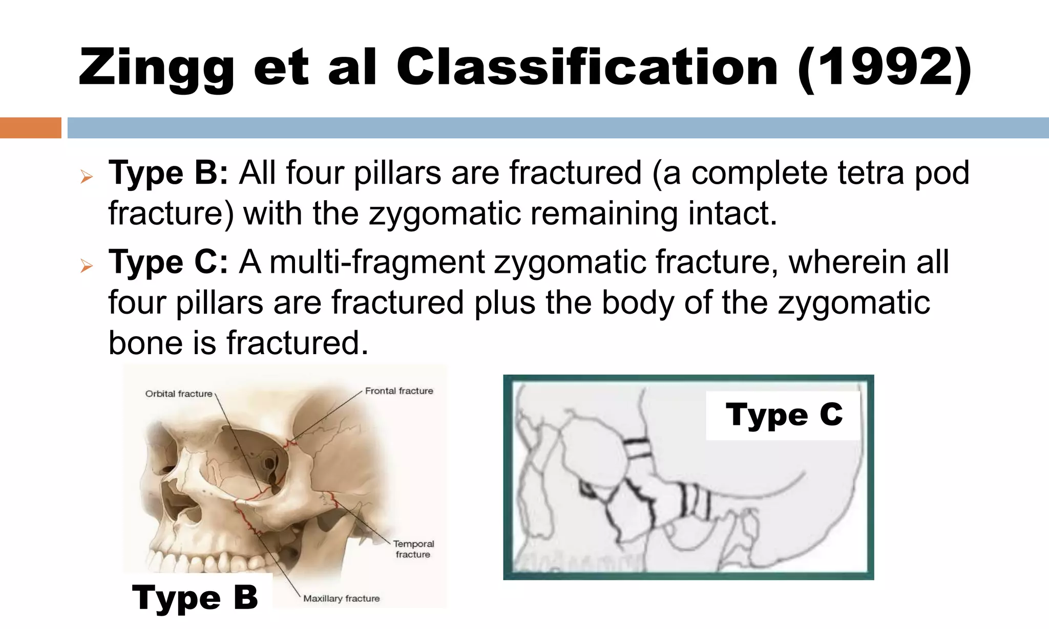
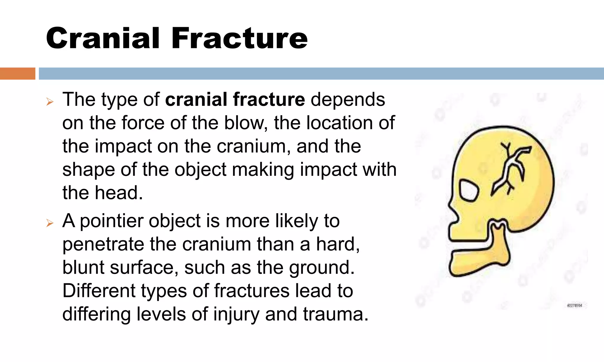
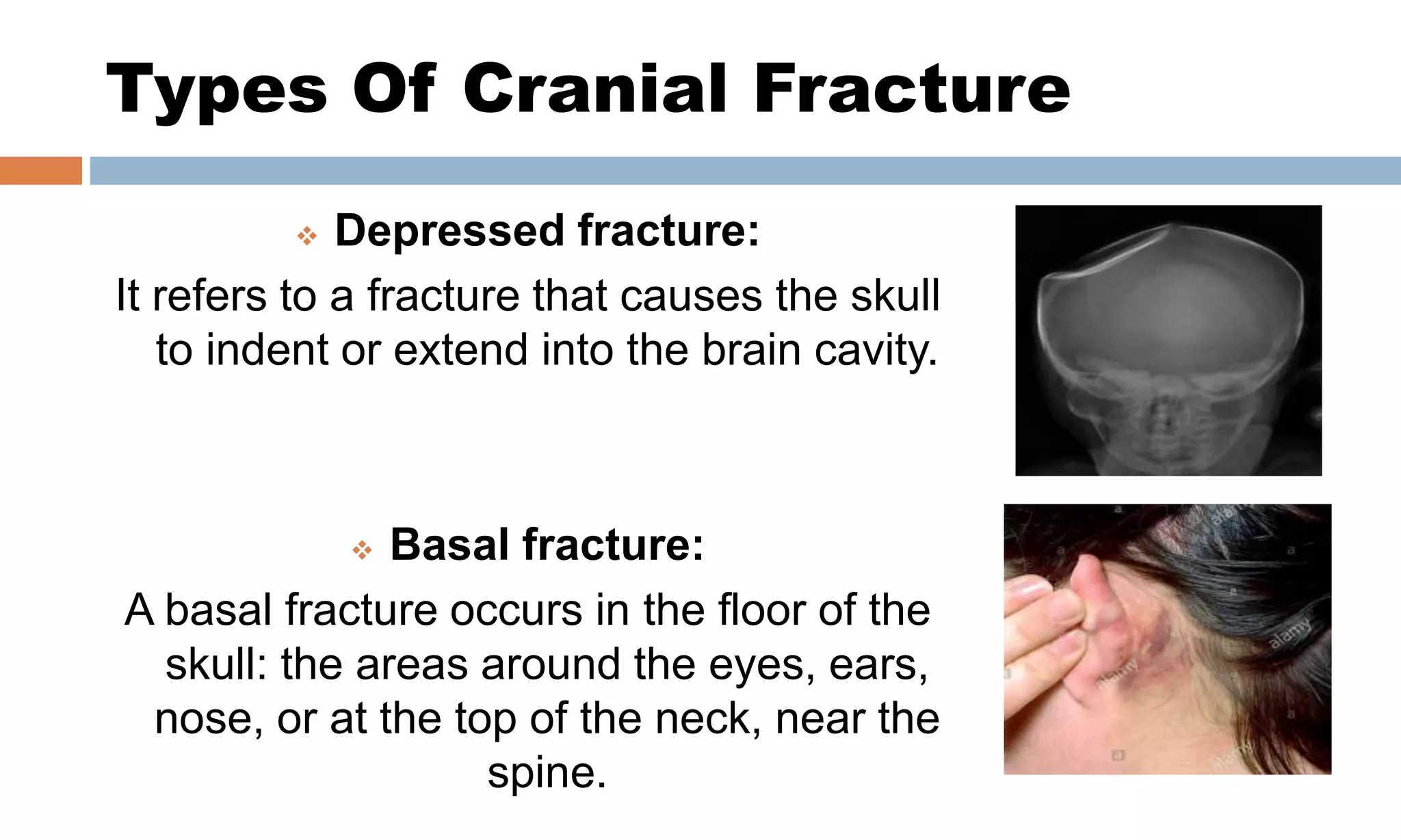
Fracture
Fracture is a break in the continuity of
bone.
 A fracture occurs when there is a
strong enough force to break the bone
hits it. This includes being hit with an
object, falling and hitting the ground,
injuring the head in a car accident, or
any other type of trauma. Get medical
attention if you have any injury
symptoms.
Fracture
 Facial Fracture
Break in the continuity of facial bones.
 Cranial Fracture
Break in the continuity of cranial bones.
Facial Fracture
 Face consists of complex bone structure. If their is a break
in the continuity of facial bones then it leads to facial
fracture.
 Facial bones consists of;
1. Frontal bone (forehead).
2. Zygomatic bone (cheekbones).
3. Orbital bones (eye sockets).
4. Nasal bones.
5. Maxillary bones (upper jaw).
6. Mandible (lower jaw).
Frontal bone
(fore head)
Nasal Bone Fracture
 Nasal bone fractures are the most common type of facial
fracture. The nasal bone is made up of two thin bones. It
takes less force to break the nasal bones than other
facial bones because they are thin and prominent.
Usually, the nose looks deformed or feels sore to the touch
after a fracture.
 Symptoms may include bleeding, swelling, bruising, and
an inability to breathe through the nose. Swelling in the
area might make it more difficult to assess how much
damage has occurred. Nosebleeds and bruising around the
nose are common symptoms of a nasal fracture.
Nasal Bone Fracture Classification
Nasal Bone Fracture
 Treatment is typically with pain medication and cold
compresses. Reduction, if needed, can typically occur after
the swelling has come down. Depending on the type of
fracture reduction may be closed or open. Outcomes are
generally good.
Mandible Bone Fracture
 The mandible holds the lower teeth in
place and moves when you are talking
or chewing. Fractures of the lower jaw
affect the sections of the lower jaw
that supports teeth (called the body),
the part where the jaw curves
upwards into the neck (the angle) or
the knob-shaped joint at the top of the
jaw bone (the condyle) or the point
where the two sides of the lower jaw
are joined (the symphysis).
Mandible Bone Fracture
 If you have a break in the lower jaw, you may also have
broken or loose teeth.
 Treatment: There are frequently several appropriate
techniques available for the
definitive management of a given mandible fracture. In
general, mandible fractures are treated either;
1) Closed fracture includes maxillomandibular fixation,
splinting and modified diet.
2) Open fracture includes plates and screws, interosseous
wiring and lag screws.
Mandible Bone Fracture
Classification
Maxillary Bone Fracture
 Blunt force trauma tends to cause fractures along three
lines of weakness in the mid-face.
 Maxillary fractures are infrequent in young children and
occur primarily in children ≥ 10 years old. The Le Fort
classification used to classify maxillary fractures in
adolescents and adults is based on the horizontal level of
the fracture.
 One common characteristic of all types of Le Fort fractures
is the fracture of the pterygoid processes, part of the
sphenoid bone.
Maxillary Bone Fracture
Classification
Maxillary Bone Fracture
Classification
 Le Fort I:
The fracture extends above the upper jaw (maxilla).
Le Fort I fractures result in separation of the maxilla from the
palate. These fractures may result from a force on the maxillary
alveolar rim in a descending direction.
 Le Fort II:
The fracture extends from the lower part of one cheek, below the
eye, across the bridge of the nose, and to the lower part of the
other cheek.
Le Fort II fractures result in separation of the cranium from the
midface.
Maxillary Bone Fracture
Classification
 Le Fort III:
The fracture extends across the bridge of the nose and the
bones surrounding the eyes.
Le Fort III is the most severe fracture and results in
complete separation of the facial bones from the cranium.
Displaced midface fractures require open reduction and
rigid internal fixation.
Zygomaticomaxillary Bone Fracture
 The zygomatic bone
(cheekbones) are attached at
several points to the upper jaw
(maxilla) and bones of the skull.
 Fractures to the cheekbone
might also involve breaks in
other facial bones nearby.
Classification Of ZMC Fractures
 Various classification systems have been used to
categorize ZMC fractures further. Below is a widely used
system of classification proposed by Zingg (1992).
 Type A: An incomplete zygomatic fracture that involves
one pillar.
1) A1: zygomatic arch fracture.
2) A2: lateral orbital wall fracture.
3) A3: infraorbital rim fracture.
Zingg et al Classification (1992)
Type
A3
Type
A2
Type
A1
Zingg et al Classification (1992)
 Type B: All four pillars are fractured (a complete tetra pod
fracture) with the zygomatic remaining intact.
 Type C: A multi-fragment zygomatic fracture, wherein all
four pillars are fractured plus the body of the zygomatic
bone is fractured.
Type C
Type B
Cranial Fracture
 The type of cranial fracture depends
on the force of the blow, the location of
the impact on the cranium, and the
shape of the object making impact with
the head.
 A pointier object is more likely to
penetrate the cranium than a hard,
blunt surface, such as the ground.
Different types of fractures lead to
differing levels of injury and trauma.
Types Of Cranial Fracture
 Closed fracture (simple fracture):
A closed fracture occurs when the
skin that covers the fracture area
isn’t broken or cut.
 Open fracture (compound
fracture):
An open fracture occurs when the
skin is broken and the bone
emerges.
Types Of Cranial Fracture
 Depressed fracture:
It refers to a fracture that causes the skull
to indent or extend into the brain cavity.
 Basal fracture:
A basal fracture occurs in the floor of the
skull: the areas around the eyes, ears,
nose, or at the top of the neck, near the
spine.
Other Classifications Of Cranial
Fracture
In addition to the above types, fractures can also classify as:
 Linear (straight line) Fracture:
Linear fractures are the most common, and usually require
no intervention for the fracture itself.
 Comminuted (broken into 3 or more sections) Fracture:
Depressed fractures are usually comminuted, with broken
portions of bone displaced inward and may require surgical
intervention to repair underlying tissue damage.
m
Linear
Fracture
Pterion Fracture
 The pterion receives its name from the Greek
root pterion, meaning wing. The pterion is
known as the weakest part of the skull.
 The pterion is located in the temporal fossa,
approximately 2.6 cm behind and 1.3 cm
above the posterolateral margin of
the frontozygomatic suture.
 It is the H shaped junction between four
bones parietal bone, temporal bone (squamous
part), sphenoid bone (greater wing) and frontal
bone.
Pterion Fracture: Clinical
Significance
 The anterior division of the middle
meningeal artery runs underneath the
pterion. Consequently, a traumatic blow to
the pterion may rupture the middle
meningeal artery causing an epidural
haematoma also termed as extradural
hematoma (EDH).
 The pterion may also be fractured indirectly
by blows to the top or back of the head that
place sufficient force on the skull to
fracture the pterion.
Cranial and facial fractures

Cranial and facial fractures

  • 2.
    Fracture Fracture is abreak in the continuity of bone.  A fracture occurs when there is a strong enough force to break the bone hits it. This includes being hit with an object, falling and hitting the ground, injuring the head in a car accident, or any other type of trauma. Get medical attention if you have any injury symptoms.
  • 3.
    Fracture  Facial Fracture Breakin the continuity of facial bones.  Cranial Fracture Break in the continuity of cranial bones.
  • 4.
    Facial Fracture  Faceconsists of complex bone structure. If their is a break in the continuity of facial bones then it leads to facial fracture.  Facial bones consists of; 1. Frontal bone (forehead). 2. Zygomatic bone (cheekbones). 3. Orbital bones (eye sockets). 4. Nasal bones. 5. Maxillary bones (upper jaw). 6. Mandible (lower jaw). Frontal bone (fore head)
  • 5.
    Nasal Bone Fracture Nasal bone fractures are the most common type of facial fracture. The nasal bone is made up of two thin bones. It takes less force to break the nasal bones than other facial bones because they are thin and prominent. Usually, the nose looks deformed or feels sore to the touch after a fracture.  Symptoms may include bleeding, swelling, bruising, and an inability to breathe through the nose. Swelling in the area might make it more difficult to assess how much damage has occurred. Nosebleeds and bruising around the nose are common symptoms of a nasal fracture.
  • 6.
    Nasal Bone FractureClassification
  • 7.
    Nasal Bone Fracture Treatment is typically with pain medication and cold compresses. Reduction, if needed, can typically occur after the swelling has come down. Depending on the type of fracture reduction may be closed or open. Outcomes are generally good.
  • 8.
    Mandible Bone Fracture The mandible holds the lower teeth in place and moves when you are talking or chewing. Fractures of the lower jaw affect the sections of the lower jaw that supports teeth (called the body), the part where the jaw curves upwards into the neck (the angle) or the knob-shaped joint at the top of the jaw bone (the condyle) or the point where the two sides of the lower jaw are joined (the symphysis).
  • 9.
    Mandible Bone Fracture If you have a break in the lower jaw, you may also have broken or loose teeth.  Treatment: There are frequently several appropriate techniques available for the definitive management of a given mandible fracture. In general, mandible fractures are treated either; 1) Closed fracture includes maxillomandibular fixation, splinting and modified diet. 2) Open fracture includes plates and screws, interosseous wiring and lag screws.
  • 10.
  • 11.
    Maxillary Bone Fracture Blunt force trauma tends to cause fractures along three lines of weakness in the mid-face.  Maxillary fractures are infrequent in young children and occur primarily in children ≥ 10 years old. The Le Fort classification used to classify maxillary fractures in adolescents and adults is based on the horizontal level of the fracture.  One common characteristic of all types of Le Fort fractures is the fracture of the pterygoid processes, part of the sphenoid bone.
  • 12.
  • 13.
    Maxillary Bone Fracture Classification Le Fort I: The fracture extends above the upper jaw (maxilla). Le Fort I fractures result in separation of the maxilla from the palate. These fractures may result from a force on the maxillary alveolar rim in a descending direction.  Le Fort II: The fracture extends from the lower part of one cheek, below the eye, across the bridge of the nose, and to the lower part of the other cheek. Le Fort II fractures result in separation of the cranium from the midface.
  • 14.
    Maxillary Bone Fracture Classification Le Fort III: The fracture extends across the bridge of the nose and the bones surrounding the eyes. Le Fort III is the most severe fracture and results in complete separation of the facial bones from the cranium. Displaced midface fractures require open reduction and rigid internal fixation.
  • 15.
    Zygomaticomaxillary Bone Fracture The zygomatic bone (cheekbones) are attached at several points to the upper jaw (maxilla) and bones of the skull.  Fractures to the cheekbone might also involve breaks in other facial bones nearby.
  • 16.
    Classification Of ZMCFractures  Various classification systems have been used to categorize ZMC fractures further. Below is a widely used system of classification proposed by Zingg (1992).  Type A: An incomplete zygomatic fracture that involves one pillar. 1) A1: zygomatic arch fracture. 2) A2: lateral orbital wall fracture. 3) A3: infraorbital rim fracture.
  • 17.
    Zingg et alClassification (1992) Type A3 Type A2 Type A1
  • 18.
    Zingg et alClassification (1992)  Type B: All four pillars are fractured (a complete tetra pod fracture) with the zygomatic remaining intact.  Type C: A multi-fragment zygomatic fracture, wherein all four pillars are fractured plus the body of the zygomatic bone is fractured. Type C Type B
  • 19.
    Cranial Fracture  Thetype of cranial fracture depends on the force of the blow, the location of the impact on the cranium, and the shape of the object making impact with the head.  A pointier object is more likely to penetrate the cranium than a hard, blunt surface, such as the ground. Different types of fractures lead to differing levels of injury and trauma.
  • 20.
    Types Of CranialFracture  Closed fracture (simple fracture): A closed fracture occurs when the skin that covers the fracture area isn’t broken or cut.  Open fracture (compound fracture): An open fracture occurs when the skin is broken and the bone emerges.
  • 21.
    Types Of CranialFracture  Depressed fracture: It refers to a fracture that causes the skull to indent or extend into the brain cavity.  Basal fracture: A basal fracture occurs in the floor of the skull: the areas around the eyes, ears, nose, or at the top of the neck, near the spine.
  • 22.
    Other Classifications OfCranial Fracture In addition to the above types, fractures can also classify as:  Linear (straight line) Fracture: Linear fractures are the most common, and usually require no intervention for the fracture itself.  Comminuted (broken into 3 or more sections) Fracture: Depressed fractures are usually comminuted, with broken portions of bone displaced inward and may require surgical intervention to repair underlying tissue damage.
  • 23.
  • 24.
    Pterion Fracture  Thepterion receives its name from the Greek root pterion, meaning wing. The pterion is known as the weakest part of the skull.  The pterion is located in the temporal fossa, approximately 2.6 cm behind and 1.3 cm above the posterolateral margin of the frontozygomatic suture.  It is the H shaped junction between four bones parietal bone, temporal bone (squamous part), sphenoid bone (greater wing) and frontal bone.
  • 25.
    Pterion Fracture: Clinical Significance The anterior division of the middle meningeal artery runs underneath the pterion. Consequently, a traumatic blow to the pterion may rupture the middle meningeal artery causing an epidural haematoma also termed as extradural hematoma (EDH).  The pterion may also be fractured indirectly by blows to the top or back of the head that place sufficient force on the skull to fracture the pterion.