This document discusses important safety topics related to magnetic resonance imaging (MRI). It begins by describing types of sentinel events that can occur during MRI scanning, such as projectile injuries from ferromagnetic objects and implant injuries. It then discusses steps to prevent sentinel events, including reviewing MRI safety policies, patient screening procedures, emergency plans, implant safety, and guidelines for administering gadolinium contrast. The remainder of the document provides more detailed information on specific safety procedures and policies regarding zone configuration, warning signs, screening patients and visitors, ensuring MRI compatibility of equipment, emergency situations and responses, and safety considerations for implants, anesthesia machines, and administering contrast to patients with impaired kidney function.
this power-point slide presentation includes lots of information like how MRI coil works. what is shimming, magnet, fringe, and design of mri coil and also magnet. this will help a lot for radiologist and technician radiographers.. thanks.
this power-point slide presentation includes lots of information like how MRI coil works. what is shimming, magnet, fringe, and design of mri coil and also magnet. this will help a lot for radiologist and technician radiographers.. thanks.
HQ Imaging helps radiology and research centers to obtain high-precision medical images, which are needed for diagnosis, therapeutic decision-making and fundamental research progress. Based on their in-depth expertise in magnetic resonance imaging (MRI), HQ Imaging offers two main services: quality assurance and protocol optimization. Whether you need a shorter scan time per patient or you would like to improve image quality, the HQ Imaging team can tune every MRI sequence to your needs. In addition, the assured image quality means less repeat scans (e.g artefact reduction) and faster radiological readings (e.g. better contrast to noise ratio, adaption of geometrical parameters).
Safety risks include translational force and torque, projectile injury, excessive specific absorption rate, burns, peripheral neurostimulation, interactions with active implants and devices, and acoustic injury. Standards for MR imaging device safety terminology were first issued in 2005 and are required by the U.S. Food and Drug Administration, with devices labeled as “MR safe,” “MR unsafe,” or “MR conditional.”
MR imaging contrast agent safety is also discussed in this article. Additional technical and safety policies relate to pediatric, unconscious, incapacitated, or pregnant patients and pregnant imaging personnel.
�
This is a much less visited and often less spoken of topic about MRI Imaging... Herein we present a compilation of the various aspects of MRI Safety regarding both the patient, precautions and any contraindications to better the understanding of magnetic resonance imaging.
Quality Assurance Programme in Computed TomographyRamzee Small
Introduction to Computed Tomography
Basic description of the components of a CT System
Introduction to Quality Assurance
Quality Assurance and Quality Control Tests in Computed Tomography base on frequency
Objective of QA/QC Test
As you age, your bones start becoming fragile and weak. There can be other reasons for weakening of bones such as prolonged illness, addiction and genetic conditions. Bone densitometry is a test suggested to determine the peripheral bone mineral density (BMD).
MRI Safety In Patients With Implanted CV Devices - Sanjoy SanyalSanjoy Sanyal
Dr Sanjoy Sanyal (then Associate Professor and Consultant Surgeon in Seychelles) presented this article in a Seychelles medical college on 29 February 2008
Latest MRI safety recommendations in patients with implanted cardiovascular devices are presented herein.
Implanted Cardiovascular Devices pose significant health risks when patients are concurrently subjected to MRI, because of the strong Magnetic Fields involved in MRI.
This article adresses some of these issues and mentions the currently accepted guidelines.
Tags: cardiovascular devices, mri safety, sanjoy sanyal, MRI, Implanted devices, Ferromagnetic substances,
MRI artifacts remains a big challenge to get a diagnostic image. This represents a practical comprehensive approach to understand MRI artifacts & how to get rid of.
Synthetic Fiber Construction in lab .pptxPavel ( NSTU)
Synthetic fiber production is a fascinating and complex field that blends chemistry, engineering, and environmental science. By understanding these aspects, students can gain a comprehensive view of synthetic fiber production, its impact on society and the environment, and the potential for future innovations. Synthetic fibers play a crucial role in modern society, impacting various aspects of daily life, industry, and the environment. ynthetic fibers are integral to modern life, offering a range of benefits from cost-effectiveness and versatility to innovative applications and performance characteristics. While they pose environmental challenges, ongoing research and development aim to create more sustainable and eco-friendly alternatives. Understanding the importance of synthetic fibers helps in appreciating their role in the economy, industry, and daily life, while also emphasizing the need for sustainable practices and innovation.
Unit 8 - Information and Communication Technology (Paper I).pdfThiyagu K
This slides describes the basic concepts of ICT, basics of Email, Emerging Technology and Digital Initiatives in Education. This presentations aligns with the UGC Paper I syllabus.
Acetabularia Information For Class 9 .docxvaibhavrinwa19
Acetabularia acetabulum is a single-celled green alga that in its vegetative state is morphologically differentiated into a basal rhizoid and an axially elongated stalk, which bears whorls of branching hairs. The single diploid nucleus resides in the rhizoid.
Model Attribute Check Company Auto PropertyCeline George
In Odoo, the multi-company feature allows you to manage multiple companies within a single Odoo database instance. Each company can have its own configurations while still sharing common resources such as products, customers, and suppliers.
Welcome to TechSoup New Member Orientation and Q&A (May 2024).pdfTechSoup
In this webinar you will learn how your organization can access TechSoup's wide variety of product discount and donation programs. From hardware to software, we'll give you a tour of the tools available to help your nonprofit with productivity, collaboration, financial management, donor tracking, security, and more.
2024.06.01 Introducing a competency framework for languag learning materials ...Sandy Millin
http://sandymillin.wordpress.com/iateflwebinar2024
Published classroom materials form the basis of syllabuses, drive teacher professional development, and have a potentially huge influence on learners, teachers and education systems. All teachers also create their own materials, whether a few sentences on a blackboard, a highly-structured fully-realised online course, or anything in between. Despite this, the knowledge and skills needed to create effective language learning materials are rarely part of teacher training, and are mostly learnt by trial and error.
Knowledge and skills frameworks, generally called competency frameworks, for ELT teachers, trainers and managers have existed for a few years now. However, until I created one for my MA dissertation, there wasn’t one drawing together what we need to know and do to be able to effectively produce language learning materials.
This webinar will introduce you to my framework, highlighting the key competencies I identified from my research. It will also show how anybody involved in language teaching (any language, not just English!), teacher training, managing schools or developing language learning materials can benefit from using the framework.
Operation “Blue Star” is the only event in the history of Independent India where the state went into war with its own people. Even after about 40 years it is not clear if it was culmination of states anger over people of the region, a political game of power or start of dictatorial chapter in the democratic setup.
The people of Punjab felt alienated from main stream due to denial of their just demands during a long democratic struggle since independence. As it happen all over the word, it led to militant struggle with great loss of lives of military, police and civilian personnel. Killing of Indira Gandhi and massacre of innocent Sikhs in Delhi and other India cities was also associated with this movement.
Biological screening of herbal drugs: Introduction and Need for
Phyto-Pharmacological Screening, New Strategies for evaluating
Natural Products, In vitro evaluation techniques for Antioxidants, Antimicrobial and Anticancer drugs. In vivo evaluation techniques
for Anti-inflammatory, Antiulcer, Anticancer, Wound healing, Antidiabetic, Hepatoprotective, Cardio protective, Diuretics and
Antifertility, Toxicity studies as per OECD guidelines
Macroeconomics- Movie Location
This will be used as part of your Personal Professional Portfolio once graded.
Objective:
Prepare a presentation or a paper using research, basic comparative analysis, data organization and application of economic information. You will make an informed assessment of an economic climate outside of the United States to accomplish an entertainment industry objective.
Palestine last event orientationfvgnh .pptxRaedMohamed3
An EFL lesson about the current events in Palestine. It is intended to be for intermediate students who wish to increase their listening skills through a short lesson in power point.
1. 4/29/2009
Important MRI Safety Topics
Sentinel events due to projectiles.
Steps to prevent sentinel events
Site Planning
Principles and Practice Review Clarian MRI Safety Policy
Clarian Health 2009 Patient and Visitor Screening Procedure
MRI Emergency Plan
Surgical Implant Safety for MRI
Review Clarian Gadolinium Contrast Safety Policy
Types of MRI Sentinel Events
The following are types of injuries that have and can
occur during the MRI Scanning process
“Missile effect” or “projectile injury” from ferromagnetic objects
Surgical Implant Injury from pacemakers, aneurysm clips, orbital
metal
Patient Burns
Acoustic Noise
MRI Contrast Agents
Cryogen/quench
Magnetic Force and Projectile Effect
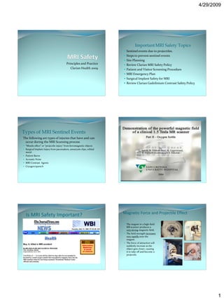
Is MRI Safety Important?
The magnet in a high-field
MR scanner produces a
very strong magnetic field.
The field strength increases
very rapidly near the
magnet.
The force of attraction will
suddenly increase as the
object gets closer, causing
it to take off and become a
projectile.
1
2. 4/29/2009
MR Zone Configuration
Preventing Sentinel
events
MR Zone Configuration MR Zones 3 and 4
Zone 1: This Zone includes all areas freely accessible by Both zones should be strictly prohibited by the general
the general public. Typically MR patient waiting. public. Key locks, passkey locking systems can be used to
Zone 2: This area is the interface from the uncontrolled restrict access to this area.
Zone 1 and the restricted areas of Zone 3 and 4. Typically Zones 3 and 4 should be marked with warning signs that
Zone 2 is the MR screening area and patients are under indicate the presence of a strong magnet fields.
the supervision of MR personnel. Zones 3 and 4 should be marked with a lighted sign that
Zone 3: Should be strictly restricted and is directly states that the “Magnet is always on”.
adjacent to the magnet room. Zone 3 is typically the MR To gain access to Zones 3 and 4 personnel are required to
control room and the entry space before entering the participate in MRI Safety Training
magnet room. All non-MRI personnel should be under constant
Zone 4: Magnet room supervision of an MRI technologist, or MRI engineer
MR Safety Warning signs MRI Magnet room
Entry
Door includes
Danger Signs in
English and
Spanish.
2
3. 4/29/2009
Zone 3 and 4 locking systems
Every MRI
Magnet room
Honeywell card reader Magnet room keypad
should have an
MRI compatible
Fire Extinguisher
MRI Compatible Equipment with labels
MRI Compatible Equipment Labels The Invivo MRI Monitor labeled with a MRI Safe sticker
The Invivo battery supply is not and must be secured to the floor with proper
warning labels.
MRI Compatible Equipment
Have a bar magnet available to double check
equipment going into the magnet room
3
4. 4/29/2009
Clarian MRI Safety Policies MRI Safety Policy
The goal of the radiology department is to make every
MRI Safety Policy effort to constantly minimize the risks of incidents
associated with MRI resulting from the magnetic field
Use of equipment in MRI Magnet Room and/or cryogens.
MRI Contrast Media Policy
All Radiology personnel will be educated in an annual MRI
Safety in-service. MRI personnel will be trained in all
aspects of MRI safety and emergency procedures.
The MRI Safety questionnaire will be completed by all
patients and visitors before entering the MRI magnet
room.
•All hospital personnel , physicians, patients, nurses, RT’s must pass MR
safety screening before entering the MR magnet room. All hospital
personnel working in the MRI environment are encouraged to complete
the MRI safety training on Pulse.
•There will no admittance of ferrous metal in the magnet room. Only MRI
compatible equipment will be used.
•All patients, visitors, and hospital personnel in the magnet room during
the scanning process are required to wear ear protection such as ear plugs,
or headphones.
•All patients will be given the MRI Technologist alert button (Squeeze Ball)
•All pregnant MRI personnel should not remain in the MRI magnet room
during the actual scanning process.
MRI Safety Training Location
MRI Safety Training
4
5. 4/29/2009
Use of equipment in MRI Policy
A policy for the safe use of medical
equipment and devices in the MRI magnet
room.
The policy states that any new medical
equipment and devices used in the magnet
room will reviewed by the Clarian MRI
Safety Committee and labeled accordingly.
Servo i Ventilator for MRI Servo i Positioned in a Magnet Room
Maquet Inc.
Servo i Ventilator will be placed outside the 20mT
(200 gauss) line. For open MR scanners it should be
placed outside the 10mT (100 gauss) line.
Brakes on
Tether secured.
MRridium
MRI Compatible Anesthesia Machines MRI
Datex Ohmeda placed outside the 30mT (300 gauss) compatible
line. IV pump
IRadimed
Corporation
http://www.iradimed.
com/en-
us/products/?sf_ses=0
f2219b54ffe09db9a58
23bcf44f196a
5
6. 4/29/2009
MR Safety Screening
Patient and Visitor Screening All patients and visitors need to complete an MRI Safety
questionnaire before entering the MR magnet room.
Greet patient confirm their name and DOB For pediatric patients the safety questionnaire needs to
be completed by a parent or guardian. Fax
Introduce yourself questionnaires to inpatient floors prior to the MRI
Procedure.
Review their MRI Safety questionnaire with them and Patients with a Pacemaker, defibrillator, or ICD should
verbally confirm that they do not have metal inside be cancelled and not imaged with MRI. The patient’s
referring physician should be notified of cancellation.
their body.
Patients with metal in their body such as bullets, BB’s or
Show patient where to get dressed and have them Shrapnel should be screened for the type of metal and
X-rays can be obtained to review the location of the
remove all loose metal such as jewelry, watches, metal. Patients with a history of orbital metal should be
hairpins, etc. screened with orbit x-rays or CT.
Start IV if needed and inform the patient if their will All Surgical devices such as a brain aneurysm clip, heart
stents, IVC filters, drug infusion pumps should be
be a short wait before their MRI begins. thoroughly researched and proven to be safe for MRI
imaging.
Prevent Patient Burning due to Induced
Currents
Insolate wires and leads.
Make sure all the coils are
plugged into the scanner.
Position patient so that the arms
MRI Imaging Safety and legs are not crossing or
touching to form current loop.
Check for metallic ECG pads and
tattoos.
Medication patches MR Unsafe Implants
All medication patches should be removed before Some implants are paramagnetic, or even ferromagnetic. These
imaging a patient. implants tend to move and align with the main magnetic field.
Medication patches contain a metal backing that can This results in a force and torque on the implant and the
cause a skin burn. implant may become dislodged, resulting in severe injury to the
patient.
Clear patches should be removed, as well. Clear
Aneurysm clips are examples of implants that can result in
medication patches may contain small amounts of death if displaced.
metal that can cause a skin burn. Clarian MRI Safety policy indicates that
http://www.fda.gov/cder/drug/advisory/transdermalp documentation and confirmation of MRI
atch.htm safety is required for all surgical implants
prior to MRI imaging.
6
7. 4/29/2009
Implant and Device Safety
Interference with other Devices Database
Some devices may not function properly in MR environment
because of the strong static magnetic field and/or radio
www.mrisafety.com
frequency field.
by Frank G. Shellock
• Pacemaker and implanted
cardiac defibrillator are Consult with the implant
manufacturer such as
typical examples of such
Medtronic, Cook, etc.
devices.
• Fringe field should be Keep a binder or folder with
less than 5 gauss (or collected implant MRI safety
0.5mT) for public area. procedures.
Gradient Switching and Nerve Stimulation Magnet Quenching Safety Concerns
As liquid cryogen becomes gas, it will displace room
Temporal variation of magnetic field (dB/dt) air and oxygen. Persons breathing in this environment
electrical potential nerve stimulation may suffocate.
The effect of stimulation varies from minor muscle Cold gas may cause skin & body injury.
twitching to severe pain.
High performance gradient and gradient intensive
sequences are more likely to cause PNST.
Ensure that arms and legs are not crossed to prevent
PNST.
Quench Safe Guards
Emergency quench button Safety
Emergency quench button should be covered by a
clear plastic cover so that it is not accidentally
pushed.
The quench button should only be depressed in
the event of a Serious Personal Injury
• Vent stack to allows cryogen gas to escape.
• Scanner room opens outward to prevent
pressure build up.
7
8. 4/29/2009
MRI Emergency Situations MRI Emergency Procedures
Fire or Smoke Alarm Call for assistance immediately.
Magnet Quench
Remove the patient from the scan room and secure the
Inadvertent Metal in the Magnet
door.
MRI Contrast Reaction Administer emergency first aid if needed until
Other Medical Emergencies assistance arrives.
Ensure that untrained personnel or ferromagnetic
material are not inadvertently allowed to enter the scan
room.
Practice MRI emergency drills with your staff
Codman Hakim Programmable Shunts
Surgical Implant Safety MRI Safety Procedure
DBS The Codman Hakim programmable shunt is MRI Conditional
according to ASTM F2503.
Vagal Nerve Stimulators MRI can be performed at any time after implantation.
Codman Programmable Shunts MRI System with a static magnetic field of 3Tesla or less.
Use an MR system with a spatial gradient of 720 gauss/cm or less.
Limit RF energy to a whole-body SAR of 3W/kg over 15min.
Verify valve pressure setting before and after the MRI procedure
Vagal Nerve Stimulator
Cyberonics
VNS is used primarily on patients with epilepsy, depression and other debilitating neurological
Medtronic Deep Brain Stimulator
disorders.
The VNS Implant delivers electrical pulsed signals to the Vagus nerve. Warnings:
A small generator is implanted in the left chest.
Read and follow the MRI information manual for the Activa Medtronic
MRI Safety Procedure:
DBS. Failure to follow all warnings and guidelines will result in serious
1. Pulse generator output programmed to 0mA before the MRI procedure.
(Neurology will set VNS to 0mA) and permanent injury.
2. After the MRI procedure the VNS will be reprogrammed to the original An MRI procedure should not be performed on a patient with an Activa
settings. (Neurology department) DBS that has a broken lead wire because higher than normal heating
3. Head coil Type: Transmit and Receive only may occur at the break
4. Static Magnetic field 2.0 Tesla or less Use only a 1.5T horizontal bore MRI
5. Specific absorption rate (SAR) <1.3 W/Kg for a 154.5lb (70kg) patient
Use only a transmit/receive head coil
6. Time-varying intensity: <10 telsa/sec
Use MRI sequence parameters that limit the Average head SAR to 0.1
7. Document MRI sequence parameters and the SAR for each sequence.
W/Kg.
Caution: MRI procedures in which the RF is transmitted by a body coil Limit the gradient dB/dt field to 20tesla/second or less
should not be done on a patient who has a VNS
Document each sequence parameters and average head SAR setting
8
9. 4/29/2009
MR Contrast Agent Safety Clarian MRI Contrast Media Policy
To inject MRI contrast you must be certified in CPR
Clarian MRI Safety form screens patients for renal impairment previous MRI
Gadolinium Contrast Reaction: contrast reactions
Headache (6.5%), Injection site coldness (3.6%), Injection site pain or Radiologist will write an order to inject MRI contrast
burning (2.5%), and Nausea (1.9%). If there are no contraindications to MRI contrast patients are given a contrast
dose from the dosing chart base on their weight
Severe allergic reactions (0.01%)
A Radiologist, a radiology PA, or other designated physician will be available
to respond to potential emergencies. If a designated physician is not available
High Risk Groups: MRI contrast will not be administered.
An emergency cart will be available in the immediate area
Pregnancy/Breast Feeding
The following MRI contrast information must be added into the RIS system:
Previous Contrast Reaction
MRI contrast brand name
Renal Deficiency Contrast Volume,
Individual performing the contrast injection
Confirm Med. Reconciliation
Any complications.
Clarian Guidelines for MRI patients with Potential NSF
Kidney Function Impairment
Nephrogenic Systemic Fibrosis or Nephrogenic Fibrosing Dermopathy
(NSF/NFD) is a rare and serious syndrome that involves fibrosis of
skin, joints, eyes, and internal organs. In NSF patients develop large
areas of hardened skin. There is no cure for NSF/NFD nor is there a
consistently successful treatment.
NSF/NFD is reported in patients with kidney failure after receiving
MR contrast containing gadolinium.
The majority of reported cases are related to Omniscan and
Magnevist, and with double and triple doses of contrast.
The FDA and ACR have issued guidance regarding the usage of
gadolinium based contrast agents in patients with impaired renal
function.
Sadowski, E. A. et al. Radiology 2007
Actions for MR Technologists
Patient Screening for MRI contrast
All patients undergoing MRI procedures will be screened for If the patient is on dialysis consult with a radiologist
impairment of renal function using the Clarian MRI Safety screening If the patient’s eGFR is less that 30 mL/min/1.73m2 then consult with
form.
a radiologist
If the patient answers “Yes” to the question on the screening form that
they have a history of decreased kidney function then current labs If the patient’s eGFR is between 30 and 60 mL/min/1.73m2 and if
must be obtained before MRI contrast is given. the accumulated dose of any gadolinium based contrast agent within
If available in the patient’s medical records eGFR (estimated the last 7 days including the current MRI contrast order is no more
Glomerular Filtration Rate) and SCr (Serum Creatinine) can be that a single dose (0.1mmol/kg). Patients can only have a single dose
obtained from Cerner or CareWeb of MRI contrast in a 7 day period.
Current labs must be used and should be within the last 30 days If the patient’s eGFR is Greater that 60 mL/min/1.73m2, proceed
iSTAT can be used for current Creatinine. The conversion chart in the with the MRI exam
policy can be used to obtain the eGFR Document the patient’s eGFR in RIS
eGFR/SCr Labs obtained
9
10. 4/29/2009
Recommended actions by attending Radiologists
NSF Remedies
If eGFR is less than 30 mL/min/1.73m2 or patient is currently on
dialysis consider alternative imaging sequences that do not require Avoid/minimize the use of Gd contrast agents in the
gadolinium. risk population:
Patient screening
If eGFR is between 30 and 60 mL/min/1.73m2 give only a Reduce dosage
single dose of gadolinium at 0.1mmol/Kg within a 7 day period of time.
Non-contrast enhanced MRI techniques
If patient is on dialysis and the benefit of giving gadolinium out Other modalities
weighs the risk then dialysis must be done within 3 hours of the Hydration
gadolinium injection. The second session of dialysis should be done
again within 24hours. A Clarian clinical consent form should be Hemodialysis
obtained by a radiologist prior to the gadolinium injection.
Clarian GBCA Safety Guidelines
Decreased Kidney
MR Safety References and Guidelines
Function AND N N N
Dialysis eGFR* < eGFR* <
Prescription of Gd patient 30 ? 60 ?
Based contrast ? MRI Safety references
Y Y Y
http://www.mayoclinic.org/nephrogenic-fibrosing-dermopathy/
*eGFR: estimate
Glomeruler Scan with original http://en.wikipedia.org/wiki/Nephrogenic_systemic_fibrosis
Total Dose** protocols
Filtration Rate >
N
and avoid
0.1mmol/kg? Omniscan &
http://www.lifenph.com/prog-shunts.asp
**Total Dose: The Magnevist
total dose of Gd
http://www.mrisafety.com/safety_article.asp?subject=175
based contrast Y http://www.codman.com/binary/org/CODMAN/PDF/VAL-10-001-
agent accumulated
in last 7 days CHPV-proc-guide.pdf
Consult with
including the Scan w/o referring
Scan w. http://www.vnstherapy.com/epilepsy/hcp/manuals/default.aspx
N N total dose
current MR exam. contrast physician /
Nephrologists <= 0.1 http://www.acr.org/SecondaryMainMenuCategories/quality_safety/
or other
mmol/kg or
modality
? other MRSafety/safe_mr07.aspx
Check by MR modality?
Technologist Y Y
Evaluation by Scan without contrast Scan with modified
Radiologist or protocols and avoid
with another modality Omniscan & Magnevist
or with another modality
Summary Thank you
Identify Zones 3 and 4. Secure both areas with a locking system.
Make sure all equipment in Zone 3 and 4 are identified, labeled and is
MRI compatible.
MRI zones 3 and 4 must be marked with MRI Warning signs
Portable O2 Tanks should never be in the MRI zones 3 and 4. Do not
mix MRI compatible O2 tanks with non-compatible tanks. Use wall
Oxygen.
Every Patient/Visitor is required to have a completed MRI Safety
Screening form prior to their MRI procedure.
Continue to educate your self on the latest MRI safety updates.
Practice MRI Safety drills with your staff.
Constantly watch non-MRI personnel working in the MRI area.
Keep your Magnet room door closed at all times.
10