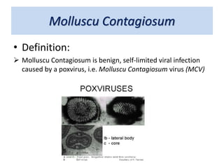
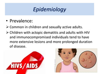
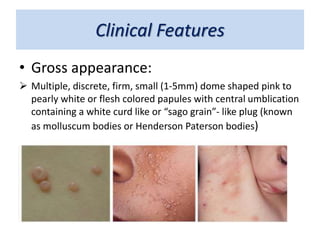
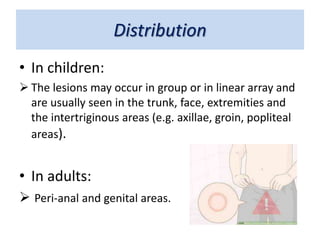
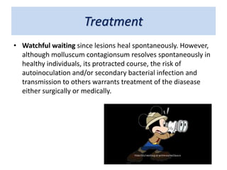
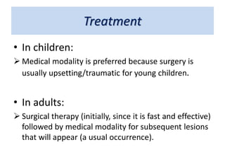
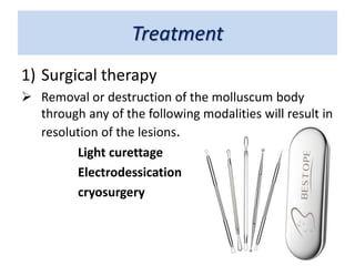
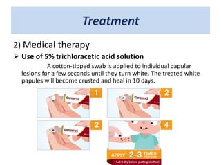
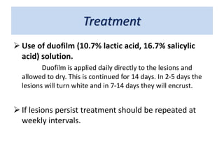
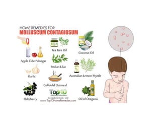
II. Common Laprosy Look-alike
Skin Viral Infection
By: Ashvini Bavda
Molluscu Contagiosum
• Definition:
 Molluscu Contagiosum is benign, self-limited viral infection
caused by a poxvirus, i.e. Molluscu Contagiosum virus (MCV)
Epidemiology
• Prevalence:
 Common in children and sexually active adults.
 Children with actopic dematitis and adults with HIV
and immunocompromised individuals tend to have
more extensive lesions and more prolonged duration
of disease.
Epidemiology
• Transmission:
 Direct skin to skin/ mucous membrane contact (particularly
during sexual activity) or through fomites.
 Close contact sports (e.g. wrestling), bath towels and
swimming pools also been reported as sources of infection.
Clinical Features
• Gross appearance:
 Multiple, discrete, firm, small (1-5mm) dome shaped pink to
pearly white or flesh colored papules with central umblication
containing a white curd like or “sago grain”- like plug (known
as molluscum bodies or Henderson Paterson bodies)
Distribution
• In children:
 The lesions may occur in group or in linear array and
are usually seen in the trunk, face, extremities and
the intertriginous areas (e.g. axillae, groin, popliteal
areas).
• In adults:
 Peri-anal and genital areas.
Distribution
• Clinical course:
 Depend upon promptness of treatment.
• Incubation period:
 2-7 days
 Lesion can spread through the autoinoculation or
koebnerization.
 Spontaneous resolution usually occures though it will
take months to years.
Distribution
• Symptoms:
 Asymptomatic unless if lesions are irritated,
inflamed or secondary infected..
• Diagnosis:
 Usually made clinically, because the characteristic
papule with central umblication is distinctive for
molluscum contagiosum.
Differential Diagnosis
• In Immunocompetent indivisuals:
 Histoid leprosy
 Verruca
 Acne
 Millia
 sebaceous hyperplasia
 syringoma
Differential Diagnosis
• In immunocompromised patients
especially those with HIV:
 Penicillosis
 Cryptococcosis
 histoplasmosi
Differential Diagnosis
• Laboratory
Diagnosis:
 Diagnosis is confirmed by
expressing the lesion to
obtain a crush preparation
of the central curd-like
material and staining with
Giemsa or Wright’s stain.
Treatment
• Watchful waiting since lesions heal spontaneously. However,
although molluscum contagionsum resolves spontaneously in
healthy individuals, its protracted course, the risk of
autoinoculation and/or secondary bacterial infection and
transmission to others warrants treatment of the diasease
either surgically or medically.
Treatment
• In children:
 Medical modality is preferred because surgery is
usually upsetting/traumatic for young children.
• In adults:
 Surgical therapy (initially, since it is fast and effective)
followed by medical modality for subsequent lesions
that will appear (a usual occurrence).
Treatment
1) Surgical therapy
 Removal or destruction of the molluscum body
through any of the following modalities will result in
resolution of the lesions.
Light curettage
Electrodessication
cryosurgery
Treatment
2) Medical therapy
 Use of 5% trichloracetic acid solution
A cotton-tipped swab is applied to individual papular
lesions for a few seconds until they turn white. The treated white
papules will become crusted and heal in 10 days.
Treatment
 Use of duofilm (10.7% lactic acid, 16.7% salicylic
acid) solution.
Duofilm is applied daily directly to the lesions and
allowed to dry. This is continued for 14 days. In 2-5 days the
lesions will turn white and in 7-14 days they will encrust.
 If lesions persist treatment should be repeated at
weekly intervals.
molluscum contagiosum

molluscum contagiosum

  • 1.
    II. Common LaprosyLook-alike Skin Viral Infection By: Ashvini Bavda
  • 3.
    Molluscu Contagiosum • Definition: Molluscu Contagiosum is benign, self-limited viral infection caused by a poxvirus, i.e. Molluscu Contagiosum virus (MCV)
  • 4.
    Epidemiology • Prevalence:  Commonin children and sexually active adults.  Children with actopic dematitis and adults with HIV and immunocompromised individuals tend to have more extensive lesions and more prolonged duration of disease.
  • 5.
    Epidemiology • Transmission:  Directskin to skin/ mucous membrane contact (particularly during sexual activity) or through fomites.  Close contact sports (e.g. wrestling), bath towels and swimming pools also been reported as sources of infection.
  • 6.
    Clinical Features • Grossappearance:  Multiple, discrete, firm, small (1-5mm) dome shaped pink to pearly white or flesh colored papules with central umblication containing a white curd like or “sago grain”- like plug (known as molluscum bodies or Henderson Paterson bodies)
  • 7.
    Distribution • In children: The lesions may occur in group or in linear array and are usually seen in the trunk, face, extremities and the intertriginous areas (e.g. axillae, groin, popliteal areas). • In adults:  Peri-anal and genital areas.
  • 8.
    Distribution • Clinical course: Depend upon promptness of treatment. • Incubation period:  2-7 days  Lesion can spread through the autoinoculation or koebnerization.  Spontaneous resolution usually occures though it will take months to years.
  • 9.
    Distribution • Symptoms:  Asymptomaticunless if lesions are irritated, inflamed or secondary infected.. • Diagnosis:  Usually made clinically, because the characteristic papule with central umblication is distinctive for molluscum contagiosum.
  • 10.
    Differential Diagnosis • InImmunocompetent indivisuals:  Histoid leprosy  Verruca  Acne  Millia  sebaceous hyperplasia  syringoma
  • 11.
    Differential Diagnosis • Inimmunocompromised patients especially those with HIV:  Penicillosis  Cryptococcosis  histoplasmosi
  • 12.
    Differential Diagnosis • Laboratory Diagnosis: Diagnosis is confirmed by expressing the lesion to obtain a crush preparation of the central curd-like material and staining with Giemsa or Wright’s stain.
  • 13.
    Treatment • Watchful waitingsince lesions heal spontaneously. However, although molluscum contagionsum resolves spontaneously in healthy individuals, its protracted course, the risk of autoinoculation and/or secondary bacterial infection and transmission to others warrants treatment of the diasease either surgically or medically.
  • 14.
    Treatment • In children: Medical modality is preferred because surgery is usually upsetting/traumatic for young children. • In adults:  Surgical therapy (initially, since it is fast and effective) followed by medical modality for subsequent lesions that will appear (a usual occurrence).
  • 15.
    Treatment 1) Surgical therapy Removal or destruction of the molluscum body through any of the following modalities will result in resolution of the lesions. Light curettage Electrodessication cryosurgery
  • 16.
    Treatment 2) Medical therapy Use of 5% trichloracetic acid solution A cotton-tipped swab is applied to individual papular lesions for a few seconds until they turn white. The treated white papules will become crusted and heal in 10 days.
  • 17.
    Treatment  Use ofduofilm (10.7% lactic acid, 16.7% salicylic acid) solution. Duofilm is applied daily directly to the lesions and allowed to dry. This is continued for 14 days. In 2-5 days the lesions will turn white and in 7-14 days they will encrust.  If lesions persist treatment should be repeated at weekly intervals.