
Recommended
Recommended
More Related Content
What's hot
What's hot (20)
Similar to Molar Root Incisor Malformation
Similar to Molar Root Incisor Malformation (20)
More from Mohamed Alkeshan
More from Mohamed Alkeshan (9)
Recently uploaded
Recently uploaded (20)
Molar Root Incisor Malformation
- 1. MOLAR ROOT-INCISOR MALFORMATION: CONSIDERATIONS OF DIVERSE DEVELOPMENTALAND ETIOLOGIC FACTORS Wright et al (2016 ) Done by R2 (Mohammad)
- 2. Introduction • ( MRIM) unique and specific phenotype Characterized by diminished and dysplastic root formation and a narrow abnormal pulp champer of the first permanent molars( 2nd primary molar and permanent anterior teeth are affected in some cases). • Genetic defects and environmental influences can cause failure of tooth development or abnormal morphologic changes of the tooth.
- 3. Introduction • Hereditary Dentin defects classified into: 1. Dentinogenesis imperfecta (DGI). 2. Dentin Dysplasia (DD). • These hereditary conditions are associated with abnormal dentin mineralization and varying degrees of changes in tooth morphology. • DGI type I is associated with osteogenesis imperfecta, whereas the clinically similar DGI type II is not associated with a syndrome and is caused by mutations in the gene encoding dentin sialophosphoprotein(DSPP).
- 4. Introduction • Individuals with DGI often have dental root structures that are diminished in size and appear sharp and “ tent peg- like” . • The phenotype in DD type I is characterized by clinically normal-appearing tooth crowns and markedly altered dentin formation (waterfall appearance), pulp chamber obliteration, and abnormal to nearly missing root development
- 5. Introduction • Truncated Root Formation mutations in the IFIH1 gene and Singleton-Merten syndrome. • Tumoral Calcinosis can have short bulbous roots. TC is caused by mutations in the GALNT3 gene. • Sponastrime Dysplasia and Bardet-Biedl Syndrome seen with Short dental roots • Kenny-Caffey syndrome caused by mutations in the TBCE gene, is associated with abnormal root formation.
- 6. Introduction • Many environmental stressors and conditions, are associated with the abnormal formation of the tooth roots. trauma, infection, radiation, and certain chemotherapeutic agents for malignancies during early childhood can result in abnormal tooth and tooth root development. • Severe infections, such as those occurring in Steven- Johnson syndrome or meningo-coccemia, can result in abnormal root formation.
- 7. Methods • The study protocol approved by SNUDH and the University of North Carolina(NC). • Radiographs taken between February 2012 and May 2013 at SNUDH . Additional cases from (NCU). • longitudinal follow-up was available. • Generalized root malformation due to anticancer therapy were excluded. And 30 cases (18 M, 12 F) were selected for this report. • Teeth were decalcified, sectioned, stained with hematoxylin and evaluated with light microscope.
- 8. RESULTS
- 9. Result
- 10. Histologic evaluation • The pulp chambers all had a greatly reduced dimension between the roof and floor of the pulp chamber. • Secondary dysplastic pulp area had frequent inclusions, which were not present in normal appearing pulp.
- 11. DISCUSSION • The first permanent molars were always affected (100%), making primary feature of this unique developmental defect of the dentition. • Secondary feature defects included second primary molars, which affected in (50%) of the cases. Affected permanent canines (13%) of cases. • Most of the MRIM cases suggesting these root malformations are caused by a secondary effect of the systemic stressors
- 12. CONCLUSIONS • In this report there is a diverse range of severity related to the teeth affected and the extent of the malformation (e.g., root/crown malformation). • Clinicians identifying cases of MRIM should carefully evaluate them for an increased risk for ectopic eruption and permanent incisor malformations that can lead to pulpal necrosis.
Editor's Notes
- Hereditary Dentin defects classified into: Dentinogenesis imperfecta (DGI). Dentin Dysplasia (DD). These hereditary conditions are associated with abnormal dentin mineralization and varying degrees of changes in tooth morphology
- Other known genetic causes of abnormal root for- mation include truncated root formation and Tumoral Calcinosis .
- The study protocol was reviewed and approved by the Institution Review Boards at the Seoul National Uni- versity Dental Hospital and the University of North Carolin
- In Table I All cases showed involvement of the first permanent molars, with all four molars being affected, as illustrated in the female subject who reported meconium aspiration at birth but no other medical problems or signifiCant illnesses in the neonatal period. Twelve cases had root malformation. Some anterior teeth had dens-in-dente and had developed periapical pathology in the absence of caries, trauma, or other discernable pulpal stressors. . seven patients reported meningomyelocel oracral dimple, six reported meningitis, four reported preterm birth, and four reported chronic renal disease. Four of the 30 affected individuals reported no major problems and were described as healthy .
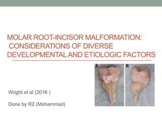
- Fig. A The permanent central incisors exhibit a marked shortening of the root with a large pulpal obstruction in the middle root area (A) The permanent central incisors show a dramatic constriction in the cervical area (B) of the clinical crown and a dens-in-dente type appearance in the pulp (arrow) Primary second molars were also affected in 15 Cases . The second primary molars also have short roots and an abnormal pulpal morphology cases, and primary first molars were affected in one case (Figure 4).
- Histologically the affected first permanent molars FIGURE (A) showed an area of pulpal tissue in the coronal area of the crown but was terminated in its apical extent Cervical to this pupal area was a region of dysplastic dentin (DP) and fibrous tissue that was poorly organized and contained areas of mineralized tissue,