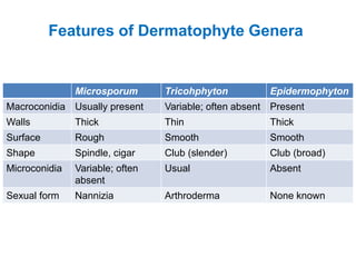
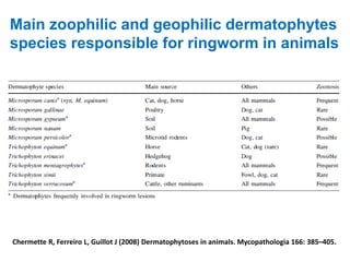
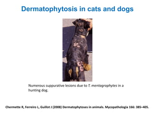
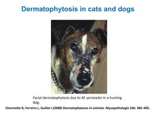
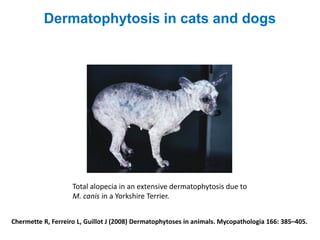
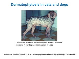
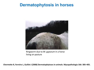
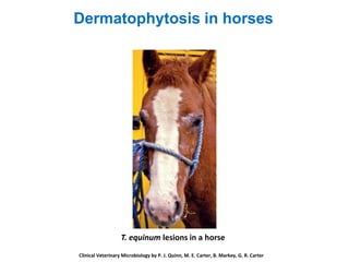
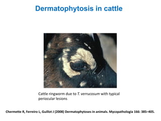
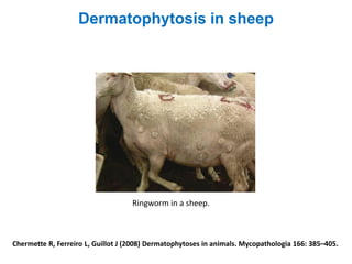
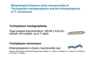
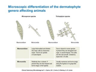
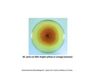
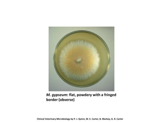
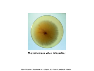
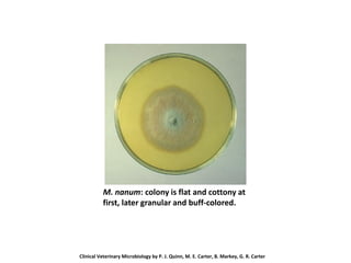
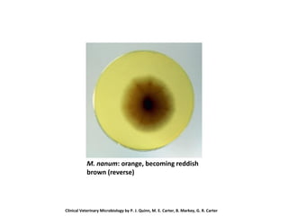
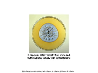
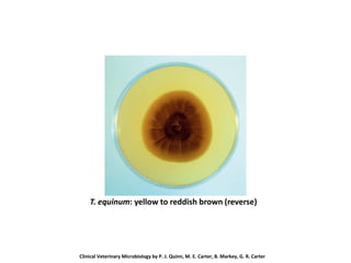
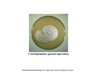
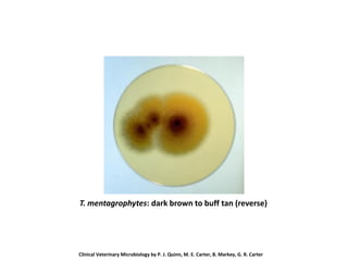
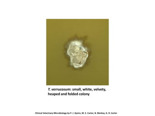
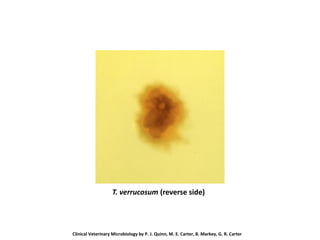
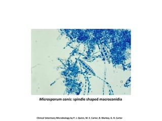
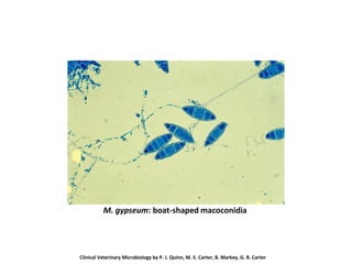
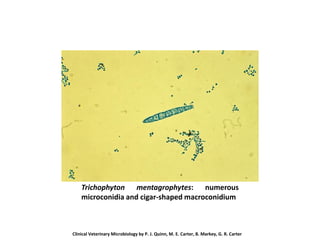
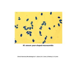
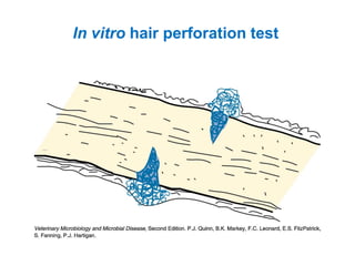
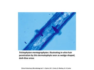
Dermatophytes
▪ Members of the phylum Ascomycota
▪ Affinity for keratinized structures; colonize and invade
skin, hair and nails
▪ Grow slowly on specially formulated laboratory media, e.g
Sabouraud dextrose agar; some require additional growth
factors
▪ Aerobic, tolerate cyclohexamide in media
▪ Colonies often pigmented
▪ Macroconidia formed in cultures
▪ Arthrospores, shed from infected animals, remain infective
for many months
▪ Cause characteristic circular skin lesions termed ringworm
Grouping of Dermatophytes
Based on host preference or habitat
▪ Zoophilic
▪ Anthropophilic
▪ Geophilic
Features of Dermatophyte Genera
Microsporum Tricohphyton Epidermophyton
Macroconidia Usually present Variable; often absent Present
Walls Thick Thin Thick
Surface Rough Smooth Smooth
Shape Spindle, cigar Club (slender) Club (broad)
Microconidia Variable; often
absent
Usual Absent
Sexual form Nannizia Arthroderma None known
Main zoophilic and geophilic dermatophytes
species responsible for ringworm in animals
Chermette R, Ferreiro L, Guillot J (2008) Dermatophytoses in animals. Mycopathologia 166: 385–405.
Virulence factors possessed by Trichophyton
▪ Endoprotease (Subtilisins, Fungalysins)
▪ Exoproteases (Serine proteases, Leucine
aminopeptidases)
▪ Kinase
▪ Mannan
▪ Sulphite
▪ LysM
Virulence factors possessed by Microsporum
▪ Endoprotease (Subtilisins, Fungalysins)
▪ Pseudokinase (inactive kinase)
▪ Exoproteases
▪ Sulphite
▪ LysM along with polysaccharide deacetylases (type I)
▪ Polycyclic aromatic prenyltransferases (pcPTases)
Important Dermatophyte Infections in Animals
Host Agent Nature of Lesions
Horse T. equinum Dry, scaly usually noninflammatory (unless secondarily infected)
M. gypseum Often suppurative under alopecic thickened areas
M. equinum Not more than mildly inflammatory, resembling T. equinum lesions
Cattle T. verrucosum Painless, thick, white, ‘asbestos’ plaques, local alopecia
Swine M. nanum Tannish, crusty, spreading centrifugally on trunk; painless, margins
slightly inflamed. No hair loss.
Dog M. canis Typically noninflammatory, scaly, alopecic patches, occasional
kerion
T. mentagrophytes Often spreading, extensively scaling to inflammatory lesions,
secondary suppuration
M. gypseum As in T. mentagrophytes
Cat M. canis Often subclinical in adults. Generally noninflammatory, except in
young kittens, may become generalized in debilitated kittens.
Occasional mycetoma (Percisan cats).
T. mentagrophytes As in dogs
Chicken M. gallinae Generally affects unfeathered portions. Whitish chalky scaling on
comb and wattles, noninflammatory.
T. simii Superficially similar to M. gallinae but often inflammatory and even
necrotizing. A poultry problem only in India.
Importance
Chermette R, Ferreiro L, Guillot J (2008) Dermatophytoses in animals. Mycopathologia 166: 385–405.
Dermatophytosis in cats and dogs
M. canis infection in a kitten with lesions on the bridge of the nose,
the ear margins and the digits
Chermette R, Ferreiro L, Guillot J (2008) Dermatophytoses in animals. Mycopathologia 166: 385–405.
Kerion due to M. canis in a dog
Dermatophytosis in cats and dogs
Chermette R, Ferreiro L, Guillot J (2008) Dermatophytoses in animals. Mycopathologia 166: 385–405.
Numerous suppurative lesions due to T. mentagrophytes in a
hunting dog.
Dermatophytosis in cats and dogs
Chermette R, Ferreiro L, Guillot J (2008) Dermatophytoses in animals. Mycopathologia 166: 385–405.
Facial dermatophytosis due to M. persicolor in a hunting
dog.
Dermatophytosis in cats and dogs
Chermette R, Ferreiro L, Guillot J (2008) Dermatophytoses in animals. Mycopathologia 166: 385–405.
Total alopecia in an extensive dermatophytosis due to
M. canis in a Yorkshire Terrier.
Dermatophytosis in cats and dogs
Chermette R, Ferreiro L, Guillot J (2008) Dermatophytoses in animals. Mycopathologia 166: 385–405.
Chronic and extensive dermatophytosis due to a mixed M.
canis and T. mentagrophytes infection in a dog
Dermatophytosis in cats and dogs
Chermette R, Ferreiro L, Guillot J (2008) Dermatophytoses in animals. Mycopathologia 166: 385–405.
Dermatophytosis in horses
Ringworm due to M. gypseum in a horse
living on pasture
T. equinum lesions in a horse
Clinical Veterinary Microbiology by P. J. Quinn, M. E. Carter, B. Markey, G. R. Carter
Dermatophytosis in horses
Chermette R, Ferreiro L, Guillot J (2008) Dermatophytoses in animals. Mycopathologia 166: 385–405.
Dermatophytosis in cattle
Cattle ringworm due to T. verrucosum with typical
periocular lesions
Chermette R, Ferreiro L, Guillot J (2008) Dermatophytoses in animals. Mycopathologia 166: 385–405.
Dermatophytosis in sheep
Ringworm in a sheep.
Chermette R, Ferreiro L, Guillot J (2008) Dermatophytoses in animals. Mycopathologia 166: 385–405.
Dermatophytosis in pigs
Dermatophytosis due to T. mentagrophytes
in a pig.
Different species of Trichophyton associated
with hair invasion
Ectothrix invasion Endothrix
invasion
Noninvasion
T. verrucosum T. schoenleinii T. rubrum
T. equinum T. violaceum T. simii
T. mentagrophytes T. tonsurans T. concentricum
▪ Hairs should be plucked from the lesions.
▪ Scab material should be obtained from the edge of the
lesion.
▪ Scrapings and clipping from claws should be taken
from as near the base as possible.
▪ Where the specimens tend to be very contaminated by
bacteria and saprophytic fungi – wiping the lesions with
70% alcohol
Specimens
Laboratory Diagnosis
Surface (ectothrix) arthrospores on a hair
shaft following clearance with 10% KOH
Chermette R, Ferreiro L, Guillot J (2008) Dermatophytoses in animals. Mycopathologia 166: 385–405.
T. verrucosum: infected bovine hair with arthrospores
Morphological features of the macroconidia of some
Microsporum species
Morphological features of the macroconidia of
Trichophyton mentagrophytes and the chlamydospores
of T. verrucosum
Microscopic differentiation of the dermatophyte
genera affecting animals
Clinical Veterinary Microbiology by P. J. Quinn, M. E. Carter, B. Markey, G. R. Carter
Species Hosts Colonial Appearance Microscopic Appearance
Microsporum
canis (var.
canis)
Cats, dogs,
important
cause of
ringworm in
humans
Growth rapid. Surface white
and silky at centre with bright
yellowvperiphery. Reverse
side bright yellow or orange
Usually abundant
macroconidia. They are
spindle-shaped and
mature spores end in a distinct
knob. Cells 6–15. Few
microconidia
M. canis var.
distortum
Dogs Growth fairly rapid. Surface
white tovtan and reverse
white or yellowish tan. Colony
is velvety to fluffy with a
tendency to form radial
grooves
Usually abundant
macroconidia that are distorted
in shape,
thick-walled and multicellular.
Numerous microconidia
M. canis (syn.
M. equinum)
Horses Slow growth. Surface white
and velvety to finely powdery.
Reverse salmon or buff
Macroconidia rare, resemble
shortened M. canis
macroconidia
M. gypseum Horses,
dogs,
rodents
Fairly rapid growth. Colony is
flat, powdery with a fringed
border. Obverse is buff to
cinnamon-brown and reverse
pale yellow to tan or
occasionally red. Odour
similar to a mouse colony
Abundant macroconidia.
Boat-shaped with rounded
ends and thick, rough walls.
Cells 4–6
Dermatophytes of Veterinary Significance
Species Hosts Colonial Appearance Microscopic Appearance
M. nanum Pigs Colony is flat, white and
cottony at first, later granular
and buff coloured
Abundant macroconidia,
pear-shaped with spiny walls.
Cells 1–3.
M. gallinae Chickens,
turkeys
Rapid growth. Surface white
to pinkish, velvety and folded.
Reverse strawberry-pink,
diffusible pigment
Abundant macroconidia,
fusiform
with blunt spatulate tips. Walls
smooth and thick. Cells 2–10.
Trichophyton
equinum
Horses Fairly rapid growth. Colony
initially flat, white and fluffy
but later velvety with central
folding. Cream to tan in
colour, reverse is yellow to
reddish-brown
Macroconidia are rare. Slightly
club-shaped, smooth, thin-
walled with 3–5 cells.
Abundant microconidia.
Chlamydospores are abundant
in old cultures
Trichophyton
equinum var.
autotrophicum
Horses Colony at first white with a
raised centre, later white to
buff with folded centre.
Reverse is yellow becoming
dark rose-red
Macroconidia not reported
Dermatophytes of Veterinary Significance – cont’d
Species Hosts Colonial Appearance Microscopic
Appearance
T. mentagrophytes
var. mentagrophytes
Rodents,
dogs,
horses
and many
other
species
Rapid growth. Two colony
forms: 1. Granular, obverse
cream, reverse buff-tan to
dark-brown; 2. Downy, white
and woolly with older colonies
becoming cream-tan, reverse
varies from white through
yellow to reddish-brown
Macroconidia
cigarshaped,
thin-walled. Cells 3–7.
Abundant
microconidia in grape-
like clusters
T. simii Monkeys,
poultry,
dogs
Rapid growth. Finely granular
colony with diffuse margin,
white to pale or rose-buff.
Reverse white and later
reddish-brown
Abundant macroconidia,
cylindrical to fusiform in
shape. Cells 3–10.
T. verrucosum Cattle Very slow-growing. Small,
white,
velvety, heaped and folded
colony. Obverse white or
whitish-grey and occasionally
yellow-ochre, reverse is white
Macroconidia very rare
but characteristic chains
of chlamydospores
Dermatophytes of Veterinary Significance – cont’d
M. canis on SDA: surface white and silky at
centre with bright yellow periphery (obverse)
Clinical Veterinary Microbiology by P. J. Quinn, M. E. Carter, B. Markey, G. R. Carter
M. canis on SDA: bright yellow or orange (reverse)
Clinical Veterinary Microbiology by P. J. Quinn, M. E. Carter, B. Markey, G. R. Carter
M. gypseum: flat, powdery with a fringed
border (obverse)
Clinical Veterinary Microbiology by P. J. Quinn, M. E. Carter, B. Markey, G. R. Carter
M. gypseum: pale yellow to tan colour
Clinical Veterinary Microbiology by P. J. Quinn, M. E. Carter, B. Markey, G. R. Carter
M. nanum: colony is flat and cottony at
first, later granular and buff-colored.
Clinical Veterinary Microbiology by P. J. Quinn, M. E. Carter, B. Markey, G. R. Carter
M. nanum: orange, becoming reddish
brown (reverse)
Clinical Veterinary Microbiology by P. J. Quinn, M. E. Carter, B. Markey, G. R. Carter
T. equinum: colony initially flat, white and
fluffy but later velvety with central folding
Clinical Veterinary Microbiology by P. J. Quinn, M. E. Carter, B. Markey, G. R. Carter
T. equinum: yellow to reddish brown (reverse)
Clinical Veterinary Microbiology by P. J. Quinn, M. E. Carter, B. Markey, G. R. Carter
T. mentagrophytes: granular type colony
Clinical Veterinary Microbiology by P. J. Quinn, M. E. Carter, B. Markey, G. R. Carter
T. mentagrophytes: dark brown to buff tan (reverse)
Clinical Veterinary Microbiology by P. J. Quinn, M. E. Carter, B. Markey, G. R. Carter
T. verrucosum: small, white, velvety,
heaped and folded colony
Clinical Veterinary Microbiology by P. J. Quinn, M. E. Carter, B. Markey, G. R. Carter
T. verrucosum (reverse side)
Clinical Veterinary Microbiology by P. J. Quinn, M. E. Carter, B. Markey, G. R. Carter
Microsporum canis: spindle shaped macroconidia
Clinical Veterinary Microbiology by P. J. Quinn, M. E. Carter, B. Markey, G. R. Carter
M. gypseum: boat-shaped macoconidia
Clinical Veterinary Microbiology by P. J. Quinn, M. E. Carter, B. Markey, G. R. Carter
Trichophyton mentagrophytes: numerous
microconidia and cigar-shaped macroconidium
Clinical Veterinary Microbiology by P. J. Quinn, M. E. Carter, B. Markey, G. R. Carter
M. nanum: pear-shaped macroconidia
Clinical Veterinary Microbiology by P. J. Quinn, M. E. Carter, B. Markey, G. R. Carter
Hair perforation test
▪ Collect hairs from a young child with fair hair.
▪ Layer the sterile hairs on a 3-5-day-old subculture of
the dermatophyte under test and incubate at 25°C.
▪ Examine the hairs daily from seventh day of
incubation onwards by mounting a few hairs in
lactophenol cotton blue and examining them
microscopically, using the low and high-dry
objectives.
In vitro hair perforation test
Trichophyton mentagrophytes: illustrating in-vitro hair
penetration by this dermatophyte seen as wedge-shaped,
dark-blue areas
Clinical Veterinary Microbiology by P. J. Quinn, M. E. Carter, B. Markey, G. R. Carter

MMA 601_Dermatophytes_2022.pdf

  • 1.
    Dermatophytes ▪ Members ofthe phylum Ascomycota ▪ Affinity for keratinized structures; colonize and invade skin, hair and nails ▪ Grow slowly on specially formulated laboratory media, e.g Sabouraud dextrose agar; some require additional growth factors ▪ Aerobic, tolerate cyclohexamide in media ▪ Colonies often pigmented ▪ Macroconidia formed in cultures ▪ Arthrospores, shed from infected animals, remain infective for many months ▪ Cause characteristic circular skin lesions termed ringworm
  • 2.
    Grouping of Dermatophytes Basedon host preference or habitat ▪ Zoophilic ▪ Anthropophilic ▪ Geophilic
  • 3.
    Features of DermatophyteGenera Microsporum Tricohphyton Epidermophyton Macroconidia Usually present Variable; often absent Present Walls Thick Thin Thick Surface Rough Smooth Smooth Shape Spindle, cigar Club (slender) Club (broad) Microconidia Variable; often absent Usual Absent Sexual form Nannizia Arthroderma None known
  • 4.
    Main zoophilic andgeophilic dermatophytes species responsible for ringworm in animals Chermette R, Ferreiro L, Guillot J (2008) Dermatophytoses in animals. Mycopathologia 166: 385–405.
  • 5.
    Virulence factors possessedby Trichophyton ▪ Endoprotease (Subtilisins, Fungalysins) ▪ Exoproteases (Serine proteases, Leucine aminopeptidases) ▪ Kinase ▪ Mannan ▪ Sulphite ▪ LysM
  • 6.
    Virulence factors possessedby Microsporum ▪ Endoprotease (Subtilisins, Fungalysins) ▪ Pseudokinase (inactive kinase) ▪ Exoproteases ▪ Sulphite ▪ LysM along with polysaccharide deacetylases (type I) ▪ Polycyclic aromatic prenyltransferases (pcPTases)
  • 7.
    Important Dermatophyte Infectionsin Animals Host Agent Nature of Lesions Horse T. equinum Dry, scaly usually noninflammatory (unless secondarily infected) M. gypseum Often suppurative under alopecic thickened areas M. equinum Not more than mildly inflammatory, resembling T. equinum lesions Cattle T. verrucosum Painless, thick, white, ‘asbestos’ plaques, local alopecia Swine M. nanum Tannish, crusty, spreading centrifugally on trunk; painless, margins slightly inflamed. No hair loss. Dog M. canis Typically noninflammatory, scaly, alopecic patches, occasional kerion T. mentagrophytes Often spreading, extensively scaling to inflammatory lesions, secondary suppuration M. gypseum As in T. mentagrophytes Cat M. canis Often subclinical in adults. Generally noninflammatory, except in young kittens, may become generalized in debilitated kittens. Occasional mycetoma (Percisan cats). T. mentagrophytes As in dogs Chicken M. gallinae Generally affects unfeathered portions. Whitish chalky scaling on comb and wattles, noninflammatory. T. simii Superficially similar to M. gallinae but often inflammatory and even necrotizing. A poultry problem only in India.
  • 8.
    Importance Chermette R, FerreiroL, Guillot J (2008) Dermatophytoses in animals. Mycopathologia 166: 385–405.
  • 9.
    Chermette R, FerreiroL, Guillot J (2008) Dermatophytoses in animals. Mycopathologia 166: 385–405.
  • 10.
    Chermette R, FerreiroL, Guillot J (2008) Dermatophytoses in animals. Mycopathologia 166: 385–405.
  • 11.
    Chermette R, FerreiroL, Guillot J (2008) Dermatophytoses in animals. Mycopathologia 166: 385–405. Dermatophytosis in cats and dogs M. canis infection in a kitten with lesions on the bridge of the nose, the ear margins and the digits
  • 12.
    Chermette R, FerreiroL, Guillot J (2008) Dermatophytoses in animals. Mycopathologia 166: 385–405. Kerion due to M. canis in a dog Dermatophytosis in cats and dogs
  • 13.
    Chermette R, FerreiroL, Guillot J (2008) Dermatophytoses in animals. Mycopathologia 166: 385–405. Numerous suppurative lesions due to T. mentagrophytes in a hunting dog. Dermatophytosis in cats and dogs
  • 14.
    Chermette R, FerreiroL, Guillot J (2008) Dermatophytoses in animals. Mycopathologia 166: 385–405. Facial dermatophytosis due to M. persicolor in a hunting dog. Dermatophytosis in cats and dogs
  • 15.
    Chermette R, FerreiroL, Guillot J (2008) Dermatophytoses in animals. Mycopathologia 166: 385–405. Total alopecia in an extensive dermatophytosis due to M. canis in a Yorkshire Terrier. Dermatophytosis in cats and dogs
  • 16.
    Chermette R, FerreiroL, Guillot J (2008) Dermatophytoses in animals. Mycopathologia 166: 385–405. Chronic and extensive dermatophytosis due to a mixed M. canis and T. mentagrophytes infection in a dog Dermatophytosis in cats and dogs
  • 17.
    Chermette R, FerreiroL, Guillot J (2008) Dermatophytoses in animals. Mycopathologia 166: 385–405. Dermatophytosis in horses Ringworm due to M. gypseum in a horse living on pasture
  • 18.
    T. equinum lesionsin a horse Clinical Veterinary Microbiology by P. J. Quinn, M. E. Carter, B. Markey, G. R. Carter Dermatophytosis in horses
  • 19.
    Chermette R, FerreiroL, Guillot J (2008) Dermatophytoses in animals. Mycopathologia 166: 385–405. Dermatophytosis in cattle Cattle ringworm due to T. verrucosum with typical periocular lesions
  • 20.
    Chermette R, FerreiroL, Guillot J (2008) Dermatophytoses in animals. Mycopathologia 166: 385–405. Dermatophytosis in sheep Ringworm in a sheep.
  • 21.
    Chermette R, FerreiroL, Guillot J (2008) Dermatophytoses in animals. Mycopathologia 166: 385–405. Dermatophytosis in pigs Dermatophytosis due to T. mentagrophytes in a pig.
  • 22.
    Different species ofTrichophyton associated with hair invasion Ectothrix invasion Endothrix invasion Noninvasion T. verrucosum T. schoenleinii T. rubrum T. equinum T. violaceum T. simii T. mentagrophytes T. tonsurans T. concentricum
  • 23.
    ▪ Hairs shouldbe plucked from the lesions. ▪ Scab material should be obtained from the edge of the lesion. ▪ Scrapings and clipping from claws should be taken from as near the base as possible. ▪ Where the specimens tend to be very contaminated by bacteria and saprophytic fungi – wiping the lesions with 70% alcohol Specimens Laboratory Diagnosis
  • 24.
    Surface (ectothrix) arthrosporeson a hair shaft following clearance with 10% KOH
  • 25.
    Chermette R, FerreiroL, Guillot J (2008) Dermatophytoses in animals. Mycopathologia 166: 385–405.
  • 26.
    T. verrucosum: infectedbovine hair with arthrospores
  • 27.
    Morphological features ofthe macroconidia of some Microsporum species
  • 28.
    Morphological features ofthe macroconidia of Trichophyton mentagrophytes and the chlamydospores of T. verrucosum
  • 29.
    Microscopic differentiation ofthe dermatophyte genera affecting animals Clinical Veterinary Microbiology by P. J. Quinn, M. E. Carter, B. Markey, G. R. Carter
  • 30.
    Species Hosts ColonialAppearance Microscopic Appearance Microsporum canis (var. canis) Cats, dogs, important cause of ringworm in humans Growth rapid. Surface white and silky at centre with bright yellowvperiphery. Reverse side bright yellow or orange Usually abundant macroconidia. They are spindle-shaped and mature spores end in a distinct knob. Cells 6–15. Few microconidia M. canis var. distortum Dogs Growth fairly rapid. Surface white tovtan and reverse white or yellowish tan. Colony is velvety to fluffy with a tendency to form radial grooves Usually abundant macroconidia that are distorted in shape, thick-walled and multicellular. Numerous microconidia M. canis (syn. M. equinum) Horses Slow growth. Surface white and velvety to finely powdery. Reverse salmon or buff Macroconidia rare, resemble shortened M. canis macroconidia M. gypseum Horses, dogs, rodents Fairly rapid growth. Colony is flat, powdery with a fringed border. Obverse is buff to cinnamon-brown and reverse pale yellow to tan or occasionally red. Odour similar to a mouse colony Abundant macroconidia. Boat-shaped with rounded ends and thick, rough walls. Cells 4–6 Dermatophytes of Veterinary Significance
  • 31.
    Species Hosts ColonialAppearance Microscopic Appearance M. nanum Pigs Colony is flat, white and cottony at first, later granular and buff coloured Abundant macroconidia, pear-shaped with spiny walls. Cells 1–3. M. gallinae Chickens, turkeys Rapid growth. Surface white to pinkish, velvety and folded. Reverse strawberry-pink, diffusible pigment Abundant macroconidia, fusiform with blunt spatulate tips. Walls smooth and thick. Cells 2–10. Trichophyton equinum Horses Fairly rapid growth. Colony initially flat, white and fluffy but later velvety with central folding. Cream to tan in colour, reverse is yellow to reddish-brown Macroconidia are rare. Slightly club-shaped, smooth, thin- walled with 3–5 cells. Abundant microconidia. Chlamydospores are abundant in old cultures Trichophyton equinum var. autotrophicum Horses Colony at first white with a raised centre, later white to buff with folded centre. Reverse is yellow becoming dark rose-red Macroconidia not reported Dermatophytes of Veterinary Significance – cont’d
  • 32.
    Species Hosts ColonialAppearance Microscopic Appearance T. mentagrophytes var. mentagrophytes Rodents, dogs, horses and many other species Rapid growth. Two colony forms: 1. Granular, obverse cream, reverse buff-tan to dark-brown; 2. Downy, white and woolly with older colonies becoming cream-tan, reverse varies from white through yellow to reddish-brown Macroconidia cigarshaped, thin-walled. Cells 3–7. Abundant microconidia in grape- like clusters T. simii Monkeys, poultry, dogs Rapid growth. Finely granular colony with diffuse margin, white to pale or rose-buff. Reverse white and later reddish-brown Abundant macroconidia, cylindrical to fusiform in shape. Cells 3–10. T. verrucosum Cattle Very slow-growing. Small, white, velvety, heaped and folded colony. Obverse white or whitish-grey and occasionally yellow-ochre, reverse is white Macroconidia very rare but characteristic chains of chlamydospores Dermatophytes of Veterinary Significance – cont’d
  • 33.
    M. canis onSDA: surface white and silky at centre with bright yellow periphery (obverse) Clinical Veterinary Microbiology by P. J. Quinn, M. E. Carter, B. Markey, G. R. Carter
  • 34.
    M. canis onSDA: bright yellow or orange (reverse) Clinical Veterinary Microbiology by P. J. Quinn, M. E. Carter, B. Markey, G. R. Carter
  • 35.
    M. gypseum: flat,powdery with a fringed border (obverse) Clinical Veterinary Microbiology by P. J. Quinn, M. E. Carter, B. Markey, G. R. Carter
  • 36.
    M. gypseum: paleyellow to tan colour Clinical Veterinary Microbiology by P. J. Quinn, M. E. Carter, B. Markey, G. R. Carter
  • 37.
    M. nanum: colonyis flat and cottony at first, later granular and buff-colored. Clinical Veterinary Microbiology by P. J. Quinn, M. E. Carter, B. Markey, G. R. Carter
  • 38.
    M. nanum: orange,becoming reddish brown (reverse) Clinical Veterinary Microbiology by P. J. Quinn, M. E. Carter, B. Markey, G. R. Carter
  • 39.
    T. equinum: colonyinitially flat, white and fluffy but later velvety with central folding Clinical Veterinary Microbiology by P. J. Quinn, M. E. Carter, B. Markey, G. R. Carter
  • 40.
    T. equinum: yellowto reddish brown (reverse) Clinical Veterinary Microbiology by P. J. Quinn, M. E. Carter, B. Markey, G. R. Carter
  • 41.
    T. mentagrophytes: granulartype colony Clinical Veterinary Microbiology by P. J. Quinn, M. E. Carter, B. Markey, G. R. Carter
  • 42.
    T. mentagrophytes: darkbrown to buff tan (reverse) Clinical Veterinary Microbiology by P. J. Quinn, M. E. Carter, B. Markey, G. R. Carter
  • 43.
    T. verrucosum: small,white, velvety, heaped and folded colony Clinical Veterinary Microbiology by P. J. Quinn, M. E. Carter, B. Markey, G. R. Carter
  • 44.
    T. verrucosum (reverseside) Clinical Veterinary Microbiology by P. J. Quinn, M. E. Carter, B. Markey, G. R. Carter
  • 45.
    Microsporum canis: spindleshaped macroconidia Clinical Veterinary Microbiology by P. J. Quinn, M. E. Carter, B. Markey, G. R. Carter
  • 46.
    M. gypseum: boat-shapedmacoconidia Clinical Veterinary Microbiology by P. J. Quinn, M. E. Carter, B. Markey, G. R. Carter
  • 47.
    Trichophyton mentagrophytes: numerous microconidiaand cigar-shaped macroconidium Clinical Veterinary Microbiology by P. J. Quinn, M. E. Carter, B. Markey, G. R. Carter
  • 48.
    M. nanum: pear-shapedmacroconidia Clinical Veterinary Microbiology by P. J. Quinn, M. E. Carter, B. Markey, G. R. Carter
  • 49.
    Hair perforation test ▪Collect hairs from a young child with fair hair. ▪ Layer the sterile hairs on a 3-5-day-old subculture of the dermatophyte under test and incubate at 25°C. ▪ Examine the hairs daily from seventh day of incubation onwards by mounting a few hairs in lactophenol cotton blue and examining them microscopically, using the low and high-dry objectives.
  • 50.
    In vitro hairperforation test
  • 51.
    Trichophyton mentagrophytes: illustratingin-vitro hair penetration by this dermatophyte seen as wedge-shaped, dark-blue areas Clinical Veterinary Microbiology by P. J. Quinn, M. E. Carter, B. Markey, G. R. Carter