This article reviews Mirizzi syndrome, including its history, classification, diagnosis, and management. Mirizzi syndrome involves gallstone impaction that causes compression or fistulization of the common hepatic duct. It was first described in 1905 but named in 1948. It is now classified into two main types depending on whether fistulization is present. Diagnosis can be challenging but involves imaging like ultrasound, CT, MRCP, and ERCP. Open surgery is the gold standard treatment and provides good outcomes, with subtotal cholecystectomy recommended for types without fistula and partial cholecystectomy with fistula closure for types with fistula. Laparoscopic treatment has shown high conversion rates and risk of bile duct injury.
Mirizzi syndrome is a condition caused by large gallstones becoming lodged in the cystic duct or gallbladder, externally compressing the common hepatic duct and causing jaundice. It can lead to stenosis of the common hepatic duct or formation of a cholecystocholedochal fistula between the gallbladder and common hepatic duct. Mirizzi syndrome is diagnosed using imaging like ultrasound or cholangiography. Treatment involves removing the gallbladder and reconstructing the common bile duct.
Choledochal cysts should be considered in the differential diagnosis in all patients with a history of biliary colic, recurrent cholangitis or pancreatitis with associated dilatation of bile duct, particularly if they are <40 years of age. Delay in the diagnosis increases the incidence of associated biliary pathology and suboptimal surgical therapy
The document discusses the management of choledocholithiasis or common bile duct stones. It covers the clinical features, investigations like ultrasound, CT, ERCP and MRCP. It discusses the diagnostic approach and various management options including endoscopic procedures like ERCP with sphincterotomy and plastic stent placement. It also discusses open CBD exploration techniques like choledochotomy and T-tube placement. Laparoscopic CBD exploration is mentioned as a minimally invasive method. Guidelines recommend ERCP as first-line treatment for CBD stones with timing based on severity of cholangitis. Sphincterotomy with balloon dilation and cholangioscopy-assisted lithotripsy are suggested for difficult stones.
This document discusses liver lesions and their appearance on various imaging modalities. It covers benign lesions like hemangioma, focal nodular hyperplasia and hepatic adenoma. Malignant primary lesions discussed are hepatocellular carcinoma and hepatoblastoma. Imaging features of hypervascular and hypovascular lesions on multiphasic CT are summarized. Hepatocellular carcinoma risk factors and clinical presentation are outlined. Imaging appearance of HCC on ultrasound, CT and MRI is described in detail. Hepatic metastases are also discussed along with hypervascular metastatic lesions.
This document provides an overview of Mirizzi's syndrome, including:
- The history and classification of Mirizzi's syndrome based on involvement of the bile duct.
- The predisposing factors, typical presentations, and diagnostic challenges of Mirizzi's syndrome, which can include obstructive jaundice.
- Treatment typically involves surgical approaches like cholecystectomy and bile duct repair, with risks of complications higher than typical cholecystectomy. Preoperative diagnosis is important for surgical planning.
This document provides information on obstructive jaundice, including:
- Causes of obstructive jaundice include gallstones, tumors, strictures.
- Clinical features include jaundice, itching, steatorrhea.
- Investigations include liver function tests, ultrasound, CT, MRCP, ERCP.
- ERCP allows both diagnosis and treatment by sphincterotomy and stone extraction.
- Surgical management of stones includes open exploration or laparoscopic removal.
- Benign strictures are often post-surgical and may require stenting or reconstruction.
This case presentation describes a 34-year-old female patient who presented with right upper quadrant pain. She had a history of epigastric pain radiating to the back for 6 days along with jaundice. Imaging showed choledocholithiasis and cholecystolithiasis. She underwent open cholecystectomy with intraoperative cholangiogram, common bile duct exploration, and T-tube placement. Her postoperative course was uncomplicated and she was discharged with a T-tube in place.
Biliary stricture is an abnormal narrowing of the bile duct. The most common cause is injury during cholecystectomy, accounting for 80% of non-malignant strictures. Bile duct injury can cause inflammation, fibrosis, scarring, and cirrhosis. Surgical treatment depends on when the injury is recognized, with immediate repair during surgery or delayed repair weeks later being options. Roux-en-Y hepaticojejunostomy is a common repair method that involves a mucosa-to-mucosa anastomosis of the bile duct to the jejunum. Factors like multiple prior repairs, proximal strictures, and surgeon inexperience can lead to poor outcomes.
This document discusses solitary liver lesions, categorizing them as benign tumours, infections, trauma, malignant tumours or other. It provides detailed information about cavernous haemangioma, including that it is the most common benign liver tumour, often appearing as a well-defined hypodense lesion on imaging with characteristic enhancement. Hepatic abscesses and hydatid cysts are also described, noting ultrasound, CT and MRI findings help differentiate bacterial vs parasitic abscesses and stages of cyst growth.
Gallbladder polyps are abnormal growths that can develop on the lining of the gallbladder. While most gallbladder polyps are non-cancerous, some polyps have the potential to become malignant. The document discusses the epidemiology, classification, pathology, diagnosis, and management of gallbladder polyps. Polyps larger than 10mm or those demonstrating changes in size have a higher risk of cancer and often require surgical removal of the gallbladder.
Emphysematous cholecystitis is a severe form of acute cholecystitis caused by gas-forming bacterial infections that can lead to gallbladder gangrene or perforation. It commonly affects men between 50-70 years old with underlying conditions like diabetes. Diagnosis can be made using imaging modalities like CT, USG, or X-ray that detect air within the gallbladder walls or lumen. Patients experience right upper quadrant pain. Without prompt treatment, emphysematous cholecystitis carries a high risk of mortality.
Choledochal cysts are congenital dilations of the bile duct that can be extrahepatic or intrahepatic. There are several proposed causes including abnormalities in the common bile duct that allow pancreatic juices to damage the duct wall. Histologically they show chronic inflammation and metaplasia. Todani classification divides choledochal cysts into 5 types based on location and extent of dilation. Clinical features include jaundice, abdominal pain, and palpable mass. Diagnosis involves imaging like ultrasound, CT, MRCP and biopsy. Treatment involves surgical excision and reconstruction, with more extensive procedures like liver transplantation needed for severe cases.
Portal hypertension is high blood pressure in the portal vein system that supplies blood to the liver. It has various clinical manifestations that may require surgical management. The surgical approach depends on factors like the underlying liver disease severity and venous anatomy. The main goals of surgery are to decrease portal pressure and prevent complications by bypassing resistance sites or directly treating complications. Procedures include devascularization to reduce variceal blood flow or decompression procedures to reduce portal pressure while maintaining hepatic blood flow. Selective shunts only decompress the variceal compartment while non-selective or partial shunts provide complete or incomplete decompression of the entire portal system. The distal splenorenal shunt is a commonly used selective shunt that
This document discusses safe laparoscopic cholecystectomy and management of bile duct injuries. It begins with an overview of laparoscopic cholecystectomy and the increased risk of bile duct injury compared to open procedures. It then covers bile duct injury mechanisms, classifications, prevention techniques such as obtaining the critical view of safety, and management strategies whether the injury is recognized intraoperatively or postoperatively. The key messages are that obtaining the correct anatomical views and following established safety procedures can help prevent bile duct injuries, and injuries need to be promptly addressed either by repair or biliary reconstruction to reestablish bile flow.
Dr Bhanupriya Singh discusses various diseases of the biliary tract. The document begins by describing the anatomy of the biliary tract and related structures. It then covers imaging findings, variants, and diseases seen on MRCP. Various pathologies are discussed such as gallstones, cholangitis, choledochal cysts, Caroli disease, and hydatid cysts. Treatment options for conditions like cholangiocarcinoma are also summarized.
Common Bile Duct Stones: A Therapeutic ChallengeKETAN VAGHOLKAR
Common bile duct stones (CBD) continue to pose the greatest challenge to the general surgeon. The morbidity associated with CBD
stones is extremely high. Management of CBD calculi requires a systematic approach to keep the morbidity and mortality associated with this
condition to a bare minimum. With the advent of advances in interventional endoscopy to manage CBD calculi, selecting the best approach is
the biggest challenge. The article discusses the complexities underlying the pathophysiology and management of CBD calculi
Imaging of the Biliary System and its DisordersAbhineet Dey
Clinical data such as history, physical examination, and laboratory tests are useful in identifying patients with biliary obstruction and biliary sources of infection. However, if intervention is planned, noninvasive imaging is needed to confirm the presence, location, and extent of the disease process.
Currently, the most commonly available and used noninvasive modalities are ultrasound (US), computed tomography (CT), magnetic resonance (MR), and nuclear medicine hepatobiliary scintigraphy (HIDA).
This document discusses obstructive jaundice and the role of various imaging modalities in evaluating it. It begins by defining jaundice and describing its types and causes. Imaging methods for investigating obstructive jaundice are then outlined, including ultrasound, CT cholangiography, MRCP, ERCP, PTC and intraoperative cholangiography. The anatomy of the biliary tree is reviewed along with normal findings and examples of obstructions on different exams. The principal role of imaging is identified as identifying and assessing major bile duct obstruction to determine if it is present, its level, cause, and whether any obstruction appears malignant.
This document discusses a case of a 46-year-old male who presented with abdominal pain after a laparoscopic cholecystectomy and was found to have a bile leak. An ERCP revealed a leak from the cystic duct stump that was treated with stent placement. Bile duct injuries are a risk of cholecystectomy and can be classified in various ways. Diagnosis involves imaging studies and treatment aims to redirect bile flow away from leak sites.
Choledochal cyst is a rare congenital dilatation of the bile duct that occurs in 1 in 100,000 people. It is more common in Asian populations and women. The pathogenesis is typically due to an anomalous pancreaticobiliary junction, where the pancreatic and biliary ducts fuse before entering the duodenum. This allows pancreatic secretions to reflux into the bile duct and cause inflammation and cystic degeneration. Choledochal cysts are classified into 5 types based on their location and extent. Patients typically present with jaundice, right upper quadrant pain, or a palpable mass. Investigations include ultrasound, CT, MRCP and ERCP to establish the diagnosis and classification. Complications include
This document discusses the management of common bile duct (CBD) stones. It begins by describing the causes, symptoms, signs, and diagnosis of CBD stones. CBD stones can be primary (formed in the duct) or secondary (passed from the gallbladder). Diagnosis involves blood tests, ultrasound, MRCP, and ERCP. Treatment depends on whether stones are detected before, during, or after cholecystectomy. Options include ERCP sphincterotomy and stone extraction, laparoscopic CBD exploration, open CBD exploration, and surgery like choledochoduodenostomy. For retained stones, additional options are extracting through a T-tube or dissolving chemically. The goal is to remove stones using the least
Bile duct injury is a rare but potentially devastating complication of cholecystectomy that can result in biliary peritonitis, sepsis, and cirrhosis. It is most commonly caused by misidentification of structures during laparoscopic cholecystectomy. Management involves controlling infection, delineating biliary anatomy, and reestablishing biliary drainage, usually through surgical hepaticojejunostomy. Prevention relies on identification of anatomical variations, achieving a "critical view of safety" before duct division, and open conversion if needed. Proper management requires a multidisciplinary approach between surgeons, radiologists, and gastroenterologists.
Hydatid cyst of the liver is very rare problem in the urban population of INDIA. However, we must know the disease its presentation, the review of literature for the same and its management with current updates.
This document discusses Mirizzi syndrome, which is defined as extrinsic compression of the common hepatic duct by an impacted gallstone in the cystic duct or gallbladder. It presents the different classifications of Mirizzi syndrome, including the original 2 types described by McSherry in 1982 and the expanded 4 types described by Csendes in 1989. It details the clinical presentation, diagnostic imaging tools including ultrasound, CT, MRCP and ERCP, as well as the surgical approaches recommended for each type of Mirizzi syndrome.
Benign Neoplasms of Liver
This document provides an overview of benign liver tumors, including their epidemiology, etiology, classification, clinical features, diagnosis and treatment. The main benign liver tumors discussed are hepatocellular adenoma, cavernous hemangioma, and focal nodular hyperplasia. Hepatocellular adenoma is more common in females using oral contraceptives and can present with pain or rupture. Cavernous hemangioma is the most common benign liver tumor and often asymptomatic. Larger hemangiomas can cause pain, jaundice or complications. Imaging such as MRI is used for diagnosis, while treatment depends on size and symptoms, and may include resection, ablation or e
This document provides an overview of gallbladder and bile duct anatomy, ultrasound techniques, and common abnormalities. It discusses the anatomy of the gallbladder and bile ducts. Key points include the normal sonographic appearance of the gallbladder and distinguishing features of various gallbladder abnormalities like stones, polyps, and wall thickening. It also reviews bile duct anatomy and variants, ultrasound technique, and pathologies that can cause bile duct dilation or wall thickening such as stones, cancer, and cystic diseases. Evaluation of both the gallbladder and bile ducts is important using ultrasound.
The document discusses various cystic lesions of the pancreas that can be identified on medical imaging. It describes the typical imaging appearance of pseudocysts, which appear as well-defined fluid collections after pancreatitis or trauma. It also covers the imaging characteristics of different cystic pancreatic neoplasms, such as serous cystadenomas, mucinous cystic neoplasms, and intraductal papillary mucinous neoplasms. The document emphasizes that cystic neoplasms often require surgical resection due to their malignant potential, whereas pseudocysts can often be managed non-operatively.
The document discusses pancreatic trauma, including relevant anatomy, epidemiology, etiology, presentation, workup, staging, and treatment. Some key points:
- The pancreas is protected by surrounding structures but can be injured by penetrating trauma or direct blunt force.
- Injury is staged based on severity from grade I (minor) to grade V (massive disruption). Treatment depends on grade and location of injury.
- Workup may include labs, CT, MRCP, ERCP. Surgical treatment ranges from observation for minor injuries to distal pancreatectomy or pancreaticoduodenectomy for more severe injuries.
- Complications include pancreatic fistula, abscess, and pseudocyst.
This document discusses Mirizzi syndrome, which refers to common hepatic duct obstruction caused by an impacted gallstone. It can occur in 0.1-2.5% of gallstone cases. Large stones can impact in the cystic duct or gallbladder neck, causing mechanical obstruction or inflammation of the common hepatic duct. Patients often present with jaundice, abdominal pain, or cholangitis. Diagnosis is difficult but can be aided by imaging like MRCP or ERCP. Surgical treatment depends on the classification and may involve cholecystectomy with possible bile duct repair or bypass. Complications can include bile duct injury, bleeding, or stricture.
Este documento describe el síndrome de Mirizzi, una complicación rara de la colecistitis crónica causada por la obstrucción del conducto biliar por un cálculo impactado. Se presentan varias clasificaciones del síndrome y sus manifestaciones clínicas, así como las pruebas de diagnóstico por imagen. El tratamiento depende del grado de compromiso del conducto biliar y la presencia de fístulas, e incluye colecistectomía parcial o total con drenaje biliar.
This document discusses the role of ultrasound in evaluating gallbladder pathologies. It begins by covering normal gallbladder anatomy and variants. Key pathological findings that can be identified on ultrasound include gallstones, sludge, cholecystitis, polyps, gallbladder cancer, and bile duct stones. Specific ultrasound findings that help characterize these various conditions are presented. The document also reviews ultrasound evaluation of the biliary tract, including assessment of the bile ducts and conditions like Mirizzi syndrome.
Gall bladder & biliary tract anomalies and variantsSanal Kumar
This document describes the normal anatomy of the gallbladder and biliary tract, as well as common anatomical variations and anomalies. It discusses the normal divisions and structures of the gallbladder and cystic duct. It then covers several anomalies including agenesis of the gallbladder, gallbladder duplication, wandering gallbladder, gallbladder torsion, and variations in gallbladder shape. The document also discusses ectopic locations of the gallbladder and variations in cystic duct insertion and bile duct anatomy.
This document discusses acute cholecystitis, including its pathogenesis, clinical manifestations, and diagnosis. It notes that acute cholecystitis is usually caused by gallstone-induced cystic duct obstruction and gallbladder wall irritation. Clinical features include right upper quadrant pain, fever, and leukocytosis. Diagnosis involves imaging like ultrasound showing gallbladder wall thickening or a positive "sonographic Murphy's sign." Cholescintigraphy can also confirm diagnosis by showing gallbladder non-filling.
This document discusses biliary pathologies and gallbladder diseases. It describes the typical anatomy of the biliary system and variations. Common gallbladder diseases are discussed such as cholecystitis, gallbladder polyps, and gallbladder carcinoma. Causes of biliary obstruction like choledocholithiasis, primary sclerosing cholangitis, Mirizzi syndrome, and cholangiocarcinoma are summarized along with their imaging appearances on ultrasound, CT, and MRCP.
This document summarizes the complications of acute and chronic cholecystitis. For acute cholecystitis, complications include perforation of the gallbladder fundus or neck, which can lead to fistula formation connecting the gallbladder to nearby structures. Other complications include peritonitis, abscesses, empyema, sepsis, and gangrenous gallbladder. Chronic cholecystitis complications include common bile duct obstruction, cholangitis, pancreatitis, and Mirizzi syndrome, where gallstones compress and damage the gallbladder and common bile duct walls.
Este documento describe el Síndrome de Mirizzi, una obstrucción del conducto hepático común causada por la impactación de un cálculo en el conducto cístico o cuello de la vesícula. Explica los cuatro componentes necesarios para el diagnóstico, los síntomas, las pruebas de imagen necesarias y la clasificación de Csendes. Finalmente, discute los desafíos quirúrgicos, la importancia de la experiencia del cirujano y el pronóstico, que es mejor para lesiones leves pero empeora con mayor
This document provides an overview of the anatomy, embryology, histology, imaging, and physiology of the gallbladder and biliary tree. It describes the components of the extrahepatic biliary system including the gallbladder, cystic duct, common hepatic duct, and common bile duct. It discusses the formation of the gallbladder during embryological development and provides details on the structure and blood supply of the gallbladder and biliary tree. It also summarizes the function of bile in digestion and the neurohormonal regulation of bile secretion and gallbladder contraction.
This document describes a case of Mirizzi syndrome in a 64-year-old man who presented with right upper quadrant pain, jaundice, and fever. Imaging revealed dilated intrahepatic and extrahepatic ducts and a large stone obstructing the common bile duct. At ERCP, the stone was found to be located within the cystic duct, compressing the common bile duct externally, consistent with Mirizzi syndrome. The patient underwent biliary stent placement via ERCP followed by laparoscopic cholecystectomy with biliary reconstruction due to damage to the common bile duct from the stone and resulting fistula. Mirizzi syndrome is a rare complication of gallstone disease where the common bile duct becomes compressed and infl
Rupture of a Hydatid Cyst into the Bile Ductasclepiuspdfs
Cholestasis secondary to a cystobiliary communication is a rare complication associated with hepatic hydatidosis. The most established surgical procedure is the evacuation of the contents of the cyst (daughter cysts) without spills, sterilization of the cyst cavity with scolicide agents to prevent the dissemination of the hydatids to the peritoneal cavity, and cavity management (capitonnage) together with the closing of the communication.
PowerPoint presentation on Choledochal Cysts, also known as biliary cyst, uploaded by Dr. Vaskar Humagain, first presented in 31st December, 2013. This presentation contains all the information about Choledochal Cysts, the original and revised Todani classification of choledochal cysts, pathogenesis, other associated congenital anomalies, clinical features in infant and adult, management of choledochal cysts. Comments are highly welcome :)
Divided Laparoscopic Cholecystectomy for Unusual Gall Stones Complication of ...wael mansy
This document describes a technique called "divided laparoscopic cholecystectomy" for treating Mirizzi's syndrome during laparoscopic cholecystectomy. The key points are:
- The gallbladder is divided above the infundibulum into two parts, with the distal part used to push the liver up and the proximal part cleared of contents.
- This allows visualization of any fistulae between the gallbladder and bile duct to determine the Mirizzi's syndrome stage.
- Management involves leaving part of the gallbladder wall adherent to the bile duct for stage I, or closing fistulae by flaps of gallbladder wall for other stages.
- The technique was used in 17
The document discusses the anatomy and variations of the extrahepatic biliary tree. It notes that the anatomy is highly variable and failure to recognize variations can result in ductal injury. It describes several common variations in drainage patterns of the hepatic ducts. It also discusses variations in the cystic duct and common bile duct, as well as arterial variations. Finally, it provides a detailed overview of choledochal cysts, including classification, presentation, diagnosis, and management considerations for different cyst types.
This case report describes a 61-year-old male patient who presented with chronic abdominal pain and was found to have xanthogranulomatous cholecystitis (XGC), a rare inflammatory disease of the gallbladder, along with gallstones. Imaging studies revealed thickening of the gallbladder wall and a mass, concerning for possible gallbladder carcinoma. The patient underwent cholecystectomy and was found to have XGC pathology, characterized by lipid-laden macrophages and chronic inflammatory cells infiltrating the gallbladder wall. XGC is a benign condition that can be confused for gallbladder cancer. The patient's surgery and recovery were uncomplicated.
Abstract
This case report describes the diagnosis and management of a large mesenteric cyst in a 55 year old lady who presented with abdominal distension & with mass in the left upper quadrant. Mesenteric cysts are rare, benign, abdominal tumors to which <1000 cases have been reported in the literature. While 40% of cases are incidental findings found either through physical examination or imaging, they can cause non-specific abdominal symptoms including pain, altered bowel habits, nausea/vomiting or anorexia. Less commonly, 10% of cases can present with bowel obstruction, volvulus, torsion or shock. In general, the lack of characteristic clinical and radiological features presents as a diagnostic difficulty.
The mainstay in imaging is computerized tomography (CT). CT identifies and helps aid the decision to pursue a laparoscopic or open laparotomy approach, where complete surgical resection is the ultimate goal. In our patient a CT Abdomen & Pelvis showed a large, loculated cystic mass measuring 30cm in cranio-caudal length and 16cm in the transverse and anterior/posterior diameter. While different approaches have been described in the literature to surgically resect such cysts, our approach was largely reflective of size and adherence to surrounding structures in this case. A laparotomy was performed using an upper mid-line 7 cm incision; 4500cc of fluid was aspirated from the cyst which was found to originate from the small bowel mesentery. A complete resection of the multi-loculated cystic sac was done that included the resection of the middle mesenteric vein. The post-operative period was uneventful. The patient was discharged on post-operative day 2. The Histopathology identified the mass as a multi-loculated peritoneal inclusion-type cyst.
This research article studied preoperative predictive factors of occult and frank intrabiliary rupture of liver hydatid cysts. The study reviewed 56 patients with 82 liver hydatid cysts who underwent surgery. Cysts were divided into three groups: no rupture, occult rupture with bile in cyst but no passage into bile duct, and frank rupture with passage into bile duct. Multivariate analysis identified jaundice, cyst size >6.5cm, and symptoms >45 days as predictors of frank rupture. Predictors of occult rupture included cyst size >6.5cm, ≥3 recurrences, type II/III cyst, leukocytosis >9,000/mm3, and eosinophilia >5.5
This case report describes a 77-year-old male who presented with acute abdominal pain and was found to have perforated diverticulitis with abscess formation based on CT imaging. He underwent partial sigmoidectomy which showed diverticulitis but no tumor. However, fragments of mucinous adenocarcinoma were found in the abscess. Further surgery revealed a moderately-differentiated mucinous adenocarcinoma arising within a diverticulum. This case illustrates the difficulty in diagnosing adenocarcinoma arising in a diverticulum due to overlapping imaging findings with diverticulitis. A high index of suspicion is needed when findings are discrepant.
Cystitis cystica and glandularis is a rare proliferative disease of the bladder epithelium characterized by foci of transitional cells that undergo glandular metaplasia. It is often found incidentally but can cause irritative voiding symptoms. While some case reports have linked cystitis glandularis to bladder cancer, larger studies found no clear association and no subsequent cancers in patients followed for several years. The role of these lesions as potential precursors to cancer is still unclear.
Isolated Splenic Metastases from Rectal Carcinoma Five Years after Surgery: C...semualkaira
Primary splenic tumors and splenic metastases are uncommon, and
metastatic splenic tumors are even rarer [1]. According to reports,
the most common source of splenic metastases include melanoma,
tumors of the breast, lung, ovary, colon, stomach, and pancreas [2-
3]. Splenic metastases after rectal cancer surgery is very rare. This
paper reports a case of a patient with splenic metastases from rectal cancer 5 years after surgery. We discuss the route of metastasis
and treatment of this case.
Incisional Hernia Occurring after Ventriculoperitoneal Shunt Fixationsemualkaira
Ventriculo-peritoneal shunt is the procedure of choice for hydrocephalus. Various complications of ventriculoperitoneal shunts
were reported. Abdominal complications involving the distal tip
of the catheter make the majority of the complications. In this case
report we present a case of incisional hernia occurring in a patient
who underwent fixation of ventriculoperitoneal shunt followed by
revision of the shunt after a while.
Simple liver cysts are congenital or acquired benign cysts formations and are commonly found incidentally. It has a prevalence of 3-5% in ultrasound studies and 18-24% in CT scans. Frequently asymptomatic, liver cysts may be associated with symptoms in 10-16% of patients. Hemoperitoneum is a far rare complication. Herein, we report a case presented at the emergency room with acute hemorrhagic rupture of a liver cyst.
Background: Sigmoid Volvulus is the third most common cause of colonic obstruction and accounts for
2-4% of intestinal obstructions. A variety of abdominal and functional factors contribute to the development of sigmoid
volvulus. The progression of pathology is extremely rapid. Hence, understanding these factors enables early diagnosis
and prompt surgical intervention. Aims: 20 cases of surgically treated sigmoid volvulus were studied retrospectively
to identify and evaluate various factors causing morbidity and mortality in these patients. Results: The condition was
commonly seen in males, especially those who were institutionalized and were using laxatives for over 5 years. The
mean age was 65.2 years. Co-morbidities were a common accompaniment. 13 patients had diabetes, 12 patients had
hypertension, 2 patients had ischemic heart disease and 9 patients had the neurological disease (Parkinson’s disease).
6 patients had single co-morbidity, 13 patients had 2 co-morbidities and 1 patient had 3 co-morbidities. A plain X-ray
of the abdomen was diagnostic in all cases. The mean time interval from the onset of symptoms to hospital admission
was 8.1 hours, the time interval from hospital admission to confirmation of diagnosis was 2.1 hours. The mean time
interval from diagnosis to surgical intervention was 3.2 hours. The surgical options exercised were resection anastomosis
with a proximal diversion in 13 patients, Hartmann’s procedure in 6 patients, and primary resection anastomosis in
1 patient. Post-operative complications included ileus in 16 patients, stomal dysfunction in 4 patients, and surgical
infections in 10 patients. The mean duration of stay in hospital ranged from 7-13 days. Only 1 patient who had 2
co-morbidities developed complications and succumbed. Conclusion: Prompt diagnosis, optimization of haemodynamic
status including co-morbidities is essential before contemplating surgical intervention. Resection anastomosis with a
proximal diverting stoma is best suited for patients who have not developed a colonic perforation whereas Hartmann’s
procedure is indicated in patients presented with perforative peritonitis.
The document discusses liver abscesses, which can be pyogenic (caused by bacteria like E. coli or K. pneumoniae) or amebic (caused by the parasite E. histolytica). A 64-year-old man presented with abdominal pain and was found to have a liver abscess based on imaging. Liver abscesses are typically treated with antibiotics and sometimes require drainage depending on their size and characteristics. Diagnosis and treatment of pyogenic vs. amebic abscesses can differ based on culture results and response to medications.
This document discusses the evaluation of choledochal cysts using ultrasonography in pediatric patients. It presents 6 case studies demonstrating the use of ultrasonography to diagnose different types of choledochal cysts according to the Todani classification system. Ultrasonography was found to be 100% sensitive in diagnosing choledochal cysts and identifying the cyst type in 20 pediatric patients. It can also be used to detect complications and guide management such as surgical excision.
Neuroendocrine Tumour in Meckel’s Diverticulum as a Cause of Acute Abdomensemualkaira
Meckel’s diverticulum is the most common congenital defect of the gastrointestinal tract, caused by an incomplete obliteration of ductus omphaloentericus (yolk sac) during intrauterine life. Given that the ductus omphaloentericus contains pluripotent cells during the intrauterine life, the diverticular mucosa may contain cell islets of different types of tissues, such as gastric and intestinal mucosa, pancreatic cells and others. However, the occurrence of neuroendocrine tumours in Meckel’s diverticulum is very rare. Causes ileus, besides its tumorous tissue, are fibrous changes in mesentery induced by the neuroendocrine tumour as well.
The paper presents a case of a 48-year-old patient with an acute abdomen, caused by perforation of Meckel’s diverticulum. Histological examination has revealed the presence of a neuroendocrine tumour spreading across muscularis propria and incipient spread into subserosa.
Non Hodgkin Lymphoma Of Caecum- A Case Reportiosrjce
This document reports a case study of a 38-year-old male who presented with abdominal pain and a mass and was diagnosed with non-Hodgkin lymphoma (NHL) of the cecum. Histopathological examination revealed diffuse large B-cell lymphoma involving the cecal wall and lymph nodes. Immunohistochemistry confirmed CD20/CD10/CD45 positivity and CD3/MPO/CD30/CD138 negativity, consistent with diffuse large B-cell lymphoma of follicular center origin. The diagnosis of colonic NHL is difficult but important to make, as chemotherapy offers excellent long-term survival outcomes.
Similar to Mirizzi syndrome history, present and (20)
This presentation explores a brief idea about the structural and functional attributes of nucleotides, the structure and function of genetic materials along with the impact of UV rays and pH upon them.
The use of Nauplii and metanauplii artemia in aquaculture (brine shrimp).pptxMAGOTI ERNEST
Although Artemia has been known to man for centuries, its use as a food for the culture of larval organisms apparently began only in the 1930s, when several investigators found that it made an excellent food for newly hatched fish larvae (Litvinenko et al., 2023). As aquaculture developed in the 1960s and ‘70s, the use of Artemia also became more widespread, due both to its convenience and to its nutritional value for larval organisms (Arenas-Pardo et al., 2024). The fact that Artemia dormant cysts can be stored for long periods in cans, and then used as an off-the-shelf food requiring only 24 h of incubation makes them the most convenient, least labor-intensive, live food available for aquaculture (Sorgeloos & Roubach, 2021). The nutritional value of Artemia, especially for marine organisms, is not constant, but varies both geographically and temporally. During the last decade, however, both the causes of Artemia nutritional variability and methods to improve poorquality Artemia have been identified (Loufi et al., 2024).
Brine shrimp (Artemia spp.) are used in marine aquaculture worldwide. Annually, more than 2,000 metric tons of dry cysts are used for cultivation of fish, crustacean, and shellfish larva. Brine shrimp are important to aquaculture because newly hatched brine shrimp nauplii (larvae) provide a food source for many fish fry (Mozanzadeh et al., 2021). Culture and harvesting of brine shrimp eggs represents another aspect of the aquaculture industry. Nauplii and metanauplii of Artemia, commonly known as brine shrimp, play a crucial role in aquaculture due to their nutritional value and suitability as live feed for many aquatic species, particularly in larval stages (Sorgeloos & Roubach, 2021).
EWOCS-I: The catalog of X-ray sources in Westerlund 1 from the Extended Weste...Sérgio Sacani
Context. With a mass exceeding several 104 M⊙ and a rich and dense population of massive stars, supermassive young star clusters
represent the most massive star-forming environment that is dominated by the feedback from massive stars and gravitational interactions
among stars.
Aims. In this paper we present the Extended Westerlund 1 and 2 Open Clusters Survey (EWOCS) project, which aims to investigate
the influence of the starburst environment on the formation of stars and planets, and on the evolution of both low and high mass stars.
The primary targets of this project are Westerlund 1 and 2, the closest supermassive star clusters to the Sun.
Methods. The project is based primarily on recent observations conducted with the Chandra and JWST observatories. Specifically,
the Chandra survey of Westerlund 1 consists of 36 new ACIS-I observations, nearly co-pointed, for a total exposure time of 1 Msec.
Additionally, we included 8 archival Chandra/ACIS-S observations. This paper presents the resulting catalog of X-ray sources within
and around Westerlund 1. Sources were detected by combining various existing methods, and photon extraction and source validation
were carried out using the ACIS-Extract software.
Results. The EWOCS X-ray catalog comprises 5963 validated sources out of the 9420 initially provided to ACIS-Extract, reaching a
photon flux threshold of approximately 2 × 10⁻⁸ photons cm−2
s
−1
. The X-ray sources exhibit a highly concentrated spatial distribution,
with 1075 sources located within the central 1 arcmin. We have successfully detected X-ray emissions from 126 out of the 166 known
massive stars of the cluster, and we have collected over 71 000 photons from the magnetar CXO J164710.20-455217.
hematic appreciation test is a psychological assessment tool used to measure an individual's appreciation and understanding of specific themes or topics. This test helps to evaluate an individual's ability to connect different ideas and concepts within a given theme, as well as their overall comprehension and interpretation skills. The results of the test can provide valuable insights into an individual's cognitive abilities, creativity, and critical thinking skills
Nucleophilic Addition of carbonyl compounds.pptxSSR02
Nucleophilic addition is the most important reaction of carbonyls. Not just aldehydes and ketones, but also carboxylic acid derivatives in general.
Carbonyls undergo addition reactions with a large range of nucleophiles.
Comparing the relative basicity of the nucleophile and the product is extremely helpful in determining how reversible the addition reaction is. Reactions with Grignards and hydrides are irreversible. Reactions with weak bases like halides and carboxylates generally don’t happen.
Electronic effects (inductive effects, electron donation) have a large impact on reactivity.
Large groups adjacent to the carbonyl will slow the rate of reaction.
Neutral nucleophiles can also add to carbonyls, although their additions are generally slower and more reversible. Acid catalysis is sometimes employed to increase the rate of addition.
ESR spectroscopy in liquid food and beverages.pptxPRIYANKA PATEL
With increasing population, people need to rely on packaged food stuffs. Packaging of food materials requires the preservation of food. There are various methods for the treatment of food to preserve them and irradiation treatment of food is one of them. It is the most common and the most harmless method for the food preservation as it does not alter the necessary micronutrients of food materials. Although irradiated food doesn’t cause any harm to the human health but still the quality assessment of food is required to provide consumers with necessary information about the food. ESR spectroscopy is the most sophisticated way to investigate the quality of the food and the free radicals induced during the processing of the food. ESR spin trapping technique is useful for the detection of highly unstable radicals in the food. The antioxidant capability of liquid food and beverages in mainly performed by spin trapping technique.
BREEDING METHODS FOR DISEASE RESISTANCE.pptxRASHMI M G
Plant breeding for disease resistance is a strategy to reduce crop losses caused by disease. Plants have an innate immune system that allows them to recognize pathogens and provide resistance. However, breeding for long-lasting resistance often involves combining multiple resistance genes
Phenomics assisted breeding in crop improvementIshaGoswami9
As the population is increasing and will reach about 9 billion upto 2050. Also due to climate change, it is difficult to meet the food requirement of such a large population. Facing the challenges presented by resource shortages, climate
change, and increasing global population, crop yield and quality need to be improved in a sustainable way over the coming decades. Genetic improvement by breeding is the best way to increase crop productivity. With the rapid progression of functional
genomics, an increasing number of crop genomes have been sequenced and dozens of genes influencing key agronomic traits have been identified. However, current genome sequence information has not been adequately exploited for understanding
the complex characteristics of multiple gene, owing to a lack of crop phenotypic data. Efficient, automatic, and accurate technologies and platforms that can capture phenotypic data that can
be linked to genomics information for crop improvement at all growth stages have become as important as genotyping. Thus,
high-throughput phenotyping has become the major bottleneck restricting crop breeding. Plant phenomics has been defined as the high-throughput, accurate acquisition and analysis of multi-dimensional phenotypes
during crop growing stages at the organism level, including the cell, tissue, organ, individual plant, plot, and field levels. With the rapid development of novel sensors, imaging technology,
and analysis methods, numerous infrastructure platforms have been developed for phenotyping.
The debris of the ‘last major merger’ is dynamically youngSérgio Sacani
The Milky Way’s (MW) inner stellar halo contains an [Fe/H]-rich component with highly eccentric orbits, often referred to as the
‘last major merger.’ Hypotheses for the origin of this component include Gaia-Sausage/Enceladus (GSE), where the progenitor
collided with the MW proto-disc 8–11 Gyr ago, and the Virgo Radial Merger (VRM), where the progenitor collided with the
MW disc within the last 3 Gyr. These two scenarios make different predictions about observable structure in local phase space,
because the morphology of debris depends on how long it has had to phase mix. The recently identified phase-space folds in Gaia
DR3 have positive caustic velocities, making them fundamentally different than the phase-mixed chevrons found in simulations
at late times. Roughly 20 per cent of the stars in the prograde local stellar halo are associated with the observed caustics. Based
on a simple phase-mixing model, the observed number of caustics are consistent with a merger that occurred 1–2 Gyr ago.
We also compare the observed phase-space distribution to FIRE-2 Latte simulations of GSE-like mergers, using a quantitative
measurement of phase mixing (2D causticality). The observed local phase-space distribution best matches the simulated data
1–2 Gyr after collision, and certainly not later than 3 Gyr. This is further evidence that the progenitor of the ‘last major merger’
did not collide with the MW proto-disc at early times, as is thought for the GSE, but instead collided with the MW disc within
the last few Gyr, consistent with the body of work surrounding the VRM.
The debris of the ‘last major merger’ is dynamically young
Mirizzi syndrome history, present and
1. REVIEW ARTICLE
MIRIZZI SYNDROME: HISTORY, PRESENT AND
FUTURE DEVELOPMENT
ERIC C. H. LAI AND WAN YEE LAU
Department of Surgery, Prince of Wales Hospital, The Chinese University of Hong Kong, Shatin,
New Territories, Hong Kong
Background: Mirizzi syndrome was reported in 0.3–3% of patients undergoing cholecystectomy. The distortion of anatomy and
the presence of cholecystocholedochal fistula increase the risk of bile duct injury during cholecystectomy.
Methods: A Medline search was undertaken to identify articles that were published from 1974 to 2004. Additional papers were
identified by a manual search of the references from the key articles.
Results: A preoperative diagnosis was made in 8–62.5% of cases. Open surgical treatment gave good short-term and long-term
results. There was a lack of good data in laparoscopic treatment. Conversion to open surgery rates was high, and bile duct injury rate
varied from 0 to 22.2%.
Conclusion: A high index of clinical suspicion is required to make a preoperative or intraoperative diagnosis, which leads to good
surgical planning to treat the condition. Open surgery is the gold standard. Mirizzi syndrome should still be considered as a contra-
indication for laparoscopic surgery.
Key words: bile duct injury, cholecystectomy, gall bladder neoplasm, laparoscopy, Mirizzi syndrome.
Abbreviations: CT, computed tomography; ERCP, endoscopic retrograde cholangiopancreatography; IOC, intraoperative
cholangiogram; MRCP, magnetic resonance cholangiopancreatography; USG, ultrasonography.
INTRODUCTION
Mirizzi syndrome was reported in 0.3–3% of patients undergoing
cholecystectomy.1–6 Mirizzi syndrome is a spectrum of disease
process evolving from gallstone impaction with biliary obstruc-
tion to cholecystocholedochal fistula to complete erosion of com-
mon hepatic duct.1 The distortion of anatomy and the presence of
cholecystocholedochal fistula increase the risk of bile duct injury
during cholecystectomy. Preoperative diagnosis of Mirizzi syn-
drome followed by good surgical planning is very important. If
preoperative diagnosis is not made, intraoperative recognition is
essential. Inadequate recognition of this condition leads to high
operative morbidity and mortality.2
This article reviews the history, classification, diagnosis and
management of Mirizzi syndrome as well as the role of laparo-
scopic management of Mirizzi syndrome.
METHODS
A Medline search was undertaken to identify articles that were
published from 1974 to 2004 using the keywords ‘Mirizzi syn-
drome’, ‘cholecystocholedochal fistula’, ‘laparoscopic cholecys-
tectomy’ and ‘bile duct injury’. Additional papers were identified
by a manual search of the references from the key articles.
HISTORY AND CLASSIFICATION
Partial ductal obstruction secondary to impacted stone and
inflammation was first described by Kehr in 1905 and Ruge in
1908.7,8 In 1948, Mirizzi described a functional hepatic syn-
drome, which consisted of a common hepatic duct obstruction
secondary to compression by the gallstone impacted at the gall
bladder neck or cystic duct, surrounding inflammation, recurrent
cholangitis and spasm of the circular muscular sphincter in the
hepatic duct.9 Mirizzi postulated that a number of factors might
trigger or predispose to the contraction of this sphincter such as
inflammation, aberrant vessels or stones impacted in the cystic
duct. Nowadays, we all know that there is no sphincter in the
common hepatic duct. Mirizzi syndrome now denotes the narrow-
ing of the common hepatic duct by gallstone impacted in the
cystic duct or in the neck of the gall bladder.
For cholecystobiliary fistula, Puestow described the first case
in 1942.10 Subsequently, more cases were reported.11,12 Mirizzi
also described four cases of cholecystocholedochal fistula in
1952.13 Corlette and Bismuth presented 24 cases of fistula in
1975.14 They classified these fistulas as type I, with a fistula
between the gall bladder and the common hepatic duct, and type
II, with a large fistula between the gall bladder and the common
duct in the ‘trajectory of the cystic duct’, such that no cystic duct
was found. The pathogenesis of cholecystocholedochal fistula
starts with a long-standing history of gallstones impacted at
the gall bladder neck or cystic duct and inflammation of the
gall bladder.15 The inflamed gall bladder adheres or even fuses
to the adjacent bile duct. The gallstone impaction causes pres-
sure necrosis of the intervening wall and further inflammation.
The stone may then erode through the wall into the adjacent
bile duct.
E. C. H. Lai MB ChB, MRCSEd; W. Y. Lau MD, FRCS, FACS,
FRACS(Hon).
Correspondence: Professor W. Y. Lau, Department of Surgery, Prince of
Wales Hospital, The Chinese University of Hong Kong, Shatin, New Territo-
ries, Hong Kong.
E-mail: josephlau@cuhk.edu.hk
Accepted for publication 24 May 2005.
ANZ J. Surg. 2006; 76: 251–257 doi: 10.1111/j.1445-2197.2006.03690.x
Ó 2006 Royal Australasian College of Surgeons
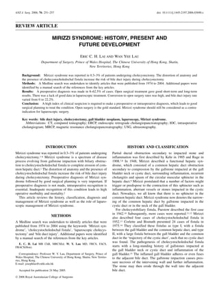
2. In the 1980s, the original type of Mirizzi syndrome and the
cholecystocholedochal fistula became recognized as different
evolving stages of the same disease process. In 1982, McSherry
et al. classified Mirizzi syndrome into two types based on endo-
scopic retrograde cholangiopancreatography (ERCP) findings
(Fig. 1).16 Type I involves the external compression of the com-
mon hepatic duct by a large stone impacted in the cystic duct or
the Hartmann’s pouch, without any lesion in the gall bladder or
the common hepatic duct wall. In type II, a cholecystcholedochal
fistula is present. It is caused by a calculus, which has eroded
partly or completely into the common duct. In 1989, Csendes
et al. classified Mirizzi syndrome into four types (Fig. 1). Their
classification further categorized the cholecystocholedochal fis-
tula according to its extent of destruction.1 Type I lesion is the
external compression of the common duct because of a stone
impacted at the neck of the gall bladder or at the cystic duct. Type
II lesion is a cholecystobiliary fistula (cholecystohepatic or chol-
ecystocholedochal) that results from the erosion of the anterior or
lateral wall of the common duct by the impacted stones, the fistula
involving less than one-third of the circumference of the common
duct. Type III lesion is a cholecystobiliary fistula with erosion of
the wall of the common duct that involves up to two-thirds of its
circumference. Type IV lesion is a cholecystobiliary fistula, with
complete destruction of the entire wall of the common duct. In the
past 20 years other authors have described different classification
systems, such as acute versus chronic, anatomic variant of cystic
duct versus no anatomic variant of cystic duct and obstruction due
to gallstones versus obstruction due to inflammation.17–19 In pub-
lished reports and in clinical practice, most clinicians would use
either the McSherry classification or the Csendes classification.
These classifications are more useful to guide surgical man-
agement. The reported incidence of McSherry type I varied from
11 to 45%, whereas that of McSherry type II (i.e. Csendes type II,
III and IV) varied from 55 to 89%. Only less than 6% of patients
with Mirizzi syndrome had Csendes type IV.
DIAGNOSIS
The preoperative diagnosis of Mirizzi syndrome is very import-
ant.20,21 In the series reported by Tan et al., bile duct injuries were
observed in four (16.7%) out of 24 patients operated. All the four
bile duct injuries occurred in patients who did not have a preop-
erative diagnosis.21
In Mirizzi syndrome, patients present with jaundice (60–100%)
and abdominal pain (50–100%).1,5,22–27 Ultrasonography (USG)
of the abdomen is widely used for the initial screening.28,29 A
typical USG finding of Mirizzi syndrome is a large, immovable
stone in the region of the neck of a shrunken gall bladder, with
dilatation of bile ducts above it and a common bile duct of normal
calibre below it. Computed tomography (CT) scan of the abdo-
men may show an irregular cavity near the gall bladder neck and
calculi outside the viscus. However, the radiological signs are not
specific.29–31 The main role of CT scan is to differentiate Mirizzi
syndrome from malignancy of the extrahepatic biliary system.
Direct cholangiography (ERCP or percutaneous transhepatic
cholangiography) is usually carried out after USG or CT scan in
order to delineate the cause, level and extent of biliary obstruc-
tion. Direct cholangiography is valuable in showing ductal ab-
normalities and fistula.32–34 Cholangiographic features of Mirizzi
syndrome are narrowing or curvilinear extrinsic compression,
usually involving the lateral portion of the distal common hepatic
duct with proximal dilatation. Magnetic resonance cholangiopan-
creatography (MRCP) has been shown to have a high sensitivity
and specificity in the detection of gallstone and bile duct steno-
sis.35,36 Magnetic resonance cholangiopancreatography can show
the typical features in Mirizzi syndrome, such as the extrinsic nar-
rowing of the common hepatic duct, a gallstone in the cystic duct,
the dilatation of the intrahepatic and common hepatic ducts and
a normal common bile duct. Magnetic resonance imaging can also
show the extent of inflammation around the gall bladder, and this
helps to differentiate this condition from other gall bladder dis-
eases.36,37 Magnetic resonance cholangiopancreatography has the
advantage of avoiding the complications of direct cholangiography.
Unfortunately, a preoperative diagnosis can be made only in
8–62.5% of patients.1,5,6,24–26 Therefore, intraoperative recognition
is essential.1,2,5,6,22–26 The presence of a shrunken gall bladder,
an obliterated Calot’s triangle, a dense fibrotic mass at the Calot’s
triangle and a dense adhesion at the subhepatic space should raise
the suspicion of Mirizzi syndrome. Baer et al. noted that if there
is a gush of bile after the removal of gall bladder stones, it
is suggestive of the presence of cholecystocholedochal fistula as
the cystic duct is always completely obliterated as a result of
chronic inflammation in patients without a fistula.2 Intraoperative
Fig. 1. Pathology and classifications of Mirizzi syndrome.
252 LAI AND LAU
Ó 2006 Royal Australasian College of Surgeons
3. cholangiogram (IOC) helps to confirm the diagnosis, determine
the location and size of the fistula, detect ductal stones and detect
whether there is any loss of integrity of the bile duct wall.1,2
However, it is sometimes difficult to have an IOC if the cystic
duct is obliterated, and persistent dissection in the Calot’s triangle
in trying to do a cholangiogram may result in bile duct injury.
Another useful investigatory tool is laparoscopic ultrasonography.
It provides real-time multiplanar images of the bile ducts from
various angles. It is useful in identifying the anatomy of the bil-
iary tree and in showing the relation of the common hepatic duct
to the cystic duct stone during dissection.38–41
MANAGEMENT
An accurate preoperative diagnosis with a careful surgical plan-
ning is very important in the management of Mirizzi syndrome.
The severe inflammatory process with dense adhesions and
oedematous tissues distort the anatomy. In addition, there may
be the presence of a cholecystocholedochal fistula. During the
operation, dissection of the Calot’s triangle may lead to bile duct
injury or excessive blood loss. Inadequate surgical planning may
lead to surgical morbidity, such as bile duct injury, delayed bile
duct stricture, secondary biliary cirrhosis of the liver, sepsis and
bleeding. In the published works in medicine, open surgical man-
agement of Mirizzi syndrome gives good short-term and long-
term results (Table 1).1,2,5,6,22–26,42,43 In some case series, there
were 0% operative morbidity and mortality.2,22–24,26,42
Mirizzi syndrome without cholecystocholedochal fistula
Conventional antegrade cholecystectomy is likely to damage the
biliary tree. Retrograde fundus first cholecystectomy cannot com-
pletely solve the problem. It can lead to a high transection of the
biliary tree if the pathology is not recognized. Subtotal cholecys-
tectomy was described by Bornman and Terblanche in 1985 to be
an easy, safe and definitive operation for a ‘difficult gall bladder’,
particularly in the presence of portal hypertension.44 This
approach is used to choose a convenient site, preferably the Hart-
mann’s pouch, to open the gall bladder. The gall bladder content is
then emptied. The gall bladder wall is partly removed by excision,
leaving a rim of wall attached to the liver. The origin of the cystic
duct and its course are identified from inside the gall bladder. Any
residual stones are removed. The cystic duct is secured without
dissection. This approach obviates the need for dangerous dissec-
tion in the Calot’s triangle, which has severe fibrosis and inflam-
mation. Since then, subtotal cholecystectomy has been used more
in the treatment of a ‘difficult gall bladder’, for example, portal
hypertension, Mirizzi syndrome.1,2,45–47 A lesser degree of chole-
cystectomy, that is, partial cholecystectomy, has also been shown
to be effective for type I Mirizzi syndrome.1,2 The common bile
duct should be investigated if necessary to exclude other causes of
obstructive jaundice. T-tube insertion may be needed for tempo-
rary decompression. Most inflammatory strictures return to nor-
mal when the inflammatory process resolves.
Mirizzi syndrome with cholecystocholedochal fistula
It is important to recognize the presence of a cholecystochole-
dochal fistula before operation. Corlette and Bismuth recom-
mended the initial removal of gallstones, followed by a partial
cholecystectomy, with a remnant of gall bladder left around the
Table1.ResultsofopensurgicalmanagementforMirizzisyndrome
StudynTypeITypeIIFollow-up
duration
Postoperative
haemorrhage
Residual
stone
Bile
leakage
Acutebile
ductinjury
External
biliaryfistula
Delaybile
ductstricture
In-hospital
mortality
Fanetal.1985224044–14months0000000
VenkateshRao
etal.198823
9092years0000000
Csendesetal.1989121923II,90;III,97;IV,9†1–13years
(mean,5.7)
42160161215
Baeretal.19902404ND0000000
Yipetal.1991245056–36months0000000
Ibrarullahetal.19935144II,7;III,3†1–27months
(mean,14)
0210000
Curetetal.199425175121–41months
(mean,30)
0221000
Xiaodongetal.199926164II,8;III,3;IV,1†78months
(mean,78)
0000000
Khanetal.19994233017–33months0000000
Shahetal.2001433482612–59months0230010
Johnsonetal.20016115II,3;III,3†1–20years1120100
†CsendesClassificationwasused;ND,nodataobtained.
MIRIZZI SYNDROME 253
Ó 2006 Royal Australasian College of Surgeons
4. fistula margins to aid in the closure of the fistula with its associ-
ated loss of part of the circumference of the bile duct wall.14
Successful choledochoplasty using the gall bladder or cystic duct
as pedicle graft has been well described in several series.48–51
A gall bladder flap has the advantage of having an independent
blood supply and a related mucosal lining. This technique has
been shown to be effective in an animal study.52 It has been widely
used in the past 20 years. Choledochoplasty using ligamentum
teres and vein patch has also been reported, but the available
outcome data were very limited.53,54 After exploring bile ducts
through a choledochotomy distal to the fistula, a T-tube is usually
placed into the bile duct through the fistula, and the gall bladder
remnant is closed around the tube. In case of a large fistula,
hepaticojejunostomy is recommended.
In the large series of 219 patients with Mirizzi syndrome
reported by Csendes et al., the authors recommended partial
cholecystectomy to remove the stones, to see the common duct,
and to define the type and location of the fistula with operative
cholangiogram.1 They recommended placing the T-tube distal to
the fistula rather than through the fistula as there were risks of
bile leakage and bile duct stricture. In Csendes’ type II Mirizzi
syndrome, suturing of the fistula using absorbable material or
choledochoplasty using the remnant of gall bladder was carried
out. In Csendes’ type III Mirizzi syndrome, choledochoplasty
rather than suturing was recommended. In Csendes’ type IV
Mirizzi syndrome, bilioenteric anastomosis was preferred. They
found that the operative mortality and morbidity increased with
the severity of the lesion. However, in the series reported by Baer
et al., high operative morbidity and mortality rates were found in
patients with cholecystocholedochal fistula managed with direct
suturing and flap closure technique.2 Baer et al. proposed that
direct repair using gall bladder flap was prone to failure because
the tissues were damaged by the inflammatory process, thus
resulting in a high rate of bile duct stenosis. Instead, they
suggested using biliary-enteric bypass to the wall of the chole-
cystocholedochal defect by using either a duodenostomy or a
Roux-en-Y jejunostomy. This technique has also been described
in other series.55,56
There is still no prospective randomized trial or large retrospec-
tive comparative trial to define whether choledochoplasty with
a gall bladder flap or choledochoduodenostomy bypass should
be used in the treatment of small-sized to moderate-sized chole-
cystocholedochal fistula and whether a T-tube should be placed at
or distal to the fistula site.
Based on the available data in the published works in medi-
cine, most authors suggest partial cholecystectomy with gall-
stone removal and exploration of the common duct through the
defect or a separate choledochotomy site. A gall bladder flap
is used to close the fistula site. A T-tube is inserted at the fistula
site or distal to the fistula site. If the gall bladder remnant tissue
is too inflamed, biliary-enteric bypass to the wall of the chole-
cystocholedochal defect in the fistula site or Roux-en-Y
hepaticojejunostomy is required. For the management of a
large cholecystocholedochal fistula with extensive destruction
of the common hepatic duct, Roux-en-Y hepaticojejunostomy is
recommended.
Non-surgical treatment of Mirizzi syndrome
Endoscopic treatment of Mirizzi syndrome comprises biliary
drainage by stent insertion and gallstone removal by using a basket
or a balloon. The alternatives are mechanical, electrohydraulic or
extracorporeal lithotripsy, or dissolution therapy.57–64 Percutane-
ous transhepatic management is reserved for patients who have
failed endoscopic treatment.65,66
The limitations of non-surgical treatment include: the need of
expertise, time-consuming procedures, multiple sessions of treat-
ment, cost of equipment and risk of complications. Complications
of non-surgical treatment include bile leakage from cystic or fis-
tula stump, sepsis and residual stones. Thus, non-surgical treat-
ment of Mirizzi syndrome should be used only in patients with
poor surgical risks. It can also be used as a temporary measure
during the preparation of patients for an elective operation.20,57,67
Mirizzi syndrome and carcinoma of gall bladder
It is difficult to differentiate Mirizzi syndrome from carcinoma of
gall bladder and from xanthogranulomatous cholecystitis.68–73 In
approximately 6–27.8% of patients with a preoperative diagnosis
of Mirizzi syndrome, carcinoma of gall bladder turned out to be
the final diagnosis. Redaelli et al. reported a high association
(27.8%) of carcinoma of gall bladder and Mirizzi syndrome.71
Computed tomography scan before an operation plays an impor-
tant role in the detection of any suspicious signs of carcinoma of
gall bladder.29 Frozen section should be obtained for all patients
with Mirizzi syndrome undergoing surgery. If an operable carci-
noma of gall bladder is diagnosed intraoperatively, open radical
cholecystectomy with porta hepatis lymph node dissection should
be carried out.74 We should not leave the diagnosis of gall bladder
carcinoma undetected at the time of surgery because of the fol-
lowing reasons. First, radical reoperation for incidental carcinoma
of gall bladder diagnosed after cholecystectomy has a poorer out-
come than one-stage radical operation.75,76 Second, for patients
who have been diagnosed to have incidental carcinoma of gall
bladder after laparoscopic cholecystectomy, port site recurrence
and peritoneal seeding are common and have been reported in 5–
20% of patients.77,78 Third, gall bladder perforation during chole-
cystectomy is associated with a high incidence of recurrence
(40%).77 All these would jeopardize the patient’s survival.79
Development in laparoscopic treatment
The incidence of iatrogenic bile duct injury is significantly higher
in laparoscopic cholecystectomy than in open cholecystectomy
for gall bladder pathologies.80–85 Three conditions predispose lap-
aroscopic cholecystectomy to an increased risk of bile duct inju-
ries: dangerous pathologies, difficult anatomy and dangerous
technique. Mirizzi syndrome is one of the dangerous pathologies.
Severe inflammation of the Calot’s triangle makes dissection of
the cystic duct and artery hazardous. The role of laparoscopic
surgery in the treatment of Mirizzi syndrome is still controversial.
Mirizzi syndrome is generally considered as a contraindication to
laparoscopic cholecystectomy.86–88 Laparoscopic subtotal chole-
cystectomy and laparoscopic fundus first cholecystectomy have
been described to decrease the chance of bile duct injury and
conversion rate to open surgery.89–93 There has been no retrospec-
tive comparative or prospective randomized trials to compare lap-
aroscopic with open surgical approach for Mirizzi syndrome.
Only small case series or case reports of laparoscopic treatment
of Mirizzi syndrome were found in the published works in medi-
cine. All these reports focused on the technical aspects and the
short-term outcomes (Table 2).41,87,88,94–103 Conversion to open
surgery rate was reported to range from 0 to 100%. If only patients
with type II Mirizzi syndrome were analysed, the conversion rate
254 LAI AND LAU
Ó 2006 Royal Australasian College of Surgeons
5. could be as high as 100%. Overall complication rate was 0–60%.
Bile duct injury rate ranged from 0 to 22.2%. In-hospital mortality
rate was reported to range from 0 to 25%.
It is evident that technically it is feasible to carry out laparo-
scopic treatment in highly selected patients with Mirizzi syn-
drome. However, the evidence in published reports showed that
laparoscopic treatment for Mirizzi syndrome is associated with
high morbidity and mortality rates. Mirizzi syndrome should
still be considered as a contraindication to laparoscopic surgery
in most centres, possibly with the exception of highly experi-
enced centres using laparoscopic treatment as a treatment under
investigation.
CONCLUSIONS
A high index of clinical suspicion is required to make a preop-
erative diagnosis of Mirizzi syndrome, which leads to good
surgical planning to treat the condition. Failing a preoperative
diagnosis, intraoperative recognition of Mirizzi syndrome is
important to avoid a high rate of bile duct injury. Intraoperative
cholangiography and ultrasound can help in the diagnosis. Open
surgical treatment is the gold standard. Mirizzi syndrome
should still be considered as a contraindication for laparoscopic
surgery.
REFERENCES
1. Csendes A, Diaz JC, Burdiles P, Maluenda F, Nava O. Mirizzi
syndrome and cholecystobiliary fistula: a unifying classifica-
tion. Br. J. Surg. 1989; 76: 1139–43.
2. Baer HU, Matthews JB, Schweizer WP, Gertsch P, Blumgart
LH. Management of the Mirizzi syndrome and the surgical
implication of cholecystocholedochal fistula. Br. J. Surg.
1990; 77: 743–5.
3. Mishra MC, Vashishitha S, Tandon R. Biliobiliary fistula: pre-
operative diagnosis and management implications. Surgery
1990; 108: 835–9.
4. Pemberton M, Wells AD. The Mirizzi syndrome. Postgrad.
Med. J. 1997; 73: 487–90.
5. Ibrarullah MD, Saxena R, Sikora SS, Kapoor VK, Saraswat VA,
Kaushik SP. Mirizzi’s syndrome: identification and management
strategy. Aust. N. Z. J. Surg. 1993; 63: 802–6.
6. Johnson LW, Sehon JK, Lee WC, Zibari GB, McDonald JC.
Mirizzi’s syndrome: experience from a multi-institutional
review. Am. Surg. 2001; 67: 11–14.
7. Kehr H. Die in neiner Klinik geubte Technik de Gallenstein
Operationen, mit einen Hinweis Auf die Indikationen und die
Dauerersolge. Munchen: JF Lehmann, 1905.
8. Ruge E. Deitrage Zur Chirurgischen Anatomie der grossen Gal-
lenwege (Ductus hepaticus, choledochus und pancreaticus).
Arch. Clin. Chir. 1908; 78: 47.
9. Mirizzi PL. Sindrome del conducto hepatico. J. Int. Chir. 1948;
8: 731–7.
10. Puestow CB. Spontaneous internal biliary fistula. Ann. Surg.
1942; 115: 1043–54.
11. Patt HH, Koontz AR. Cholecystocholedochal fistula; a report of
two cases. Ann. Surg. 1951; 134: 1064–5.
12. Behrend A, Cullen ML. Cholecysto-choledochal fistula: an
unusual internal biliary fistula. Ann. Surg. 1950; 132: 297–303.
13. Mirizzi PL. Les fistules biliobiliares internes spontanees.
J. Chir. 1952; 68: 23–8.
14. Corlette MB, Bismuth H. Biliobiliary fistula. A trap in the sur-
gery of cholelithiasis. Arch. Surg. 1975; 110: 377–83.
15. Tanaka N, Nobori M, Furuya T et al. Evolution of Mirizzi’s
syndrome with biliobiliary fistula. J. Gastroenterol. 1995; 30:
117–21.
16. McSherry CK, Ferstenberg H, Virship M. The Mirizzi syn-
drome: suggested classification and surgical therapy. Surg. Gas-
troenterol. 1982; 1: 219–25.
17. Morelli A, Narducci F, Ciccone R. Can Mirizzi syndrome be
classified into acute and chronic form? An endoscopic retro-
grade cholangiography (ERC) study. Endoscopy 1978; 10:
109–12.
18. Starling JR, Matallana RH. Benign mechanical obstruction of
the common hepatic duct (Mirizzi syndrome). Surgery 1980; 88:
737–40.
19. Nagakawa T, Ohta T, Kayahara M, Ueno K, Konishi I, Sanada H.
A new classification of Mirizzi syndrome from diagnostic and
therapeutic viewpoints. Hepatogastroenterology 1997; 44:
63–7.
20. Gomez G. Mirizzi syndrome. Curr. Treat. Options Gastroen-
terol. 2002; 5: 95–9.
Table 2. Results of laparoscopic management for Mirizzi syndrome
Study n Type I Type II Follow-up
duration
Conversion
rate (%)
Overall complication
rate (%)
Residual
stone
Bile
leakage
Acute bile
duct injury
In-hospital
mortality
Rust et al. 199187 1 1 0 ND 100 0 0 0 0 0
Paul et al. 199294 1 1 0 ND 0 0 0 0 0 0
Binnie et al. 199295 1 0 1 ND 0 0 0 0 0 0
Meng et al. 199541 1 1 0 ND 0 0 0 0 0 0
Posta et al. 199588 1 0 1 ND 100 0 0 0 0 0
Targarona et al. 199796 4 0 4 3–36 months
(mean, 19)
100 0 0 0 0 0
Sare et al. 199897 4 3 1 ND 25† 25 0 1 0 1
Kok et al. 199898 6 3 3 8–17 months
(mean, 12)
16.7 0 0 0 0 0
Chowbey et al. 200099 27 12 15 2.1 years
(mean, 2.1)
22 0 0 0 0 0
Vezakis et al. 2000100 5 2 3 26–61 months 0 60 3 0 0 1
Bagia et al. 2001101 9 8 1 ND 22.2 33.3 1 1 1 0
Schafer et al. 2003102 39 34 5 ND 74† 18 0 1 0 0
Yeh et al. 2003103 11 10 1 ND 36.4† 9.1 1 0 0 0
†All patients were Mirizzi type II; ND, no data obtained.
MIRIZZI SYNDROME 255
Ó 2006 Royal Australasian College of Surgeons
6. 21. Tan KY, Ching HC, Chen CYY, Tan SM, Poh BK, Hoe MNY.
Mirizzi syndrome: noteworthy aspects of a retrospective study
in one centre. ANZ J. Surg. 2004; 74: 833–7.
22. Fan ST, Lau WY, Lee MJR, Wong KK. Cholecystohepaticodo-
chal fistula: the value of pre-operative recognition. Br. J. Surg.
1985; 72: 743–4.
23. Venkatesh Rao PS, Tandon RK, Kapur BML. Biliobiliary
fistula: review of nine cases. Am. J. Gastroenterol. 1988; 83:
652–7.
24. Yip AW, Chow WC, Chan J, Lam KH. Mirizzi syndrome with
cholecystocholedochal fistula: preoperative diagnosis and man-
agement. Surgery 1992; 111: 335–8.
25. Curet MJ, Rosendale DE, Congilosi S. Mirizzi syndrome
in a Native American population. Am. J. Surg. 1994; 168:
616–21.
26. Xiaodong H, Hongsheng L, Chaoji Z, Zhenhuan Z, Jianxi Z.
Diagnosis and treatment of the Mirizzi syndrome. Chin. Med.
Sci. J. 1999; 14: 246–8.
27. Lubbers EJ. Mirizzi syndrome. World J. Surg. 1983; 7: 780–5.
28. Dewbury KC. The features of the Mirizzi syndrome on ultra-
sound examination. Br. J. Radiol. 1979; 52: 990–92.
29. Becker CD, Hassler H, Terrier F. Preoperative diagnosis of the
Mirizzi syndrome: limitations of sonography and computed
tomography. AJR Am. J. Roentgenol. 1984; 143: 591–6.
30. Pedrosa CS, Casanova R, de la Torre S, Villacorta J. CT findings
in Mirizzi syndrome. J. Comput. Assist. Tomogr. 1983; 7:
419–25.
31. Salloum RM, Koniaris L. Image of the month. Mirizzi syn-
drome. Arch. Surg. 2004; 28: 254–7.
32. Cornud F, Grenier P, Belghiti J, Breil P, Nahum H. Mirizzi
syndrome and biliobiliary fistulas: roentgenologic appearance.
Gastrointest. Radiol. 1981; 6: 265–8.
33. Ravo B, Epstein H, La Mendola S, Ger R. The Mirizzi syn-
drome: preoperative diagnosis by sonography and transhepatic
cholangiography. Am. J. Gastroenterol. 1986; 81: 688–90.
34. Cruz FO, Barriga P, Tocornal J, Burhenne HJ. Radiology of the
Mirizzi syndrome: diagnostic importance of the transhepatic
cholangiogram. Gastrointest. Radiol. 1983; 8: 249–53.
35. Becker CD, Grossholz M, Becker M, Mentha G, de Peyer R,
Terrier F. Choledocholithiasis and bile duct stenosis: diagnostic
accuracy of MR cholangiopancreatography. Radiology 1997;
205: 523–30.
36. Becker CD, Grossholz M, Mentha G, de Peyer R, Terrier F. MR
cholangiopancreatography: technique, potential indications, and
diagnostic features of benign, postoperative, and malignant con-
ditions. Eur. Radiol. 1997; 7: 865–74.
37. Kim PN, Outwater EK, Mitchell DG. Mirizzi syndrome:
evaluation by MRI imaging. Am. J. Gastroenterol. 1999; 94:
2546–50.
38. Tranter SE, Thompson MH. Potential of laparoscopic ultraso-
nography as an alternative to operative cholangiography in the
detection of bile duct stones. Br. J. Surg. 2001; 88: 65–9.
39. Tomonaga T, Filipi CJ, Lowham A, Martinez T. Laparoscopic
intracorporeal ultrasound cystic duct length measurement:
a new technique to prevent common bile duct injuries. Surg.
Endosc. 1999; 13: 183–5.
40. Kelly SB, Remedios D, Lau WY, Li AK. Laparoscopic ultraso-
nography during laparoscopic cholecystectomy. Surg. Endosc.
1997; 11: 67–70.
41. Meng WCS, Kwok SPY, Kelly SB, Lau WY, Li AKC. Manage-
ment of Mirizzi syndrome by laparoscopic cholecystectomy and
laparoscopic ultrasonography. Br. J. Surg. 1995; 82: 396.
42. Khan TF, Muniandy S, Hayat FZ, Sherazi ZA, Nawaz MH.
Mirizzi syndrome: a report of 3 cases with a review of the
present classifications. Singapore Med. J. 1999; 40: 171–3.
43. Shah OJ, Dar MA, Wani MA, Wani NA. Management of Mirizzi
syndrome: a new surgical approach. ANZ J. Surg. 2001; 71:
423–7.
44. Bornman PC, Terblanche J. Subtotal cholecystectomy: for the
difficult gallbladder in portal hypertension and cholecystitis.
Surgery 1985; 98: 1–6.
45. Douglas PR, Ham JM. Partial cholecystectomy. Aust. N. Z. J.
Surg. 1990; 60: 595–7.
46. Cottier DJ, McKay C, Anderson JR. Subtotal cholecystectomy.
Br. J. Surg. 1991; 78: 1326–8.
47. Katsohis C, Prousalidis J, Tzardinoglou E et al. Subtotal chole-
cystectomy. HPB Surg. 1996; 9: 133–6.
48. Sandblom P, Tabrizian M, Rigo M, Fluckiger A. Repair of
common bile duct defects using the gallbladder or cystic
duct as a pedicled graft. Surg. Gynecol. Obstet. 1975; 140:
425–32.
49. Philippakis M, Apostolidis N, Androulakakis P, Doundoulakis
N, Legakis N. Use of the gallbladder neck in the reconstruction
of the bifurcation of the main hepatic ducts. Am. Surg. 1975; 41:
103–5.
50. Chourdakis CN, Androulakakis PA, Lekakos NL. Repair of
cholecystocholedochal fistulas using gallbladder patching.
Arch. Surg. 1976; 111: 197–9.
51. Strugnell NA, Sali A. Choledochoplasty for cholecystocholedo-
chal fistula (Mirizzi syndrome type II): a case report and liter-
ature review. Aust. N. Z. J. Surg. 1995; 65: 285–8.
52. Mortensen FV, Ishibashi T, Hojo N, Yasuda Y. A gallbladder
flap for reconstruction of the common bile duct. An experimen-
tal study on pigs. J. Hepatobiliary Pancreat. Surg. 2004; 11:
112–5.
53. Settaf A, Balafrej S. Biliary surgery using the ligamentum teres.
Technique for repairing loss of substance in the common bile
duct. Ann. Chir. 1993; 47: 529–33.
54. Rutledge RH. Methods of repair of noncircumferential bile duct
defects. Surgery 1983; 93: 333–42.
55. Karakoyunlar O, Sivrel E, Koc O, Denecli AG. Mirizzi’s syn-
drome must be ruled out in the differential diagnosis of any
patients with obstructive jaundice. Hepatogastroenterology
1999; 46: 2178–82.
56. Karademir S, Astarcioglu H, Sokmen S et al. Mirizzi’s syn-
drome: diagnostic and surgical considerations in 25 patients.
J. Hepatobiliary Pancreat. Surg. 2000; 7: 72–7.
57. England RE, Martin DF. Endoscopic management of Mirizzi’s
syndrome. Gut 1997; 40: 272–6.
58. Tsuyuguchi T, Saisho H, Ishihara T, Yamaguchi T, Onuma EK.
Long-term follow-up after treatment of Mirizzi syndrome
by peroral cholangioscopy. Gastrointest. Endosc. 2000; 52:
639–44.
59. Benninger J, Rabenstein T, Farnbacher M, Keppler J, Hahn EG,
Schneider HT. Extracorporeal shockwave lithotripsy of gall-
stones in cystic duct remnants and Mirizzi syndrome. Gastro-
intest. Endosc. 2004; 60: 454–9.
60. Vakil N, Sawyer R. Endoscopic drainage of the gallbladder in
a septic variant of the Mirizzi syndrome. Gastrointest. Endosc.
1994; 40: 236–8.
61. Adam A, Roddie ME, Benjamin IS. Case report: Mirizzi syn-
drome: treatment with metallic endoprosthesis. Clin. Radiol.
1993; 48: 198–201.
62. Delcenserie R, Joly JP, Dupas JL. Endoscopic diagnosis and
treatment of Mirizzi syndrome. J. Clin. Gastroenterol. 1992;
15: 343–6.
63. Martin DF, Tweedle DEF, Rao PN. Endoscopic gallbladder
catheterization and extracorporeal shockwave lithotripsy in
the management of Mirizzi’s syndrome. Endoscopy 1988; 20:
321–2.
64. Binmoeller KF, Thonke F, Soehendra N. Endoscopic treat-
ment of Mirizzi’s syndrome. Gastrointest. Endosc. 1993; 39:
532–6.
65. Cairns SR, Watson GN, Lees WR, Salmon PR. Percutaneous
lithotripsy and endoprosthesis: a new treatment for obstructive
jaundice in Mirizzi’s syndrome. Br. Med. J. 1987; 295: 1448.
256 LAI AND LAU
Ó 2006 Royal Australasian College of Surgeons
7. 66. Oxtoby JW, Yeong CC, West DJ. Mirizzi syndrome treated by
percutaneous stone removal. Cardiovasc. Intervent. Radiol.
1994; 17: 207–9.
67. Hazzan D, Golijanin D, Reissman P, Adler SN, Shiloni E. Com-
bined endoscopic and surgical management of Mirizzi syn-
drome. Surg. Endosc. 1999; 13: 618–20.
68. Guzman-Valdivia G. Xanthogranulomatous cholecystitis: 15
years’ experience. World J. Surg. 2004; 28: 254–7.
69. Lee KC, Yamazaki O, Horii K et al. Mirizzi syndrome caused
by xanthogranulomatous cholecystitis: report of a case. Surg.
Today 1997; 27: 757–61.
70. Sharma AK, Rangan HK, Choubey RP, Thakur SK, Kumar A.
Pitfalls in the management of Mirizzi’s syndrome. Trop. Gastro-
enterol. 1998; 19: 72–4.
71. Redaelli CA, Buchler MW, Schilling MK, Krahenbuhl L, Ruchti
C, Blumgart LH. High coincidence of Mirizzi syndrome and
gallbladder carcinoma. Surgery 1997; 121: 58–63.
72. Principe A, Del Gaudio M, Grazi GL, Paolucci U, Cavallari A.
Mirizzi syndrome with cholecysto-choledocal fistula with a high
CA 199 level micmicking biliary malignancies: a case report.
Hapatogastroenterology 2003; 50: 1259–62.
73. Nishimura A, Shirai Y, Hatakeyama K. High coincidence of
Mirizzi syndrome and gallbladder carcinoma. Surgery 1999;
126: 587–8.
74. Shirai Y, Yoshida K, Tsukada K, Muto T, Watanabe H. Radical
surgery for gallbladder carcinoma: long-term results. Ann. Surg.
1992; 216: 565–8.
75. Shirai Y, Yoshida K, Tsukada K, Muto T. Inapparent carcinoma
of the gallbladder. An appraisal of a radical second operation
after simple cholecystectomy. Ann. Surg. 1992; 215: 326–31.
76. Toyonaga T, Chijiiwa K, Nakano K et al. Completion radical
surgery after cholecystectomy for accidentally undiagnosed
gallbladder carcinoma. World J. Surg. 2003; 27: 266–71.
77. Z’graggen K, Birrer S, Maurer CA, Wehrli H, Klaiber C, Baer
HU. Incidence of port site recurrence after laparoscopic chole-
cystectomy for preoperatively unsuspected gallbladder carci-
noma. Surgery 1998; 124: 831–8.
78. Fong Y, Brennan MF, Turnbull A, Coit DG, Blumgart LH. Gall-
bladder cancer discovered during laparoscopic surgery. Poten-
tial for iatrogenic tumour dissemination. Arch. Surg. 1993; 128:
1054–6.
79. Lai EC, Lau WY. Aggressive surgical resection for carcinoma of
gallbladder. ANZ J. Surg. 2005; 75: 441–4.
80. Davidoff AM, Pappas TN, Murray EA et al. Mechanisms of
major biliary injury during laparoscopic cholecystectomy. Ann.
Surg. 1992; 215: 196–202.
81. Moossa AR, Easter DW, van Sonnenberg E, Casola G,
D’Agostino H. Laparoscopic injuries to the bile duct: a cause
for concern. Ann. Surg. 1992; 215: 203–8.
82. Deziel DJ, Millikan KW, Economou SG, Doolas A, Ko ST,
Airan MC. Complication of laparoscopic cholecystectomy:
a national survey of 4,292 hospitals and an analysis of 77,604
cases. Am. J. Surg. 1993; 165: 9–14.
83. Strasberg SM, Hertl M, Soper NJ. An analysis of the problem of
biliary injury during laparoscopic cholecystectomy. J. Am. Coll.
Surg. 1995; 180: 101–25.
84. Adamsen S, Hansen OH, Funch-Jensen P, Schulze S, Stage JG,
Wara P. Bile duct injury during laparoscopic cholecystectomy:
a prospective nationwide series. J. Am. Coll. Surg. 1997; 184:
571–8.
85. Fletcher DR, Hobbs MS, Tan P et al. Complications of chole-
cystectomy: risks of the laparoscopic approach and protective
effects of operative cholangiography. Ann. Surg. 1999: 229:
449–57.
86. Contini S, Dalla Valle R, Zinicola R, Botta GC. Undiagnosed
Mirizzi’s syndrome: a word of caution for laparoscopic sur-
geons: a report of three cases and review of the literature. J. Lap-
aroendosc. Adv. Surg. Tech. A. 1999; 9: 197–203.
87. Rust KR, Chancy TV, Warren G, Mertesdorf J, Maxwell JG.
Mirizzi syndrome: a contraindication to coelioscopic cholecys-
tectomy. J. Laparoendosc. Surg. 1991; 1: 133–7.
88. Posta CG. Unexpected Mirizzi anatomy: a major hazard to the
common bile duct during laparoscopic cholecystectomy. Surg.
Laparosc. Endosc. 1995; 5: 412–14.
89. Crosthwaite G, Mckay C, Anderson JR. Laparoscopic subtotal
cholecystectomy. J. R. Coll. Surg. Edinb. 1995; 40: 20–1.
90. Ransom KJ. Laparoscopic management of acute cholecystitis
with subtotal cholecystectomy. Am. Surg. 1998; 64: 955–7.
91. Michalowski K, Bormman PC, Krige JE, Gallagher PJ,
Terblanche J. Laparoscopic subtotal cholecystectomy in patients
with complicated acute cholecystitis or fibrosis. Br. J. Surg.
1998; 85: 904–6.
92. Chowbey PK, Sharama A, Khullar R, Mann V, Baijal M, Vashis-
tha A. Laparoscopic subtotal cholecystectomy: a review of
56 procedures. J. Laparoendosc. Adv. Surg. Tech. A. 2000; 10:
31–4.
93. Mahmud S, Masaud M, Canna K, Nassar AH. Fundus-first lap-
aroscopic cholecystectomy. Surg. Endosc. 2002; 16: 581–4.
94. Paul MG, Burris DG, McGuire AM, Thorfinnson HD, Schone-
kas H. Laparoscopic surgery in the treatment of Mirizzi syn-
drome. J. Laparoendosc. Surg. 1992; 2: 157–63.
95. Binnie NR, Nixon SJ, Palmer KR. Mirizzi syndrome managed
by endoscopic stenting and laparoscopic cholecystectomy. Br. J.
Surg. 1992; 79: 647.
96. Targarona EM, Andrade E, Balague C, Ardid J, Trias M. Mir-
izzi’s syndrome diagnostic and therapeutic controversies in the
laparoscopic era. Surg. Endosc. 1997; 11: 842–5.
97. Sare M, Gurer S, Taskin V, Aladag M, Hilmioglu F, Gurel M.
Mirizzi syndrome: choice of surgical procedure in the laparo-
scopic era. Surg. Laparosc. Endosc. 1998; 8: 63–7.
98. Kok KY, Goh PY, Ngoi SS. Management of Mirizzi’s syndrome
in the laparoscopic era. Surg. Endosc. 1998; 12: 1242–4.
99. Chowbey PK, Sharma A, Mann V, Khullar R, Baijal M,
Vashistha A. The management of Mirizzi syndrome in the lap-
aroscopic era. Surg. Laparosc. Endosc. Percutan. Tech. 2000;
10: 11–14.
100. Vezakis A, Davides D, Birbas K, Ammori BJ, Larvin M,
McMahon MJ. Laparoscopic treatment of Mirizzi syndrome.
Surg. Laparosc. Endosc. Percutan. Tech. 2000; 10: 15–18.
101. Bagia JS, North L, Hunt DR. Mirizzi syndrome: an extra hazard
for laparoscopic surgery. ANZ J. Surg. 2001; 71: 394–7.
102. Schafer M, Schneiter R, Krahenbuhl L. Incidence and manage-
ment of Mirizzi syndrome during laparoscopic cholecystec-
tomy. Surg. Endosc. 2003; 17: 1186–90.
103. Yeh CN, Jan YY, Chen MF. Laparoscopic treatment for Mirizzi
syndrome. Surg. Endosc. 2003; 17: 1573–8.
MIRIZZI SYNDROME 257
Ó 2006 Royal Australasian College of Surgeons