

Recommended
More Related Content
What's hot
What's hot (20)
Similar to OBS Jaundice.pptx
Similar to OBS Jaundice.pptx (20)
Recently uploaded
Recently uploaded (20)
OBS Jaundice.pptx
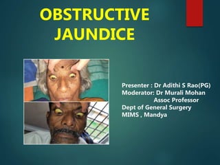
- 1. OBSTRUCTIVE JAUNDICE Presenter : Dr Adithi S Rao(PG) Moderator: Dr Murali Mohan Assoc Professor Dept of General Surgery MIMS , Mandya
- 2. Contents Anatomy Physiology Bilirubin metabolism Patho-physiology of Obstructive Jaundice Classification Clinical features Investigations Individual Diseases
- 3. Definition Cholestasis is defined as failure of normal amounts of bile to reach duodenum. Extrahepatic biliary disease which is often amenable to surgical treatment is called “SURGICAL JAUNDICE” or “OBSTRUCTIVE JAUNDICE”.
- 4. Anatomy
- 5. Physiology Bile is composed of water, electrolytes, bile salts, proteins, lipids, and bile pigments. The primary bile salts, cholate and chenodeoxycholate, are synthesized in the liver by cholesterol. Bile salts are formed from bile acids aid in the digestion and absorption of fats in the intestines. About 95% of the bile acid pool is reabsorbed and returned through the portal venous system to the liver, also known as the enterohepatic circulation
- 6. … The color of bile is due to the presence of the pigment bilirubin diglucuronide, which is the metabolic product from the breakdown of hemoglobin Once in the intestine, bacteria convert it into urobilinogen, a small fraction of which is absorbed and secreted into the bile.
- 8. Pathophysiology-Physical effects of biliary obstruction Increased pressure Bile reflux across the hepatocyte Hepatocellular damage Reduced hepatic blood flow
- 9. Biochemical effect Conjugated hyperbilirubinemia Elevation of alkaline phosphatase Reduced blood fibrinogen, prothrombin and factor VIII leading to impaired blood coagulation process Reduced serum albumin & prealbumin
- 10. Patho physiology Systemic effect Abnormal serum lipids- cell membrane defects Bile acids, bilirubin- exert direct toxic effects on cells ENDOTOXEMIA d/t raised absorption of endotoxins from intestine d/t absent bile salts
- 11. Systemic effects of long standing obstructive Jaundice Impaired wound healing Coagulopathies- vit K def, platelet dysfunction Reduced immunity- sepsis Acute renal failure Hypotension
- 12. Site of obstruction A. In the lumen of duct • Choledocholithiasis • Parasitic infestation due to: Hydatid disease, Ascariasis • Hemobilia Classification
- 13. B. In the wall of duct 1. Congenital: Biliary atresia,Choledochol cyst 2. Acquired: Papillary stenosis,Strictures ,Mirrizzi’s syndrome 3. Malignant causes: Ca Gall bladder, Cholangiocarcinoma
- 14. C. Outside the wall 1. Benign: Pseudocyst of pancreas 2. Malignant: • Ca head of pancreas • Enlarged lymph nodes at porta hepatitis • Periampullary Ca • Extra biliary malignancy
- 15. Type of obstruction Type I: Complete obstruction, producing overt jaundice. e.g. Ca head of pancreas Cholangio carcinoma
- 16. Type II: Intermittent obstruction, which produces symptoms and typical biochemical changes, but may or may not be associated with attacks of clinical jaundice. e.g. Cbd stones Periampullary tumor Duodenal diverticula Choledochal cyst Intra biliary parasites Hemobilia
- 17. Type III: Chronic incomplete obstruction, with or without classic symptoms or the observation of biochemical changes which will eventually produce pathological changes in the bile duct or the liver. e.g. s Strictures of the CBD Stenosed biliary enteric anastamosis Cystic fibrosis Chronic pancreatitis Stenosis of the Sphincter of Oddi
- 18. Type IV: Segmental obstruction, in which one or more anatomical segments of the intrahepatic biliary tree are obstructed. Traumatic , Hepato docho lithiasis = intra hepatic stones, Sclerosing cholangitis
- 19. Clinical features SYMPTOMS Jaundice: Itching : Frequent with malignant obstruction- cause is obscure ??Bile acids which cause injury to the membrane of skin cells and a release of pruritogenic proteases.
- 20. Steatorrhea: Malabsorption of fats due to absence of bile salts- pale, bulky, offensive stools. Biliary pain : Epigastrium, radiates to right hypochondrium and to right sub-scapular region, most commonly constant in nature with only minor fluctuation in intensity.
- 21. Hepatic failure Long standing cholestasis- months to years. Edema, hypoalbuminemia, deep jaundice, bleeding unresponsive to parenteral vit K, decrease of itching.
- 22. ■ Signs of liver failure: Late stages
- 23. General examination 1. Icterus 2. Xanthomas : are due to increased serum cholesterol for > 3 months. Appear as whitish macular lesions due to cholesterol deposition in skin. Xanthelesma are raised and seen around the eyes. 3. Per abdomen: 1. HEPATOMEGALY: Liver enlarged – SHARP EDGE Palpable gall bladder (courvoisier’s law). 2. Ascites may be related to liver disease, hypoalbuminemia with portal hypertension or malignancy.
- 24. Courvoisier’s law In the presence of Jaundice , a palpable Gall bladder is seldom due to stones. Virchow’s node • Left supraclavicular lymph nodes . • Troisier’s sign
- 25. Investigations BIOCHEMICAL investigations LFT : Sclera 2.5mg/dl
- 29. USG GALLBLADDER 1. IOC for GB pathology 2. After fasting for 6 hour- distend GB 3. Gallstones: acoustic shadow, postural variation 4. Cholecystitis: wall > 5 mm, distended GB, peri cholecystic fluid, sludge , subserosal edema , sonographic murphy’s 5. Polyps 6. Ca
- 31. Endoscopic Ultrasound Requires a special endoscope with an ultrasound transducer at its tip. It offers noninvasive imaging of the bile ducts and adjacent structures. It is of particular value in the evaluation of STAGE of tumors and their resectability. The ultrasound endoscope has a biopsy channel, allowing needle biopsies of a tumor under ultrasonic guidance.
- 32. Computed Tomography (CT) One disadvantage is gallstones and bile appear nearly isodense on CT; that is, it is difficult to distinguish gallstones from bile, unless the stones are heavily calcified. CT identifies gallstones within the biliary tree and gallbladder with a sensitivity of only about 55% to 65%.
- 33. Conversely, CT is more accurate at identifying the site and cause of extrahepatic biliary obstruction. Abdominal CT is a powerful tool for evaluating hepatobiliary or pancreatic neoplasm, liver abscess, or hepatic parenchymal disease (e.g., biliary cirrhosis, organ atrophy). Use of CT cholangiogram provides improved definition of the biliary tract comparable to magnetic resonance cholangiography
- 35. ERCP INVASIVE IOC for distal CBD obstruction Pt is kept NPO for 6 hrs Under sedation Side viewing duodenoscope Cannula is inserted into papilla & contrast (IOPROMIDE/IOGLYCAMATE) is injected Films taken fluoroscopy
- 36. ERCP: Diagnostic Inspection of stomach, duodenum, pancreatic ducts, biliary tract in one sitting Manometry of sphincter of oddi Bile / pancreatic juice for culture Stricture / growths – brush cytology / biopsy
- 37. ERCP- Therapeutic Endoscopic sphincterotomy- with/without stone extraction Endoscopic biliary endoprosthesis Endoscopic NBD ERCP Failure • Ampullary diverticulum/stricture/tumor • Billroth 2 surgery
- 38. ERCP complications 2-5% Technical errors, unsterile Pancreatitis: M/C ,raised serum amylase, esp if pancreatic duct cannulated Cholangitis: 2nd M/C Bleeding – procedure Perforation– procedure
- 39. ERCP-CBD STONES
- 40. PTC- percutaneous trans hepatic cholangiogram Proximal biliary obstruction /ERCP is not technically possible. PTC involves advancement of a 21- or 22-gauge needle from the right MAL into the expected location of the central intrahepatic biliary tree using fluoroscopic or ultrasound guidance. When a bile duct is engaged, it is filled with contrast. Images are obtained from multiple projections PTBD/anatomical landmarks during surgical reconstruction, /access for nonoperative dilation of strictures.
- 41. PTC
- 42. Criterion ERCP PTC Success rate 80-90% 80-90% Complications 5% 5% Mortality 0.2% 0.2% Conditions for use skilled personnel distal lesion failed PTC skilled personnel proximal lesion abnormal anatomy failed ERCP
- 43. MRCP MRCP v/s ERCP without the added risk of pancreatitis, sedation, and perforation. Image quality is less with MRCP Intervention not possible MRCP is of value in patients with a low probability of gallstones or obstruction in the bile ducts or pancreas, or in patients who are too sick for the anesthesia required for ERCP .
- 44. Magnetic resonance cholangiopancreatography (MRCP). Liver metastasis (M) common bile duct (CBD) pancreatic duct (PD).
- 45. MRCP ERCP
- 46. FDG-PET Well established for differentiation of benign from malignant lesions, staging malignant lesions, detection of malignancy recurrence, and monitoring therapy for various malignancies Accurate in predicting the presence of nodular cholangiocarcinoma (mass >1 cm) and gallbladder carcinoma (sensitivity, 78%).
- 48. MEDICAL TREATMENT PRURITIS Bile drainage CHOLESTYRAMINE: DOC binds bile salts in intestine Doubtful value ?? URSODIOL, RIFAMPIN
- 49. NUTRITION Calorie + Protein supplementation FATS : (Restrict other fats-Steatorrhea) Medium Chain Triglycerides MCT rich in coconut oil supplemented – 40 g/day Vit K-10mg/day Vit D-4000 U /day Vit A-25000U/day
- 50. Choledocholithiasis PRIMARY : Stones are formed in CBD. - associated with biliary stasis and infection and are more commonly seen in Asian populations. The causes of biliary stasis that lead to the development of primary stones include biliary stricture, papillary stenosis, or tumors.
- 51. … SECONDARY: Stones formed primarily in gallbladder & migrate into CBD. More common than primary
- 52. … IOC is ERCP, also therapeutic :cannulation of the ampulla of Vater and diagnostic cholangiography are achieved in more than 90% of cases. MRC provides excellent anatomic detail
- 53. Management of CBD stones Endoscopic Cholangiography Endoscopic clearance of stones from the common bile duct can avoid the need for an open surgery Patients with worsening cholangitis, ampullary stone impaction, biliary pancreatitis, multiple co morbidities, and cirrhosis are considered good candidates for endoscopic therapy. Endoscopic sphincterotomy is done followed by extraction of stones using Dormia basket. If the stones are large or impacted mechanical lithotripsy can be done. Prompt cholecystectomy after endoscopic clearance of the common bile duct should be performed to prevent recurrences
- 54. Laparoscopic CBD Exploration : Lap CBDE comb with lap cholecystectomy - through the cystic (5mm choledohoscope) or with formal choledochotomy allows the stones to be retrieved during the same procedure. If a choledochotomy is performed, a T tube is left in place.
- 57. Open Common Bile Duct Exploration CHOLEDOCHOTOMY & stone extraction Stones impacted in the ampulla may be difficult for complete ductal clearance and common bile duct exploration. In these cases, transduodenal sphincteroplasty and stone extraction should be performed; alternatively, if this is not successful, a choledochoduodenostomy or a Roux-en-Y choledochojejunostomy should be performed.
- 60. Choledochojejunostomy using a loop of jejunum to the distal common hepatic duct. A loop of jejunum just distal to the ligament of Treitz is brought over the (antecolic) to the dilated bile duct. A side- to-side or end-to-side choledochojejunostomy is performed using a two-layer anastomosis. The jejunum is usually secured to the liver capsule to tension on the anastomosis. A jejunojejunostomy is performed to minimize reflux into the bile duct.
- 61. Choledochojejunostomy with a Roux-en-Y loop. The jejunum is transected about cm distal to the ligament of and the distal end is oversewn and mobilized to the bile duct. end-to-side anastomosis is performed between the dilated bile duct and the antimesenteric border of the jejunum. Intestinal continuity is established with a jejunojejunostomy 45 cm from choledochojejunostomy to prevent reflux into the biliary tree. . This drainage procedure is preferred to a loop because reflux of enteric contents into the bile ducts rarely occurs.
- 62. BILIARY STRICTURES-BENIGN CAUSES BILE DUCT INJURIES Postoperative bile duct strictures Cholecystectomy :exploration of common bile duct Other operative procedures: Biliary enteric anastamosis, liver transplant Strictures Related to Endoscopic or Percutaneous Biliary Manipulations Stricture after blunt or penetrating injury POST-INFLAMMATORY STRUCTURE: Cholangitis, Pancreatitis, Duodenal ulcer Primary Sclerosing Cholangitis Radiation-induced Cholangitis
- 63. Iatrogenic bile duct injury SURGERY- Commonest cause of biliary stricture Factors - Anatomical Variation, Use of diathermy near Calot’s triangle, Technical errors, Bile duct ischemia, Pathological factors.
- 64. Most benign strictures follow iatrogenic bile duct injury, most commonly during laparoscopic cholecystectomy. Long-term sequel may lead to recurrent cholangitis, secondary biliary cirrhosis, and portal hypertension.
- 65. Ways of injuring the extrahepatic biliary ducts at operation. A, Quick clamping at a hemorrhage from the cystic artery area in a field obscured by blood. B, Too much traction on the gallbladder with knuckling of the common duct and the forceps applied too low. C, Cystic duct clamped too close to the common duct. A tie will then completely obstruct the duct. D, Inadvertent clamping of long cystic duct closely adherent to the common hepatic duct.
- 68. Primary end-to-end repair of a bile duct injury over a T- tube.
- 69. Choledochal Cyst Congenital cystic dilation of the biliary ducts ? Cause Weakness of the wall/distal obstruction /Reflux of pancreatic enzymes into the CBD secondary to an anomaly of the pancreaticobiliary junction (APBJ)
- 70. C/F Infantile group consisting of babies younger than 1 year, with or without obvious hepatomegaly, with prolonged jaundice and acholic stools. adult form of choledochal cyst - classic triad: pain, jaundice, and a palpable mass.
- 71. Choledochal Cyst- Classification Type I - Cystic or fusiform dilatation of the common bile duct (CBD); most frequent type (90-95% of the cases). Type II - Diverticulum of the CBD, with normal size CBD Type III - Choledochocele, a cystic dilatation of the distal intramural portion of the CBD, typically protruding into the second portion of the duodenum Type IV - Cystic or fusiform dilatation of the CBD associated with cystic, fusiform, or saccular dilatation of intrahepatic bile ducts, also termed form fruste Type V - Cystic, fusiform, or saccular dilatation of the intrahepatic bile ducts associated with a normal CBD; may be associated with hepatic fibrosis (referred to as Caroli disease)
- 72. Todani modification of the classic Alonso-Lej classification
- 73. … Ultrasonography – dilated ducts & type 1 cysts ERCP is the standard diagnostic study. It clearly shows the anatomy of the pancreaticobiliary junction CT scanning may also be useful to delineate the cyst and its relationship to surrounding structures.
- 74. CT scan shows a large cyst with wall thickening.
- 75. Rx of choledochal cysts Total excision of the cyst in types I, II, and IV followed by reconstruction of the biliary tree with hepaticojejunostomy in a Roux-en-Y fashion Internal drainage, either with cystoduodenostomy or cystojejunostomy. :high incidence of calculi, recurrent cholangitis, anastomotic strictures, and carcinoma arising from the cyst. Lilly’s technique: inflammation – entire cyst cant be removed-resection of the anterolateral part of the cyst followed by an endocystic resection of the lining, leaving the back wall adjacent to the portal vein in place.
- 76. … Type III choledochal cysts, lateral duodenotomy with unroofing of the choledochocele followed by ductoplasty Type V choledochal cysts, patients with localized disease may benefit from a hepatic lobectomy. If the disease is diffuse, involving both lobes of the liver, treatment is palliative and liver transplantation may be required.
- 77. Extra hepatic Biliary atresia Failure of vacuolization of the solid embryonic bile ducts. most common surgically treatable cause of cholestasis encountered during the newborn period Causes prolonged neonatal jaundice, for more than 2 weeks.
- 78. … There are three main types of extrahepatic biliary atresia:- Type I: atresia restricted to the common bile duct. Type II: atresia of the common hepatic duct. Type III: atresia of the right and left hepatic duct.
- 79. TREATMENT OF BILIARY ATRESIA The Kasai procedure consists of mobilizing the extrahepatic ducts and anastomosing a jejunal Roux en-Y loop to the liver hilum. Complications include progressive biliary cirrhosis, ascending cholangitis, and portal hypertension. Liver transplantation is indicated in cases of failed portoenterostomy, progressive fibrosis, or biliary cirrhosis. In fact, biliary atresia is the most common cause of end-stage liver disease in infants and a leading indication for liver transplantation.
- 80. Ca GALLBLADDER One of the most aggressive malignancy – survival poor Jaundice : Direct invasion into CBD Nodes at porta hepatis or pressure effect on CBD
- 81. Rx of Ca GALLBLADDER T1 – Cholecystectomy T2/T3 - Extended cholecystectomy – GB +lymphadenectomy of the cystic duct, pericholedochal, portal, right celiac, and posterior pancreatoduodenal lymph nodes +hepatic parenchyma - at least a 2-cm margin OR extended right hepatectomy
- 82. PALLIATIVE TREATMENT FOR UNRESECTABLE GB Ca Endoscopic or percutaneously placed biliary stent to relieve obstruction
- 83. Cholangiocarcinoma ? Etio: Long standing Primary sclerosing cholangitis, choledochal cysts, and hepatolithiasis Cholangiocarcinoma is best classified anatomically into three broad groups: 1. Intrahepatic 2. Perihilar 3. Distal Most commonly at the hepatic duct bifurcation (60%-80% of cases) – PERI HILAR also known as KLATSKIN TUMOR
- 84. Perihilar cholangiocarcinoma / Klatskin s tumor’s Bismuth – Corlette classification : Type I – involving common hepatic duct Type II – involving bifurcation of common hepatic duct Type III a – involving common hepatic duct with right hepatic duct Type III b – involving common hepatic duct with left hepatic duct Type IV – involving common hepatic duct with right and left hepatic duct
- 86. (TNM) classification Stage IA tumors are limited to the bile duct, Stage IB tumors invade periductal tissues. Stage IIA tumors are locally advanced without lymph node metastases, and stage IIB tumors have regional lymph node metastases. Stage III tumors are locally advanced and unresectable, Stage IV tumors have distant metastases.
- 87. Treatment of peri hilar cholangio carcinoma For type I and II lesions, the procedure is en bloc resection of the extrahepatic bile ducts and gallbladder with 5- to 10-mm bile duct margins, and regional lymphadenectomy with Roux-en-Y hepaticojejunostomy.
- 88. … Type III and IV tumors are amenable to potentially curative resection in centers with expertise in these procedures. Aggressive techniques such as hepatectomy and portal vein resection to achieve negative margins are now routine in specialized centers. Palliative : Stenting to relieve obstruction
- 89. MALIGNANT PANCREATIC TUMORS Ductal adenocarcinoma and its variants account for 80% to 90% of all pancreatic neoplasms 70% of ductal cancers arise in the pancreatic head or uncinate process. 80% of pts with pancreatic head ca present with jaundice & pruritis Palpable gallbladder in a patient with painless jaundice (i.e., Courvoisier's sign)
- 90. Investigations for pancreatic Ca CECT, MPCP, EUS Biopsy to confirm the presence and identify the type of cancer is usually required before chemoradiation therapy of unresectable pancreatic tumors or neoadjuvant treatment of resectable tumors.
- 91. Endoscopic ultrasound of pancreatic adenocarcinoma of the head of the pancreas
- 92. Stage I and II cancers are amenable to resection WHIPPLE s Procedure – Pancreatico duodenectomy
- 93. Modified Whipples – Pylorus sparing pancreaticoduodenectomy Resection of head of pancreas, distal bile duct,gall bladder and distal duodenum with reconstruction – Hepatico jejunostomy+ duodenojejunostomy +pancreaticojejunostomy
- 94. PALLIATION OF UNRESECTABLE PANCREATIC Ca ERCP - transpapillary stent is placed across the obstructed segment of bile duct. For lesions that are not amenable to stents, surgical cholecysto jejunostomy or choledocho jejunostomy may be required.
- 95. Peri ampullary carcinoma Periampullary cancers can be broadly considered as tumors arising within 1 cm of the ampulla of Vater and include ampullary, distal bile duct, pancreatic, and duodenal cancers ,it is difficult to differentiate the tumor type. Jaundice is observed in 80% of cases. Unlike the jaundice observed with cancer of the pancreas, jaundice produced by papillary cancers may fluctuate, especially early in the course of the obstructive process.
- 96. ERCP and duct cytology (91%) Percutaneous transhepatic cholangiography (100%) – mainstay in diagnosis Treatment –Pancreaticoduodenectomy if resectable Palliative – stenting / surgical bypass
- 97. Rare causes of obstructive jaundice Chronic pancreatitis: Duodenal ulcer - perforation with adhesive peritonitis Duodenal diverticulum Hemobilia – bleed into biliary tract Intra biliary parasites Papillary stenosis,Strictures , Mirrizzi’s syndrome “TREAT THE CAUSE”
- 98. Bibiliography Bailey and Love 27th ed Sabiston Textbook of Surgery 21st ed Schakleford’s Surgery of Ailmentary Tract ,6th ed Maingot’s Abdominal Operations
Editor's Notes
- Best prelim imaging study BILE DUCTS Intrahepatic: < 2 mm CHD < 4 mm CBD < 7 mm Dilated s/o obstruction
- CT cholangiogram shows enhanced imaging of the biliary system comparable to MRC. Intrahepatic and extrahepatic biliary ducts are clearly seen in this patient.
- Fluoroscopic image of multiple common bile duct stones seen at the time of ERCP and (DACP). The stone was impacted in the distal common bile duct and was crushed with intracorporeal lithotripsy.
- (FDG-PET) imaging in a patient undergoing surveillance after treatment for cholangiocarcinoma. The FDG-PET images demonstrate FDG uptake corresponding to the hilum on the respective CT image, indicating local recurrence and metastatic spread.
- A transduodenal sphincterotomy can be used to remove distal bile duct stones that may be affected. An incision is made enlarging the papilla along the long axis of the duct, and the calculus is either expressed or removed using stone forceps.