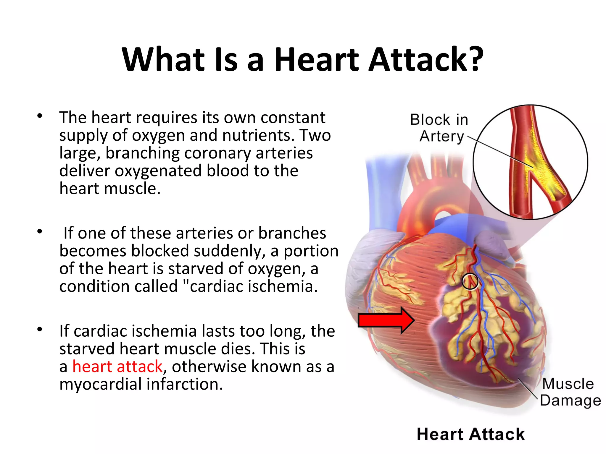
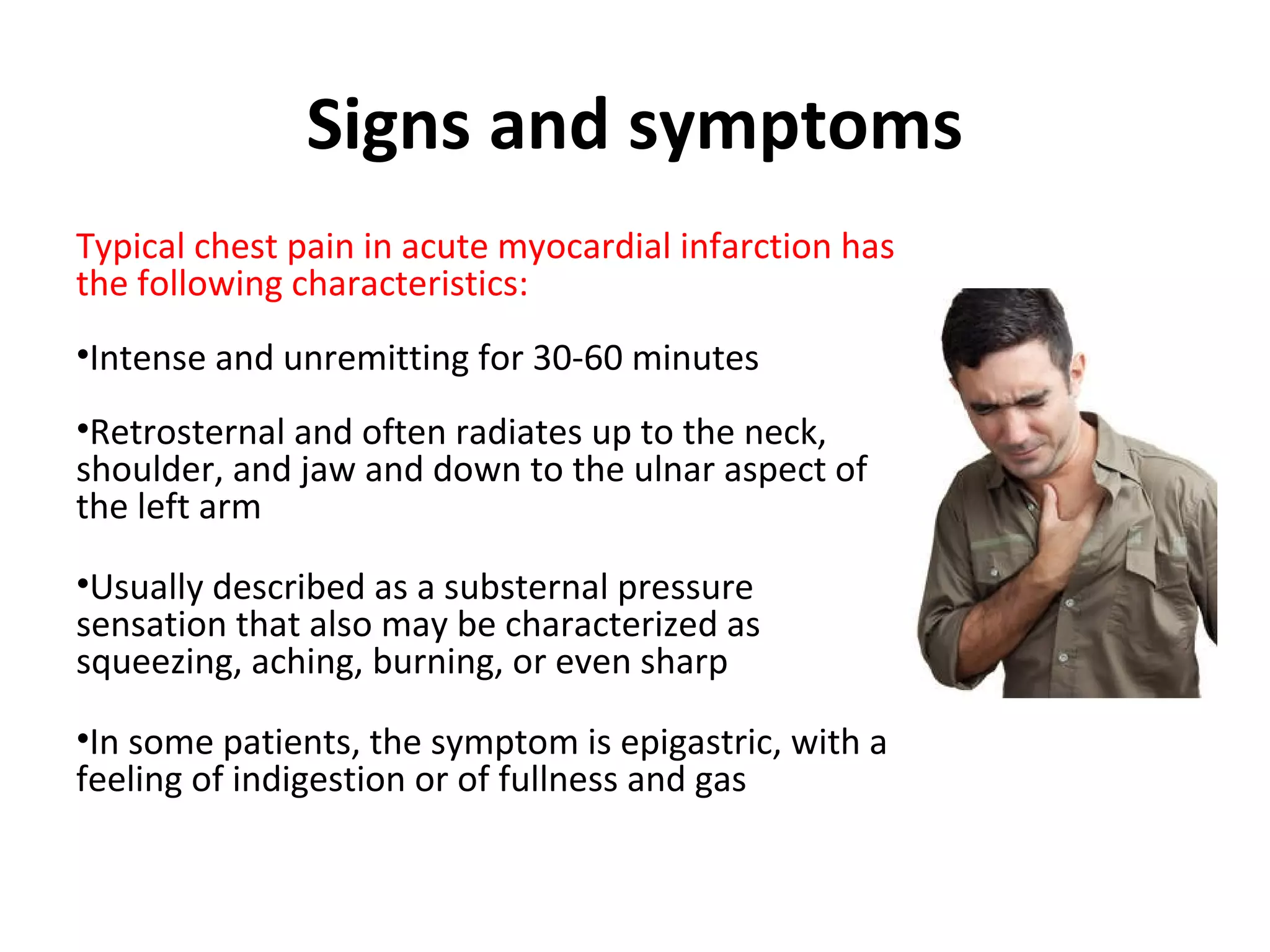
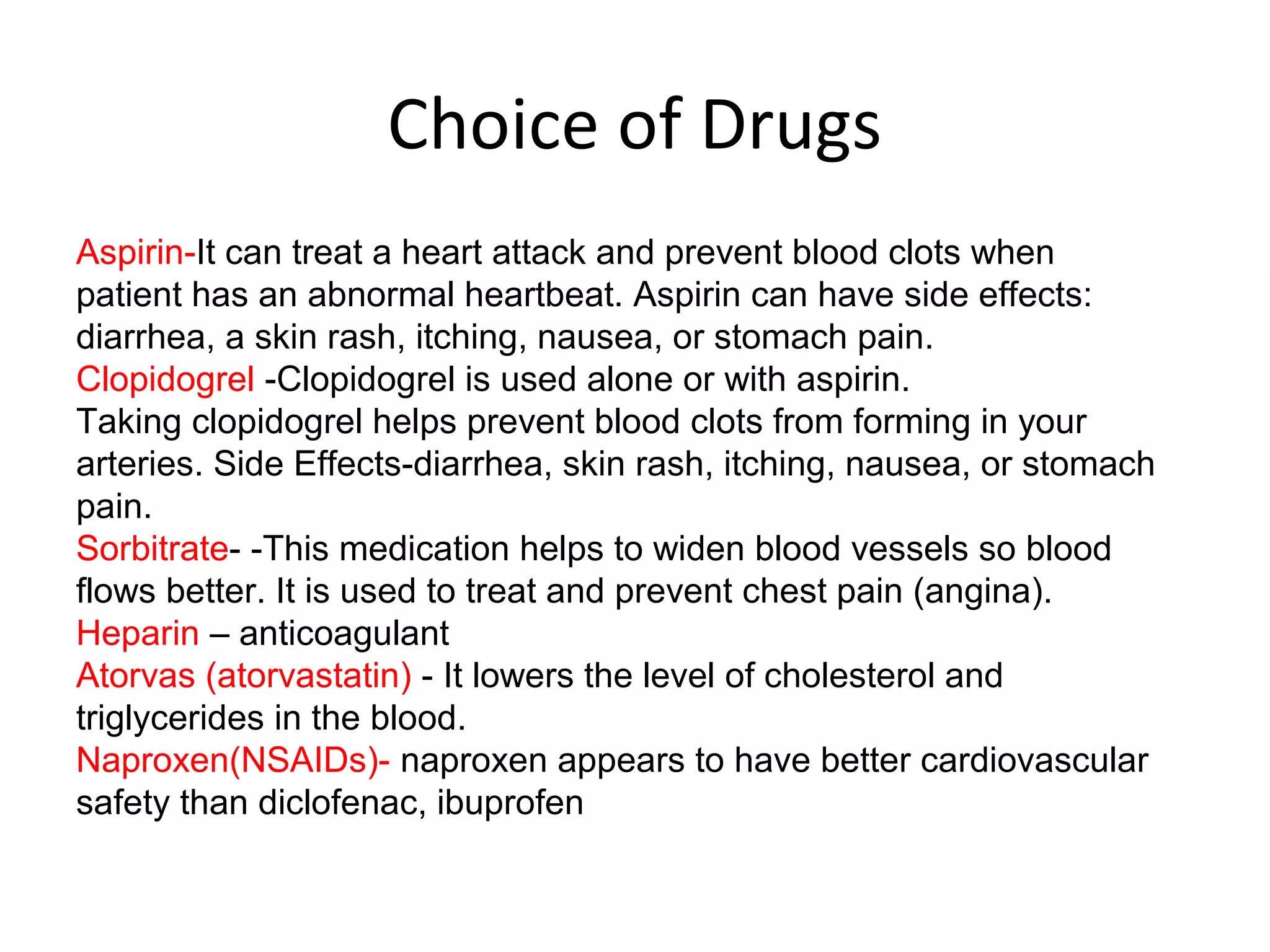
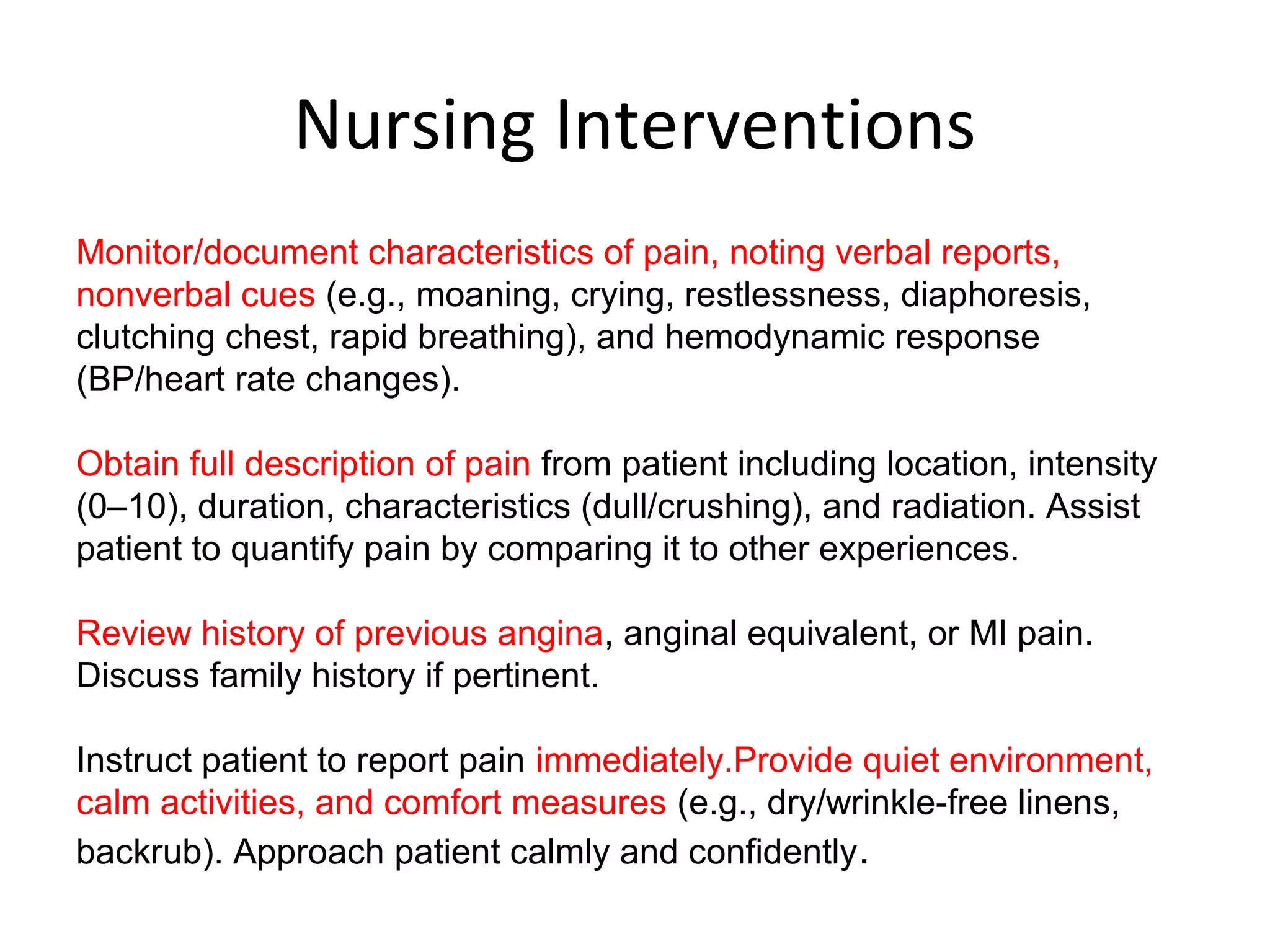
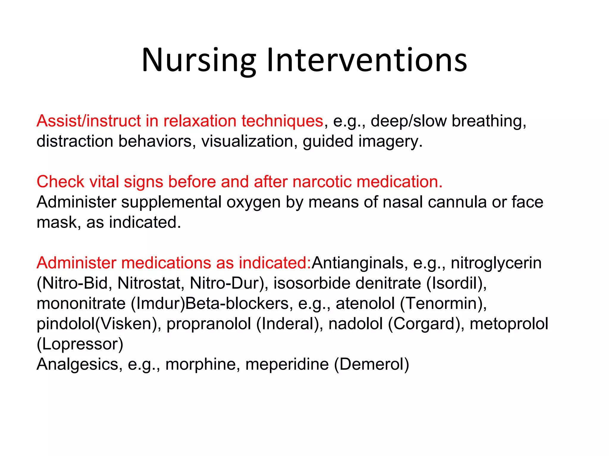
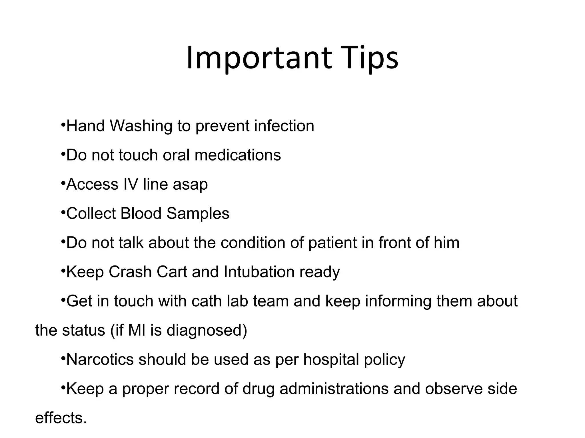
This document provides information about acute myocardial infarction (AMI) or heart attack. It defines AMI as a condition where a portion of the heart muscle dies due to lack of oxygen from a blocked artery. It describes the signs and symptoms of AMI such as chest pain and changes in vital signs. It also lists common risk factors for AMI like age, smoking, high cholesterol, and diabetes. The diagnosis involves laboratory tests, electrocardiography, and imaging of the coronary arteries. Emergency treatment focuses on oxygen, pain management, and drugs to reduce workload on the heart. Nurses monitor symptoms and vital signs, educate patients, administer medications, and ensure crash carts are prepared.
cardiac arrest is the sudden cessation of heart beat and normal cardiac function resulting in loss of effective circulation. cardiopulmonary resuscitation (CPR) is the immediate first aid treatment in case of a cardiac arrest. CPR has to be initiated within 10 seconds after cardiac arrest. cardiac arrest can be determined by palpating for carotid pulse. carotid pulse is absent in case of cardiac arrest.
will help you in understanding myocardial infarction in more detail with its management and therapy with complications and with graphical knowledge you can understand it better and some laboratry test are also included in it .
Ventricular tachycardia are difficult to understand. it is classified in to two types. 1. VT in structurally normal heart, 2. VT in heart with structural diseases. I have tried to simplify the VT in structurally normal heart, which may be helpful to many students and learners.
Idiopathic VT refers to VT occurring in structurally normal hearts in the absence of myocardial scarring. Classification of monomorphic idiopathic VT includes outflow tract VT, fascicular VT, papillary muscle VT,annular VT, and miscellaneous (VT from the body of the RV and crux of
the heart). It is commonly seen in young patients and usually has a benign course. The 12-lead lectrocardiogram is critical in distinguishing the specific form and locations of idiopathic VT. Treatment options include medical therapy specific to the underlying mechanism of VT or catheter
ablation.
Myocardial Infarction - Case Presentation and an OverviewAbubakkar Raheel
Case Presented by Final Year MBBS sudents of Frontier Medical College at the 1st Clinico-Pathological Conference for the year 2015.The Presentation is divided into two parts. First part is about a case of an Acute ST Segment elevated Myocardial Infarction with. Its management at the Hospital and the findings. Second part is about the pathophysiology, Cinical signs and symptoms and an effective gold standard treatment of MI.
Professor DR Md . TOUFIQUR RAHMAN , FCPS, MD
Professor & Head, Cardiology, CMMC, Manikganj
drtoufiq19711@yahoo.com; drtoufiq1971@gmail.com
What are the causes of sinus bradycardia? (DU-04Ja)
Sinus bradycardia is a condition where the sinus node in the heart beats slower than the normal range of 60-100 beats per minute. Some common causes of sinus bradycardia include:
Vagal stimulation: This occurs due to an increased activity of the vagus nerve, which is responsible for slowing down the heart rate.
Medications: Certain medications like beta-blockers, calcium channel blockers, and digoxin can cause sinus bradycardia.
Hypothyroidism: Inadequate production of thyroid hormones can cause a decrease in metabolic rate and lead to bradycardia.
Increased intracranial pressure: High pressure within the skull due to conditions like head injury, brain tumors or bleeding can affect the autonomic nervous system and cause bradycardia.
Obstructive sleep apnea: Repeated episodes of apnea during sleep can cause bradycardia due to decreased oxygen supply to the body.
Aging: As the body ages, the electrical activity of the heart can slow down, leading to sinus bradycardia.
Other causes of sinus bradycardia include viral infections, genetic disorders, and certain electrolyte imbalances.
2. A 25 years old female presented with palpitation, on examination her pulse was irregularly irregular. How will you assess and investigate her? (DU- 05Ja)
The patient's presentation suggests the possibility of atrial fibrillation, which is a common arrhythmia characterized by an irregularly irregular pulse. The following are the steps that can be taken to assess and investigate the patient:
History taking: Obtain a detailed history of the patient's symptoms, including the onset, duration, and frequency of palpitations, associated symptoms, and any relevant medical history.
Physical examination: Conduct a thorough physical examination, including a cardiovascular examination, to assess the patient's heart sounds, rhythm, and rate. Check for any signs of heart failure or underlying heart disease.
Electrocardiogram (ECG): Perform an ECG to confirm the diagnosis of atrial fibrillation and to determine the heart rate and rhythm. An ECG will also help rule out other arrhythmias or underlying heart conditions.
Blood tests: Check the patient's thyroid function, electrolyte levels, and other relevant blood tests to identify any underlying conditions that may be causing the arrhythmia.
Echocardiography: Perform an echocardiogram to assess the structure and function of the heart and to identify any underlying heart disease.
Holter monitor: Use a Holter monitor to monitor the patient's heart rate and rhythm over a 24-hour period to identify any episodes of atrial fibrillation that may not be captured during a routine ECG.
Other tests: Consider other tests, such as a stress test or electrophysiology study, if necessary, to further evaluate the patient's heart funct
It contains meaning, pathophysiology, types, risk factors, lab and diagnostic procedures and tests, Rx goals, appropriate medications for ANGINA PECTORIS ..... Enjoy and Learn from it!!!!
A brief description for 2nd year MBBS students about IHD- MI,Unstable Angina by Dr Sabu Augustine. content from other presentations (ppts)and text books
Angina pectoris is a clinical syndrome usually characterized by episodes of pain or pressure in the anterior chest . The cause is usually insufficient coronary blood flow which results in a decreased oxygen supply to meet an increased myocardial demand for oxygen in response to physical exertion or emotional stress.
Angina also known as angina pectoris is a medical condition characterized by chest pain usually left sided due to inadequate blood supply (ischemia) to the heart muscles due to obstruction (like presence of blood clot), narrowing or contraction (vasospasm) of the supplying coronary arteries.
2. What Is a Heart Attack?
• The heart requires its own constant
supply of oxygen and nutrients. Two
large, branching coronary arteries
deliver oxygenated blood to the
heart muscle.
• If one of these arteries or branches
becomes blocked suddenly, a portion
of the heart is starved of oxygen, a
condition called "cardiac ischemia.
• If cardiac ischemia lasts too long, the
starved heart muscle dies. This is
a heart attack, otherwise known as a
myocardial infarction.
4. Signs and symptoms
Typical chest pain in acute myocardial infarction has
the following characteristics:
•Intense and unremitting for 30-60 minutes
•Retrosternal and often radiates up to the neck,
shoulder, and jaw and down to the ulnar aspect of
the left arm
•Usually described as a substernal pressure
sensation that also may be characterized as
squeezing, aching, burning, or even sharp
•In some patients, the symptom is epigastric, with a
feeling of indigestion or of fullness and gas
5. Signs and symptoms
The patient’s vital signs may demonstrate the following in myocardial
infarction:
The patient’s heart rate is often increased secondary to sympatho-adrenal
discharge
The pulse may be irregular because of ventricular ectopy, In general, the patient's
blood pressure is initially elevated because of peripheral arterial vasoconstriction
resulting from an adrenergic response to pain and ventricular dysfunction
However, with right ventricular myocardial infarction or severe left ventricular
dysfunction, hypotension is seen
The respiratory rate may be increased in response to pulmonary congestion or
anxiety
Coughing, wheezing, and the production of frothy sputum may occur
Fever is usually present within 24-48 hours, Body temperature may occasionally
exceed 102°F
6. Causes/Risk Factors
Rupture of high-risk plaque in the coronary arteries is a primary
causative factor in the development of AMI.
Increasing age is considered the most significant risk factor for CAD.
Individuals aged older than 45 years have an eight times greater risk
for AMI (less than 10% of patients who have AMI are aged younger
than 45 years). The risk for mortality after AMI is higher for older
individuals
Cigarette smoking is a major risk factor for atherosclerosis and
hence AMI.
Dyslipidemia:Elevated serum levels of low-density lipoprotein
cholesterol (LDL-C) and non–high-density lipoprotein cholesterol
(HDL-C) (other triglyceride-rich lipoproteins) significantly increase
the risk of AMI
Hypertension
Diabetes mellitus
family history of ischemic heart disease (IHD) in a first-degree
relative have a higher risk for AMI
7. Diagnosis
Laboratory studies
Troponin levels: Troponin is a contractile protein that normally is not
found in serum; it is released only when myocardial necrosis occurs
Creatine kinase (CK) levels: CK-MB levels increase within 3-12 hours
of the onset of chest pain, reach peak values within 24 hours, and
return to baseline after 48-72 hours
Myoglobin levels: Myoglobin is released more rapidly from infarcted
myocardium than is troponin; urine myoglobin levels rise within 1-4
hours from the onset of chest pain
Complete blood count
Chemistry profile
Lipid profile
C-reactive protein and other inflammation markers
8. Diagnosis
Radiology Investigations
Electrocardiography
The ECG is the most important tool in the initial evaluation and
triage of patients in whom an acute coronary syndrome (ACS),
such as myocardial infarction, is suspected. It is confirmatory
of the diagnosis in approximately 80% of cases.
Cardiac imaging
For individuals with highly probable or confirmed ACS, a
coronary angiogram can be used to definitively diagnose or
rule out coronary artery disease.
9. Emergency Management of MI
Immediate management priorities
Intravenous access must be available for effective administration of
emergency drug
Keep your crash cart (including defibrillator) and intubation trolley
ready for emergency use.
Administration of oxygen
An ECG should be arranged rapidly.
Analgesia
Anti-emetics
Nitrates -- Nitrates reduce myocardial workload and hence
myocardial oxygen demand by reducing preload (venodilatation) and
afterload (reduced peripheral resistance and blood pressure).
10. Choice of Drugs
Aspirin-It can treat a heart attack and prevent blood clots when
patient has an abnormal heartbeat. Aspirin can have side effects:
diarrhea, a skin rash, itching, nausea, or stomach pain.
Clopidogrel -Clopidogrel is used alone or with aspirin.
Taking clopidogrel helps prevent blood clots from forming in your
arteries. Side Effects-diarrhea, skin rash, itching, nausea, or stomach
pain.
Sorbitrate- -This medication helps to widen blood vessels so blood
flows better. It is used to treat and prevent chest pain (angina).
Heparin – anticoagulant
Atorvas (atorvastatin) - It lowers the level of cholesterol and
triglycerides in the blood.
Naproxen(NSAIDs)- naproxen appears to have better cardiovascular
safety than diclofenac, ibuprofen
11. Nursing Interventions
Monitor/document characteristics of pain, noting verbal reports,
nonverbal cues (e.g., moaning, crying, restlessness, diaphoresis,
clutching chest, rapid breathing), and hemodynamic response
(BP/heart rate changes).
Obtain full description of pain from patient including location, intensity
(0–10), duration, characteristics (dull/crushing), and radiation. Assist
patient to quantify pain by comparing it to other experiences.
Review history of previous angina, anginal equivalent, or MI pain.
Discuss family history if pertinent.
Instruct patient to report pain immediately.Provide quiet environment,
calm activities, and comfort measures (e.g., dry/wrinkle-free linens,
backrub). Approach patient calmly and confidently.
12. Nursing Interventions
Assist/instruct in relaxation techniques, e.g., deep/slow breathing,
distraction behaviors, visualization, guided imagery.
Check vital signs before and after narcotic medication.
Administer supplemental oxygen by means of nasal cannula or face
mask, as indicated.
Administer medications as indicated:Antianginals, e.g., nitroglycerin
(Nitro-Bid, Nitrostat, Nitro-Dur), isosorbide denitrate (Isordil),
mononitrate (Imdur)Beta-blockers, e.g., atenolol (Tenormin),
pindolol(Visken), propranolol (Inderal), nadolol (Corgard), metoprolol
(Lopressor)
Analgesics, e.g., morphine, meperidine (Demerol)
13. Important Tips
•Hand Washing to prevent infection
•Do not touch oral medications
•Access IV line asap
•Collect Blood Samples
•Do not talk about the condition of patient in front of him
•Keep Crash Cart and Intubation ready
•Get in touch with cath lab team and keep informing them about
the status (if MI is diagnosed)
•Narcotics should be used as per hospital policy
•Keep a proper record of drug administrations and observe side
effects.