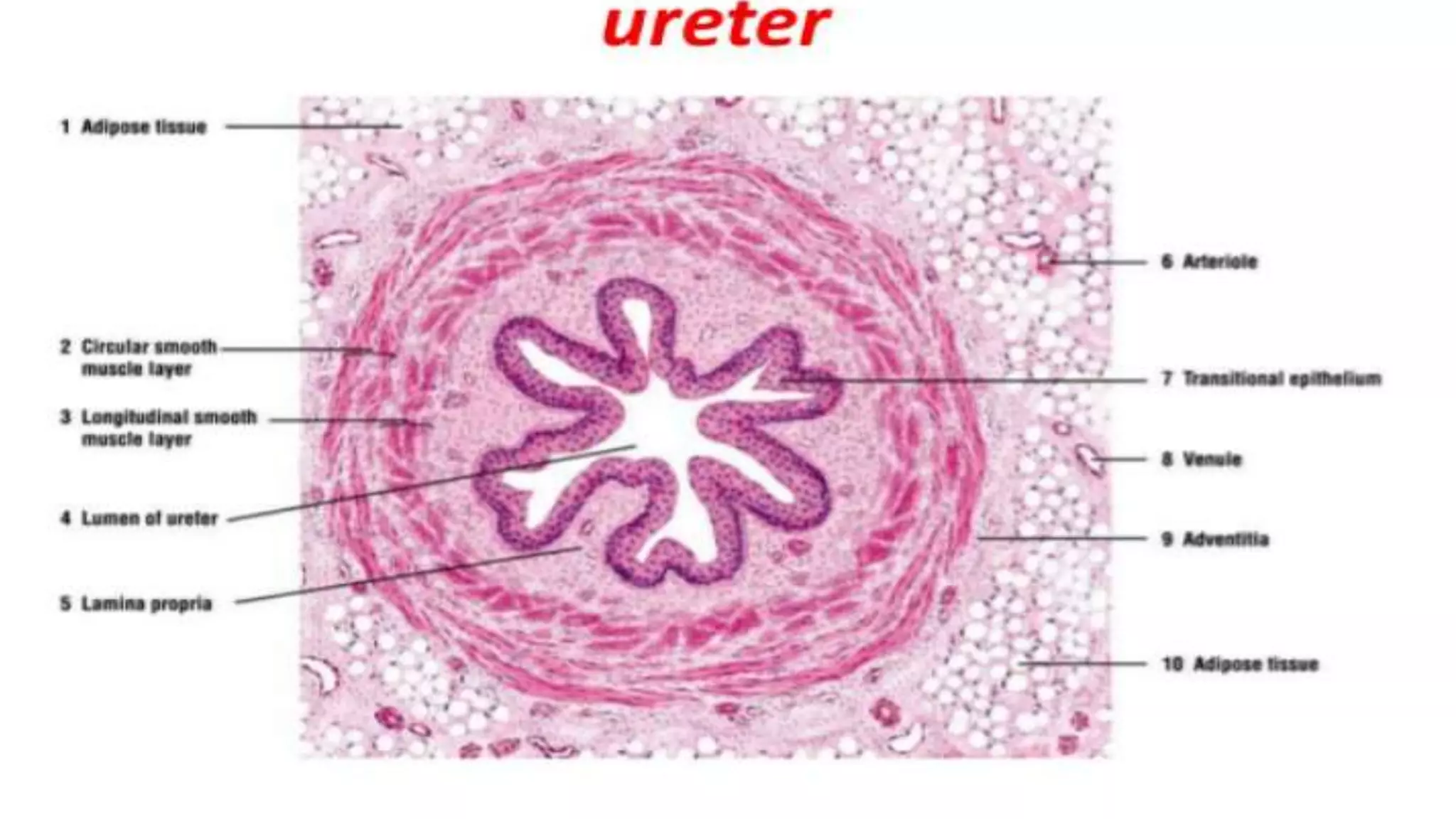
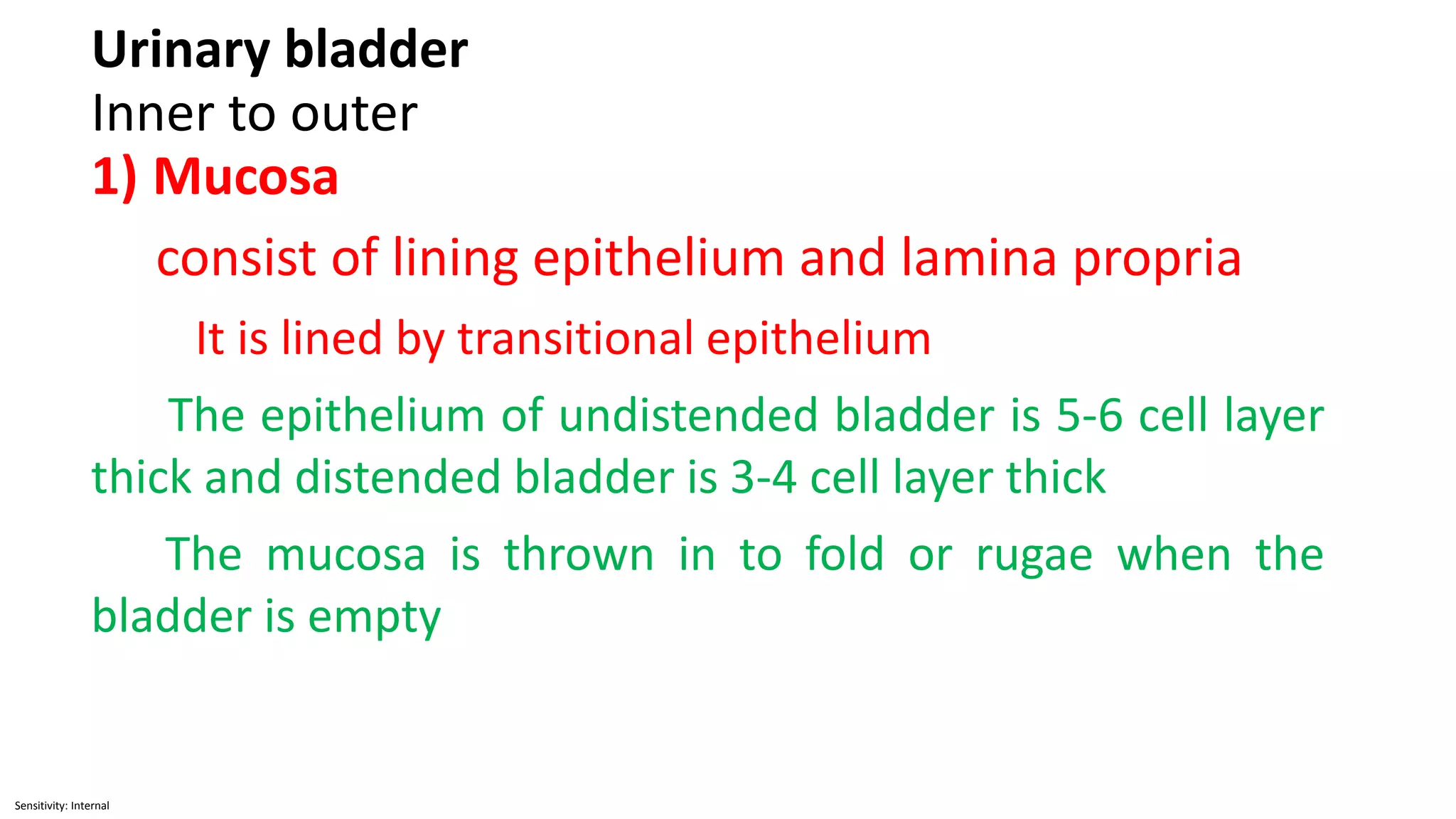
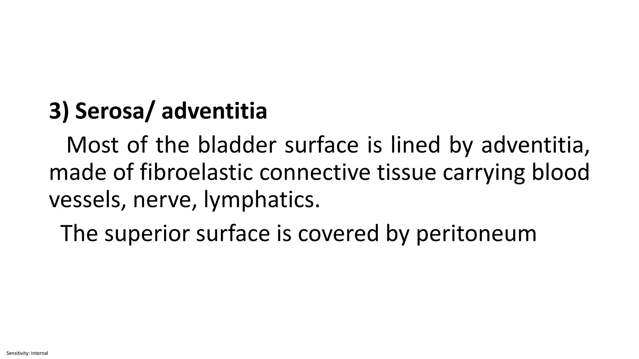
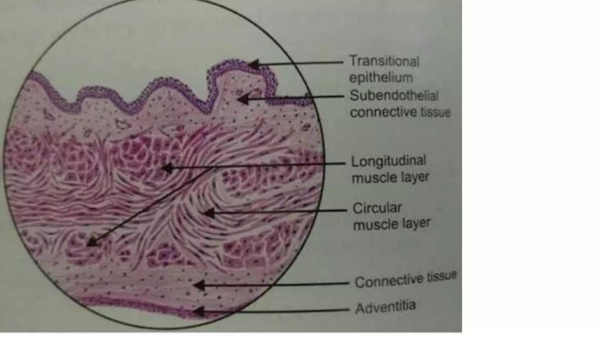
Sensitivity: Internal
Ureter, urinary bladder, urethra
Sensitivity: Internal
Ureter:
The wall of ureter has 3 layer
from inner to outer: Mucosa
Muscle layer
Adventitia
A) Mucosa:
- Mucosa consist of lining epithelium and lamina
propria
- Mucous membrane is lined by transitional
epithelium or urothelium
Sensitivity: Internal
- In transverse section the lumen has a star shaped
appearance, that is due to longitudinal folds of
the mucosa. This fold disappear when the ureter
is distended.
- The urothelium composed of following three
layer:
1) A single layer of small basal cell resting on a
very thin basement membrane. These cells are
cuboidal or columner
Sensitivity: Internal
2) Intermediate cell layer
-contain one to several layer of polyhedral cells
-the thickness of these layer varies with the
state of urinary tract expansion, which in human may
reach up to five layer thick
3) Superficial layer
- The layer contain large, polyhedral or bulbous
cell called umbrella cells which are occassionally bi or
multinucleated and are highly differentiated to
protect underlying cell against the cytotoxic effect of
hypertonic urine
Sensitivity: Internal
- The shape of these epithelial cells depends on the
filling state of the excretory passage
- In empty urinary bladder, dome shaped cells
are roughly cuboidal, when bladder is filled, they are
stretched and appear as flat or squamous
Sensitivity: Internal
Lamina propria
Fibroelastic connective tissue with more
fibroblast
Diffuse lymphatic tissue and occasional small
lymphatic nodule may be observe in lamina propria
Sensitivity: Internal
B) Muscular
- smooth muscle present here
- In upper 2/3 : inner longitudinal
outer circular
- In lower 1/3 : inner longitudinal
middle circular
outer longitudinal
Sensitivity: Internal
C) Adventitia
Fibroelastic connective tissue
contain numerous blood vessels and fat cells
Sensitivity: Internal
Function of transitional epithelium
- Prevent reabsorption of urine
- Allow free distension and contraction
Sensitivity: Internal
Urinary bladder
Inner to outer
1) Mucosa
consist of lining epithelium and lamina propria
It is lined by transitional epithelium
The epithelium of undistended bladder is 5-6 cell layer
thick and distended bladder is 3-4 cell layer thick
The mucosa is thrown in to fold or rugae when the
bladder is empty
Sensitivity: Internal
Lamina propria
connective tissue fiber, Rich in blood vessels
2) Muscular layer
The muscularis consist of three poorly delinated
layer collectively called the detrusor muscle, which
contract to empty the bladder. These are inner and
outer longitudinal and middle circular. Smooth
muscle bundle of the detrusor muscle are irregularly
arrange.
Sensitivity: Internal
smooth muscle of urinary passage is mixed with connective tissue (
collagen fiber) , so that it form parallel bundle rather than pure
muscular sheet.
Sensitivity: Internal
3) Serosa/ adventitia
Most of the bladder surface is lined by adventitia,
made of fibroelastic connective tissue carrying blood
vessels, nerve, lymphatics.
The superior surface is covered by peritoneum
Sensitivity: Internal
Urethra
-Urethra is a tube that carries the urine from
bladder to the exterior
-The urethral mucosa has large longitudinal folds,
giving it a distinctive appearance in cross section
Sensitivity: Internal
-Male urethra is much longer 20 cm than female 4-
5 cm
-The basic structure of the two is same
-The wall has three layered-
Mucosa
Submucosa
Muscular
Sensitivity: Internal
1) Mucosa
-consist of lining epithelium and lamina propria
-Both male and female the greater part of urethra
is lined by pseudostratified columner epithelium. A
short part adjoining the urinary bladder is lined by
transitional epithelium, while the part near the
external orifice is lined by stratified epithelium.
Lamina propria
Here mucous secreting tubular gland present
Sensitivity: Internal
2) Submucosa
3) Muscular
inner longitudinal
outer circular
This arrangement is better in female urethra
In male urethra it is well defined only in
membranous and prostatic part
Sensitivity: Internal
Male urethra
- Male urethra is a common tubular passage for the
elimination of urine and semen
- It extends from the internal urethral orifice at the
apex of trigone of urinary bladder to the external
urethral orifice close to the tip of glans penis
- 3 parts: prostatic
membranous
spongy
Sensitivity: Internal
Prostatic part:
- Lined by transitional epithelium
- 3-4 cm in length
- It is the widest and most dilatable part of male
urethra
- On cross section it present crescentic lumen
- It runs almost vertically downwards through the
prostate from the base to slightly in front of the
apex, at the junction of anterior one third and
posterior two third of the gland
Sensitivity: Internal
Membranous urethra
- Lined by stratified or pseudostratified columner
epithelium
- Its anterior wall 2 cm and posterior wall 1.25 cm
- On cross section it is stellate in appearance
- It extend from the prostate to the upper surface of
bulb of penis
- It is surrounded by sphincter urethrae muscle (
striated muscle) to form external sphincter
- Pierce the deep perineal pouch
Sensitivity: Internal
Spongy urethra
- Lined by pseudostratified columner epithelium
except at its distal ends where it is lined with
stratified squamous epithelium
- 15 cm long
- It passes through the bulb, body and glans penis
and terminate at the external urethral orifice close
to the tip of glans
- In its course it present two dilatation- intrabulbar
fossa within bulb of penis and navicular fossa in the
glans penis
Sensitivity: Internal
Female urethra
- 4-5 cm long
- It begins from the internal urethral orifice of the
bladder and passes downwards and forwards
embedded in the anterior wall of vagina
-lined initially nearer to internal urethral orifice is
transitional epithelium then by stratified squamous
epithelium and some areas of pseudostratified
columner epithelium

MICROSCOPIC STRUCTURE OF URETER, URINARY BLADDER ,URETHRA.pptx

  • 1.
  • 2.
    Sensitivity: Internal Ureter: The wallof ureter has 3 layer from inner to outer: Mucosa Muscle layer Adventitia A) Mucosa: - Mucosa consist of lining epithelium and lamina propria - Mucous membrane is lined by transitional epithelium or urothelium
  • 3.
    Sensitivity: Internal - Intransverse section the lumen has a star shaped appearance, that is due to longitudinal folds of the mucosa. This fold disappear when the ureter is distended. - The urothelium composed of following three layer: 1) A single layer of small basal cell resting on a very thin basement membrane. These cells are cuboidal or columner
  • 4.
    Sensitivity: Internal 2) Intermediatecell layer -contain one to several layer of polyhedral cells -the thickness of these layer varies with the state of urinary tract expansion, which in human may reach up to five layer thick 3) Superficial layer - The layer contain large, polyhedral or bulbous cell called umbrella cells which are occassionally bi or multinucleated and are highly differentiated to protect underlying cell against the cytotoxic effect of hypertonic urine
  • 5.
    Sensitivity: Internal - Theshape of these epithelial cells depends on the filling state of the excretory passage - In empty urinary bladder, dome shaped cells are roughly cuboidal, when bladder is filled, they are stretched and appear as flat or squamous
  • 6.
    Sensitivity: Internal Lamina propria Fibroelasticconnective tissue with more fibroblast Diffuse lymphatic tissue and occasional small lymphatic nodule may be observe in lamina propria
  • 7.
    Sensitivity: Internal B) Muscular -smooth muscle present here - In upper 2/3 : inner longitudinal outer circular - In lower 1/3 : inner longitudinal middle circular outer longitudinal
  • 8.
    Sensitivity: Internal C) Adventitia Fibroelasticconnective tissue contain numerous blood vessels and fat cells
  • 9.
    Sensitivity: Internal Function oftransitional epithelium - Prevent reabsorption of urine - Allow free distension and contraction
  • 10.
  • 11.
  • 12.
  • 13.
  • 14.
    Sensitivity: Internal Urinary bladder Innerto outer 1) Mucosa consist of lining epithelium and lamina propria It is lined by transitional epithelium The epithelium of undistended bladder is 5-6 cell layer thick and distended bladder is 3-4 cell layer thick The mucosa is thrown in to fold or rugae when the bladder is empty
  • 15.
    Sensitivity: Internal Lamina propria connectivetissue fiber, Rich in blood vessels 2) Muscular layer The muscularis consist of three poorly delinated layer collectively called the detrusor muscle, which contract to empty the bladder. These are inner and outer longitudinal and middle circular. Smooth muscle bundle of the detrusor muscle are irregularly arrange.
  • 16.
    Sensitivity: Internal smooth muscleof urinary passage is mixed with connective tissue ( collagen fiber) , so that it form parallel bundle rather than pure muscular sheet.
  • 17.
    Sensitivity: Internal 3) Serosa/adventitia Most of the bladder surface is lined by adventitia, made of fibroelastic connective tissue carrying blood vessels, nerve, lymphatics. The superior surface is covered by peritoneum
  • 18.
  • 19.
    Sensitivity: Internal Urethra -Urethra isa tube that carries the urine from bladder to the exterior -The urethral mucosa has large longitudinal folds, giving it a distinctive appearance in cross section
  • 20.
    Sensitivity: Internal -Male urethrais much longer 20 cm than female 4- 5 cm -The basic structure of the two is same -The wall has three layered- Mucosa Submucosa Muscular
  • 21.
    Sensitivity: Internal 1) Mucosa -consistof lining epithelium and lamina propria -Both male and female the greater part of urethra is lined by pseudostratified columner epithelium. A short part adjoining the urinary bladder is lined by transitional epithelium, while the part near the external orifice is lined by stratified epithelium. Lamina propria Here mucous secreting tubular gland present
  • 22.
    Sensitivity: Internal 2) Submucosa 3)Muscular inner longitudinal outer circular This arrangement is better in female urethra In male urethra it is well defined only in membranous and prostatic part
  • 23.
    Sensitivity: Internal Male urethra -Male urethra is a common tubular passage for the elimination of urine and semen - It extends from the internal urethral orifice at the apex of trigone of urinary bladder to the external urethral orifice close to the tip of glans penis - 3 parts: prostatic membranous spongy
  • 24.
    Sensitivity: Internal Prostatic part: -Lined by transitional epithelium - 3-4 cm in length - It is the widest and most dilatable part of male urethra - On cross section it present crescentic lumen - It runs almost vertically downwards through the prostate from the base to slightly in front of the apex, at the junction of anterior one third and posterior two third of the gland
  • 25.
    Sensitivity: Internal Membranous urethra -Lined by stratified or pseudostratified columner epithelium - Its anterior wall 2 cm and posterior wall 1.25 cm - On cross section it is stellate in appearance - It extend from the prostate to the upper surface of bulb of penis - It is surrounded by sphincter urethrae muscle ( striated muscle) to form external sphincter - Pierce the deep perineal pouch
  • 26.
    Sensitivity: Internal Spongy urethra -Lined by pseudostratified columner epithelium except at its distal ends where it is lined with stratified squamous epithelium - 15 cm long - It passes through the bulb, body and glans penis and terminate at the external urethral orifice close to the tip of glans - In its course it present two dilatation- intrabulbar fossa within bulb of penis and navicular fossa in the glans penis
  • 27.
    Sensitivity: Internal Female urethra -4-5 cm long - It begins from the internal urethral orifice of the bladder and passes downwards and forwards embedded in the anterior wall of vagina -lined initially nearer to internal urethral orifice is transitional epithelium then by stratified squamous epithelium and some areas of pseudostratified columner epithelium