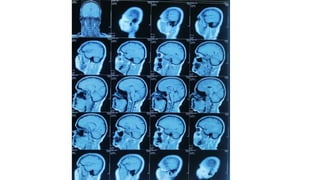
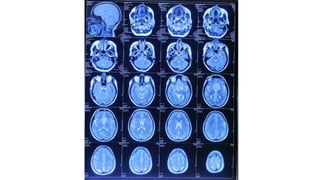
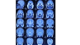
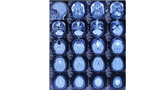
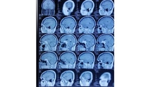
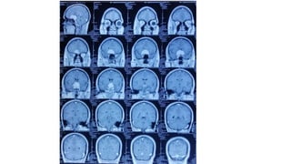
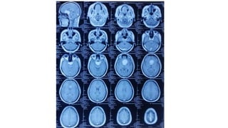
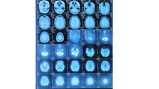
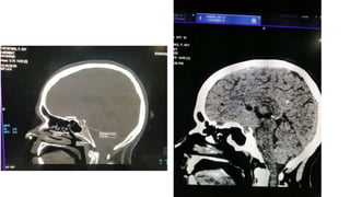
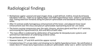
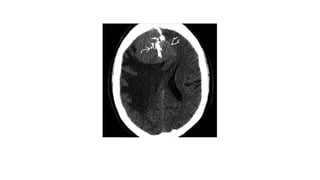
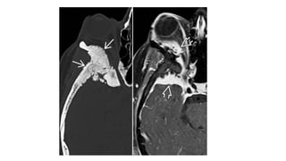
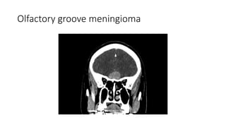
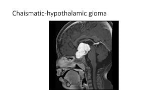
Case presentation
Dr. Rubel Chakma
MD resident , Phase-B ,3rd year
Department of Radiology and Imaging
BSMMU
Patient particulars
• 40 years old female patient,hailing from Manikgonj came with the
complains of progressive impairment of vision on both sides (more
on right side) for 6 months . She also complains occasional headache
but gives no history of vomiting , convulsion and focal neurological
deficit. She had no history of DM ,hypertention ,TB , any primary
malignacncy or hematological disorder. With this complains she went
to see doctor at eye outdoor department, BSMMU and respective
doctor adviced to do MRI scan of brain in our radiology department
Radiological findings
• Multiplaner sagital ,coronal and axial images show a well defined ,midline, broad dural based,
lobulated T1WI iso , T2WI iso to slightly hyper and FlAIR hyperintense mass noted in suprasellar
area having extension forwards along the planum sphenoidale and backwards along the dorsum
sellae.
• After IV contrast avidly homogenous enhancement of the lesion and adjacent linear dural
enhancement over the clivus and planum sphenoidale representing dural tail are noted.
• The lesion causing compression over the optic chiasma, pituitary gland and floor of 3rd ventricle,
and encasing the supraclinoid ICA, ACA and MCA of both sides.
• The mass effect is evidenced by obliteration of Suprasellar & interpeduncular cysterns and
symmetrical indentation of underlying cerebral cortex.
• No perilsional edema is noted
• However lateral , 3rd and 4rth ventricles appear normal
• Complementary CT cut was taken and showing iso to slightly hyperdense lesion. No calcification is
noted. Bone CT shows bony hyperostosis at planum sphenoidale .Sellar size is within normal limit
Radiological diagnosis
• Suprasellar meningioma with planum sphenonale and dorsum sallar
extension
Intracranial Meningioma
• Defintion
A tumour originating from arachnoid cell rest(which are related to
arachnoid mater granulations)
Demographics
• Age
Middle decades. A peak prevalence during 5th and 6th decades
• Gender
Female : Male= 2:1
• Epidemiology
The commonest non-glial intracranial neoplasm
20% of all primary intracranial tumours
Grading according to WHO
WHO grade-I: The majority (90%) and representing a typical benign
tumour.
WHO grade-II: Demonstrating atypical features( increased mitotic
activity and necrosis).10% of all meningioma(frequency rising)
WHO grade-III: An anaplastic (malignant) tumour.1-3% of all
meningioma(rare)
Pathology
• Etiology
 Arise from arachnoid meningothelial (“cap”) cells
 Loss of chromosome 22(40-60% of sporadic meningiomas)
 NF2 meningioma
genomic instability
more likely to be atypical
 Non NF2 meningioma
nearly always benign
chromosomal stability
• Associated abnormalities
 Neurofibromatosis type2(NF2)
Multiple inherited schwannomas, meningiomas and
ependymomas.
50-75% of patients with NF2 develop meningioma
10% of patients with multiple meningiomas have NF2
Location
Supratentorial (90%)
Parasagital region(falx) and cerebral convexities( 45%)
Sphenoid ridge(15-20%)
Olfactory grove(5-10%)
Parasellar (5-10%)
Other(5%): intraventricular, optic nerve sheath, pineal region
 Infratentorial (8-10%)
Cerebellopontine angle, clivus
Multiple meningioma in1-9% cases
Morphology
• Two basic morphologies
 Globose=globular , well demarcated neoplasm with wide dural
attachement
60% of tumors demonstrates local invasion into dural tail
En plaque=sheet like extension covering dura without parenchymal
invagination
Clinical presentation
• The majority are asymptomatic
• <10% of all meningiomas are symptomatic
• Symtomps depend on tumor site
Imaging
• NECT
Sharply cercumscribed, smooth mass abutting the dura
Hyperdense(70-75%), isodense(25%)
Hypodense(1-5%), fat density(rare lipoblastic subtype)
• Calcified(20-25%)
Can be diffuse, focal ,sand lie(“psammomatous”), sunburst,
globular rim patterns
• Necrosis, cysts, hemorrhage(8-23%)
• Trapped CSF pools, cysts in adjacent brain common
• Peritumoral hypodense vasogenic oedema(60%)
• Bone CT
Hyperostosis, irregular cortex( Particularly common when the skull
base or anterior cranial fossa is involved)
• CECT
>90% enhance homogenously and strongly
MRI
• T1WI
Typically iso-to slightly hypointense with cortex
Necrosis, cyst, hemorrhage(8-23%)
Look for gray matter buckling
• T2WI
Frequently isointense
Best sequence for visualizing CSF/vascular cleft between tumour and
brain
identifying vascular flow void(80%)
• FLAIR
Hyperintense peritumoral vasogenic oedema
Vasogenic oedema does not correlate with tumour size
• T1WI C+(best imaging tool)
>95% enhance homogenously ,intensely
Dural tail sign(35-80% of cases) non specific
A linear contrast –enhancing “dural tail” extending from the tumour
along the dura matter
En plaque: sessile thickened enhancing dura
• MRS
Alanine peak(chareterstic) but seen in 50%
• DWI
DWI, ADC maps for TM variable in appearance
Lower ADC in MM and AM compared to TM
• T2* GRE
Ca++ common, Hemorrhage
• MRV:
Evaluate sinus involvement
Perfusion MRI
• High rCVB in peritumoral edema of anaplastic meningioma
• Angiographic findings
“ sunburst”or radial appearance
dural vessels supply lesion core
Pial vessels supply pheriphery
Prolonged vascular stain
Venous phase vital to evaluate sinus involvement
Falx meningioma
Cerebral convexity meningioma
Sphenoid wing meningioma
Olfactory groove meningioma
CPA meningioma
Intraventricular meningioma
Multiple meningioma with NF2
Atypical and Malignant Meningioma
• General features
Dural based , locally invasive lesion with areas of necrosis ,marked
brain edema
Location
may occur anywhere in neuraxis (brain>> spine)
AM frequent in CPA, along tentorium
MM frequent in parasagittal (44%),cerebral convexities(16%)
• Pathology
• AM: High mitotic activity
• MM: AM features+ findings of frank malignancy
Imaging
• Difficult to predict meningioma tumor grade on imaging
• Imaging findings of typical meningioma do not exclude atypical, malignant
variant
• NECT
CT triad of MM: extracranial mass, osteolysis , intracranial tumour
Marked perifocal edema
• CECT
Enhancing tumour mass
Prominent tumour pannus extending away from mass=mushrooming
MRI
• T1WI
Indistinct tumour margin
Infiltrating tumour interdigitates with brain
• T1WI+C
Enhacing tumour mass
May extend into brain, skull scalp
• FAIR
marked peritumoral edema
• DWI
markedly hyperintense on DWI , hypointense in ADC map
Differential diagnosis
• Papillary Craniopharyngeoma
• Chaismatic-hypothalamic glioma
Differtiating points Suprasellar meninigioma Papillary
Craniophayngioma
Chaismatic Hypothalamic
glioma
Definition A tumour originating from
arachnoid cell rest(which
are related to dura mater
arachnoid granulations)
A benign mostly solid
suprasellar tumor arising
from squamous epithelial
remnants of rathke’s
pouch
An astrocytic tumor
Age Middle decades Middle decades
Usually 4rth to 6th decade
Childhood
Location and extention Tebercular , dorsal or
diapgragm sellae
Often shows forward
extension along dura
mater of anterior cranial
fossa
Commonly located within
the suprasellar region
A purely intrasellar
location is uncommon
optic chiasm and optic
tract. Chaismal tumor
tumors may into
hypothalamus
Differentiatig points Suprasellar meningioma Papiilary
Craniopharyngioma
Chiasmatic Hypothamic
glioma
NECT Usullally broad dural
based, hyperdense lesion
Calcification is seen 20%
cases
Usuully solid suprasellar
tumour
Small cysts but not a
significant feature
calcification is absent
Often large and lobulated
when at chiasm and can
extend into hypothalamus
No calcification
CECT/MRC+ Avidly homogenous
enhancement
Soild component-intense
contrast inhencement
Variable inhancement
T2WI Frequently isointense to
cortex
High signal intesity High signal intensity
Bony hyperostosis Usually present Absent Absent
Dural tail sign Usually present Absent Absent
Papillary craniopharyngeoma
Chaismatic-hypothalamic gioma
Treatment
• Preoperative embolization
• Surgical goals
Resection of tumor and involved dura / dural tail(with tumor free
margin)
Resection of involved or hyperostotic bone
Radiotherapy : frequently used for AM, MM
Prognosis
• Typical benign meningioma –only 9% recurrence
• AM
recurrence 28%
5 years survival :86%
• MM
recurrence 75%
5 years survival :35%
Take home massage
• A tumour originating from arachnoid cell rests
• The majority (90%) are typical benign tumour
• Common age 4th to 6th decades and slight female predominance
• Two common morphological types: a spherical well circumscribed
mass or a flat ,infiltrating (en plaque)lesion
• 90% are supratentorial location
• Best imaging protocol T1WI+c showing avid homogenous
enhancement of the lesion with dural tail sign(supportive sign)
• Bone CT to see hyperostosis and calcification(supportive sign)
meningioma rubel.pptx

meningioma rubel.pptx

  • 1.
    Case presentation Dr. RubelChakma MD resident , Phase-B ,3rd year Department of Radiology and Imaging BSMMU
  • 2.
    Patient particulars • 40years old female patient,hailing from Manikgonj came with the complains of progressive impairment of vision on both sides (more on right side) for 6 months . She also complains occasional headache but gives no history of vomiting , convulsion and focal neurological deficit. She had no history of DM ,hypertention ,TB , any primary malignacncy or hematological disorder. With this complains she went to see doctor at eye outdoor department, BSMMU and respective doctor adviced to do MRI scan of brain in our radiology department
  • 12.
    Radiological findings • Multiplanersagital ,coronal and axial images show a well defined ,midline, broad dural based, lobulated T1WI iso , T2WI iso to slightly hyper and FlAIR hyperintense mass noted in suprasellar area having extension forwards along the planum sphenoidale and backwards along the dorsum sellae. • After IV contrast avidly homogenous enhancement of the lesion and adjacent linear dural enhancement over the clivus and planum sphenoidale representing dural tail are noted. • The lesion causing compression over the optic chiasma, pituitary gland and floor of 3rd ventricle, and encasing the supraclinoid ICA, ACA and MCA of both sides. • The mass effect is evidenced by obliteration of Suprasellar & interpeduncular cysterns and symmetrical indentation of underlying cerebral cortex. • No perilsional edema is noted • However lateral , 3rd and 4rth ventricles appear normal • Complementary CT cut was taken and showing iso to slightly hyperdense lesion. No calcification is noted. Bone CT shows bony hyperostosis at planum sphenoidale .Sellar size is within normal limit
  • 13.
    Radiological diagnosis • Suprasellarmeningioma with planum sphenonale and dorsum sallar extension
  • 14.
    Intracranial Meningioma • Defintion Atumour originating from arachnoid cell rest(which are related to arachnoid mater granulations)
  • 15.
    Demographics • Age Middle decades.A peak prevalence during 5th and 6th decades • Gender Female : Male= 2:1 • Epidemiology The commonest non-glial intracranial neoplasm 20% of all primary intracranial tumours
  • 16.
    Grading according toWHO WHO grade-I: The majority (90%) and representing a typical benign tumour. WHO grade-II: Demonstrating atypical features( increased mitotic activity and necrosis).10% of all meningioma(frequency rising) WHO grade-III: An anaplastic (malignant) tumour.1-3% of all meningioma(rare)
  • 17.
    Pathology • Etiology  Arisefrom arachnoid meningothelial (“cap”) cells  Loss of chromosome 22(40-60% of sporadic meningiomas)  NF2 meningioma genomic instability more likely to be atypical  Non NF2 meningioma nearly always benign chromosomal stability
  • 18.
    • Associated abnormalities Neurofibromatosis type2(NF2) Multiple inherited schwannomas, meningiomas and ependymomas. 50-75% of patients with NF2 develop meningioma 10% of patients with multiple meningiomas have NF2
  • 19.
    Location Supratentorial (90%) Parasagital region(falx)and cerebral convexities( 45%) Sphenoid ridge(15-20%) Olfactory grove(5-10%) Parasellar (5-10%) Other(5%): intraventricular, optic nerve sheath, pineal region  Infratentorial (8-10%) Cerebellopontine angle, clivus Multiple meningioma in1-9% cases
  • 21.
    Morphology • Two basicmorphologies  Globose=globular , well demarcated neoplasm with wide dural attachement 60% of tumors demonstrates local invasion into dural tail En plaque=sheet like extension covering dura without parenchymal invagination
  • 22.
    Clinical presentation • Themajority are asymptomatic • <10% of all meningiomas are symptomatic • Symtomps depend on tumor site
  • 23.
    Imaging • NECT Sharply cercumscribed,smooth mass abutting the dura Hyperdense(70-75%), isodense(25%) Hypodense(1-5%), fat density(rare lipoblastic subtype) • Calcified(20-25%) Can be diffuse, focal ,sand lie(“psammomatous”), sunburst, globular rim patterns • Necrosis, cysts, hemorrhage(8-23%) • Trapped CSF pools, cysts in adjacent brain common • Peritumoral hypodense vasogenic oedema(60%)
  • 24.
    • Bone CT Hyperostosis,irregular cortex( Particularly common when the skull base or anterior cranial fossa is involved) • CECT >90% enhance homogenously and strongly
  • 27.
    MRI • T1WI Typically iso-toslightly hypointense with cortex Necrosis, cyst, hemorrhage(8-23%) Look for gray matter buckling • T2WI Frequently isointense Best sequence for visualizing CSF/vascular cleft between tumour and brain identifying vascular flow void(80%)
  • 28.
    • FLAIR Hyperintense peritumoralvasogenic oedema Vasogenic oedema does not correlate with tumour size • T1WI C+(best imaging tool) >95% enhance homogenously ,intensely Dural tail sign(35-80% of cases) non specific A linear contrast –enhancing “dural tail” extending from the tumour along the dura matter En plaque: sessile thickened enhancing dura • MRS Alanine peak(chareterstic) but seen in 50%
  • 29.
    • DWI DWI, ADCmaps for TM variable in appearance Lower ADC in MM and AM compared to TM • T2* GRE Ca++ common, Hemorrhage • MRV: Evaluate sinus involvement Perfusion MRI • High rCVB in peritumoral edema of anaplastic meningioma
  • 30.
    • Angiographic findings “sunburst”or radial appearance dural vessels supply lesion core Pial vessels supply pheriphery Prolonged vascular stain Venous phase vital to evaluate sinus involvement
  • 31.
  • 32.
  • 33.
  • 34.
  • 35.
  • 36.
  • 37.
  • 38.
    Atypical and MalignantMeningioma • General features Dural based , locally invasive lesion with areas of necrosis ,marked brain edema Location may occur anywhere in neuraxis (brain>> spine) AM frequent in CPA, along tentorium MM frequent in parasagittal (44%),cerebral convexities(16%)
  • 39.
    • Pathology • AM:High mitotic activity • MM: AM features+ findings of frank malignancy
  • 40.
    Imaging • Difficult topredict meningioma tumor grade on imaging • Imaging findings of typical meningioma do not exclude atypical, malignant variant • NECT CT triad of MM: extracranial mass, osteolysis , intracranial tumour Marked perifocal edema • CECT Enhancing tumour mass Prominent tumour pannus extending away from mass=mushrooming
  • 41.
    MRI • T1WI Indistinct tumourmargin Infiltrating tumour interdigitates with brain • T1WI+C Enhacing tumour mass May extend into brain, skull scalp • FAIR marked peritumoral edema • DWI markedly hyperintense on DWI , hypointense in ADC map
  • 43.
    Differential diagnosis • PapillaryCraniopharyngeoma • Chaismatic-hypothalamic glioma
  • 44.
    Differtiating points Suprasellarmeninigioma Papillary Craniophayngioma Chaismatic Hypothalamic glioma Definition A tumour originating from arachnoid cell rest(which are related to dura mater arachnoid granulations) A benign mostly solid suprasellar tumor arising from squamous epithelial remnants of rathke’s pouch An astrocytic tumor Age Middle decades Middle decades Usually 4rth to 6th decade Childhood Location and extention Tebercular , dorsal or diapgragm sellae Often shows forward extension along dura mater of anterior cranial fossa Commonly located within the suprasellar region A purely intrasellar location is uncommon optic chiasm and optic tract. Chaismal tumor tumors may into hypothalamus
  • 45.
    Differentiatig points Suprasellarmeningioma Papiilary Craniopharyngioma Chiasmatic Hypothamic glioma NECT Usullally broad dural based, hyperdense lesion Calcification is seen 20% cases Usuully solid suprasellar tumour Small cysts but not a significant feature calcification is absent Often large and lobulated when at chiasm and can extend into hypothalamus No calcification CECT/MRC+ Avidly homogenous enhancement Soild component-intense contrast inhencement Variable inhancement T2WI Frequently isointense to cortex High signal intesity High signal intensity Bony hyperostosis Usually present Absent Absent Dural tail sign Usually present Absent Absent
  • 46.
  • 47.
  • 48.
    Treatment • Preoperative embolization •Surgical goals Resection of tumor and involved dura / dural tail(with tumor free margin) Resection of involved or hyperostotic bone Radiotherapy : frequently used for AM, MM
  • 49.
    Prognosis • Typical benignmeningioma –only 9% recurrence • AM recurrence 28% 5 years survival :86% • MM recurrence 75% 5 years survival :35%
  • 50.
    Take home massage •A tumour originating from arachnoid cell rests • The majority (90%) are typical benign tumour • Common age 4th to 6th decades and slight female predominance • Two common morphological types: a spherical well circumscribed mass or a flat ,infiltrating (en plaque)lesion • 90% are supratentorial location • Best imaging protocol T1WI+c showing avid homogenous enhancement of the lesion with dural tail sign(supportive sign) • Bone CT to see hyperostosis and calcification(supportive sign)