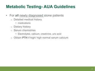
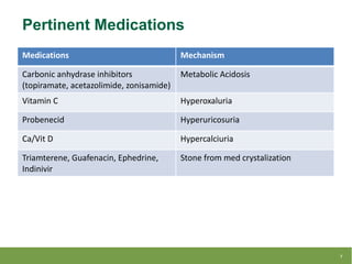
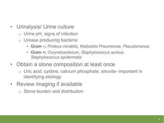
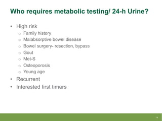
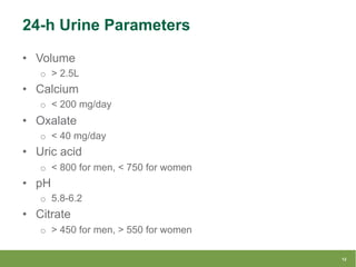
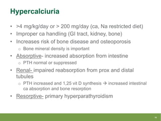
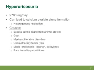
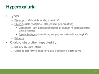
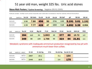
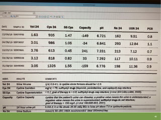
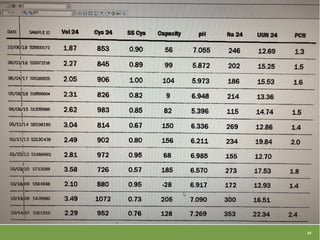
Metabolic Stone Work-Up: Focus on
the 24-hr Urine Evaluation
Kevan M. Sternberg
Associate Professor, Division of Urology
University of Vermont
@kevanms42
• Nothing to disclose
• Content from:
o AUA Core Curriculum
o AUA Guidelines
o Dr. John Asplin Stone Camp
Disclosures
2
• Background
• Guidelines and basics
• Specific diagnoses/metabolic abnormalities
• Litholink case- reviews
Objectives
3
• Prevalence= 1:11 (8.8%) adults in the U.S
o Likely underestimated
• Stone disease is increasing
o With children as well, particularly adolescents
• Resulting increase cost associated with nephrolithiasis
o $ 4.5 Billion
• Gender gap narrowing
Background
4
• Chronic, systemic condition w/multiple contributing
factors:
o Genetics
o Diet
o Lifestyle
o Medical comorbidities
• Associated systemic conditions:
o Obesity
o Metabolic syndrome
o Type 2 DM
o HTN
Nephrolithiasis
5
• For all newly diagnosed stone patients
o Detailed medical history
• medications
o Dietary history
o Serum chemistries
• Electrolytes, calcium, creatinine, uric acid
o Obtain PTH if high/ high normal serum calcium
Metabolic Testing- AUA Guidelines
6
Medications Mechanism
Carbonic anhydrase inhibitors
(topiramate, acetazolimide, zonisamide)
Metabolic Acidosis
Vitamin C Hyperoxaluria
Probenecid Hyperuricosuria
Ca/Vit D Hypercalciuria
Triamterene, Guafenacin, Ephedrine,
Indinivir
Stone from med crystalization
Pertinent Medications
7
• Urinalysis/ Urine culture
o Urine pH, signs of infection
o Urease producing bacteria:
• Gram –; Proteus mirabilis, Klebsiella Pneumonae, Pseudomanas
• Gram +; Corynebacterium, Staphylococcus aureus,
Staphylococcus epidermidis
• Obtain a stone composition at least once
o Uric acid, cystine, calcium phosphate, struvite- important in
identifying etiology
• Review imaging if available
o Stone burden and distribution
8
• High risk
o Family history
o Malabsorptive bowel disease
o Bowel surgery- resection, bypass
o Gout
o Met-S
o Osteoporosis
o Young age
• Recurrent
• Interested first timers
Who requires metabolic testing/ 24-h Urine?
9
• Goal- To help guide medical and dietary intervention to
decrease risk of stone recurrence
• 1 vs. 2 tests- no clear answer
• Patients should be instructed to do what they were doing
at the time the stone was formed
(diet/medication/supplement/etc.)
• Collections likely differ when done at home vs. work.
o Most do their test on a weekend/Sunday for convenience.
o May be best to have 2 tests/patient- one home and on work?
24-hr Urine Testing
10
11
• Volume
o > 2.5L
• Calcium
o < 200 mg/day
• Oxalate
o < 40 mg/day
• Uric acid
o < 800 for men, < 750 for women
• pH
o 5.8-6.2
• Citrate
o > 450 for men, > 550 for women
24-h Urine Parameters
12
• Creatinine- accuracy of collection
o Weight-base values for men (20-25 mg/kg) and
o Women (15-20 mg/kg)
• Sodium
• Potassium
• Protein parameters:
o PCR (protein catabolic rate)
o UUN (reflects total protein intake)
o Sulfate (sulfa containing Amino Acids/net acid load =
methionine)
• Super saturation indices
o monitor effectiveness of tx
13
• Hypercalciuria
• Hyperoxaluria
• Hyperuricosuria
• Hypocitraturia
• Low Urine pH
• Low urine volume
Specific 24-h Urine Abnormalities
14
• >4 mg/kg/day or > 200 mg/day (ca, Na restricted diet)
• Improper ca handling (GI tract, kidney, bone)
• Increases risk of bone disease and osteoporosis
o Bone mineral density is important
• Absorptive- increased absorption from intestine
o PTH normal or suppressed
• Renal- impaired reabsorption from prox and distal
tubules
o PTH increased and 1,25 vit D synthesis à increased intestinal
ca absorption and bone resorption
• Resorptive- primary hyperparathyroidism
Hypercalciuria
15
• Main players- Calcium, Sodium, and animal protein.
• Stone formers- more Na sensitive
• Na goal should be 120
• When patients are instructed to drink more fluids, often
there is a tendency to increase salt intake à
hypercalciuria.
• Sugar and caffeine can also affect calcium output.
• Protein loading/protein shakes à significant
hypercalciuria
Hypercalciuria and Diet
16
• Consider primary HPT even if high normal PTH
• If borderline results, have blood work done fasting-
accounts for the diurnal variation.
• Draw Ca at same time as PTH
• Take patient off of thiazide (~2 weeks) if working up
Primary HPT
o Thiazides can cause hypercalcemia
Hypercalcemia
17
• High serum Ca, low/suppressed PTH à
o Consider malignancy (PTHrp, bony metastasis),
o Granulomatous disease-
• sarcoid (extrarenal source of 1,25 Vit D from alveolar
macrophages/granulomas)
• Tuberculosis
• Silicosis
o thiazides, Ca supplements
o Thyrotosicosis
o Vit D intoxication
o 24-hydroxylase deficiency (can’t metabolize 1-25 Vit D resulting
in hypercalcemia)
Additional Hypercalcemia Causes
18
• If elevated serum Ca and low PTH (its not HPT)- check
1-25 Vit D level
• Primary HPT can occur in the setting of normocalcemia
• If PTH elevated and normocalcemic-
o check renal function (PTH increases in CKD) and
o vit D levels (25 OH) -make sure no deficiency (>30) also can
increase PTH
19
• Don’t start a thiazide if any level of hypercalcemia
• Thiazides great for hypercalciuria + HTN
o Typical doses of HCTZ or the thiazide component in combo pills
are typically too low to impact Ca excretion.
o Chlorthalidone and Indapamide
• Greater % fall in urine ca
Thiazide Diuretics
20
• *** Be mindful of hypokalemic effects of thiazides. Can
place on K immediately or follow BMP.
o Hypokalemia can lead to hypocitraturia
• Hyperglycemia (rare) affecting diabetic management
• Gout
o Increased uric acid
• ED
Thiazide Side Effects
21
• >700 mg/day
• Can lead to calcium oxalate stone formation
o Heterogenous nucleation
• Causes:
o Excess purine intake from animal protein
o Gout
o Myeloproliferative disorders
o Chemotherapy/tumor lysis
o Meds- probenecid, losartan, salicylates
o Rare hereditary conditions
Hyperuricosuria
22
• Types:
o Dietary- oxalate rich foods, vitamin C
o Enteric- malabsorption (IBD, celiac, pancreatitis)
• Mechanism- fatty acid saponification of calcium à increased free
luminal oxalate
• Typical findings- low volume, low pH, low ca/Na/citrate, high Ox
o Primary
• Oxalate absorption impacted by:
o Dietary calcium intake
o Oxalobacter formigenes (oxalate degrading bacterium)
Hyperoxaluria
23
• Autosomal recessive genetic disorder of glyoxalate
metabolism
• Endogenous overproduction of oxalate
• Recurrent nephrolithiasis
• Renal damage à renal failure
• 3 types (type 1 most common and most sever)
• Young age of presentation
• Urine oxalate levels > 80-100 mg/day
• Liver/kidney transplant
Primary Hyperoxaluria
24
• <320 mg/day
• Citrate excretion depends on systemic acid-base status
• Hypocitraturia occurs with acidosis
• Causes of hypocitraturia-
o RTA type 1 (distal)- impaired H ion secretion in distal tubule à
can’t excrete acid load à systemic acidosis
• Hypokalemia, high urine pH, very low citrate
o Chronic diarrhea- loss of bicarbonate à systemic acidosis
o Medications- thiazides (hypokalemia, intracellular acidosis),
carbonic anhydrase inhibitors (prevent bicarb reabsorption in
nephron)
o Idiopathic
Hypocitraturia
25
• Can’t excrete acid load
o NH4 (ammonium) will not be high on 24-h testing
• Treatment with Urocit K
o Ph rises but citrate goes up and ca excretion goes down
• Citrate complexes ca and is a direct crystal inhibitor
• Topiramate, acetazolamide, zonisamide (carbonic
anhydrase inhibitors) à stones
o Hard to get citrate to go up with Urocit-K
RTA and CaP Stones
26
• Urine pH <5.5
• Main factor leading to uric acid stone formation
o Low volume and hyperuricosuria also contribute
• Can have heterogenous nucleation à CaOx stones
• Uric acid stones more common in DM type 2
o Insulin resistance à impaired renal ammonium excretion
Low Urine pH
27
• Acid production- from methionine/cysteine (sulfate
excretion/H2SO4) and lysine/arginine
o Higher sulfate excretion = higher acid load
• Alkali production- aspartate/glutamate and organic
anions
• Diets are net acid producing
• SO4 (sulfate) and NH4 (ammonium) should travel
together
o Sulfate is our dietary acid load
o Ammonium is our acid excretion/what we pee out
Acid-Base
28
• Alkali therapy
o High pH, suppressed NH4 secretion
• Urease activity
o High pH, high NH4
• Met syndrome, hyperkalemia, inadequate alkali tx
o Low pH (can’t get rid of acid as NH4, suppressed NH4 secretion
(insulin resistance)
• GI alkali loss, diarrhea (looses alkali in the gut and
kidneys must secrete acid), hypokalemia
o Low pH, ammonium >> sulfate
• If both sulfate and ammonium are high- that is a normal
response- to a high protein diet (should trend together)
29
• Need an alkaline urine to solubilize cystine
• Want SS cystine to be <0.8
• Capacity – Unique parameter to Litholink
o assess risk of further cystine stone formation
o particularly when on a thiol
• Neg capacity= bad (supersaturated);
• Pos capacity= good (undersaturated)
• Cystine stone formers have high pH at baseline
Cystinuria
30
• Not all cystine forming pts need meds- do not need to
start Thiola immediately
• Initial management = diet
o Hydration
o Low salt diet
o Low protein diet- not a huge benefit to protein limitation in terms
of cystine excretion
31
• Alkalinization- solubility goes up in general with
increasing pH
• Thiol drugs: pH must be high for meds to work
• Thiol binding to cystine occurs quicker with higher pH
• Need pH of 7.5 for thiol drugs to work
• Make sure you test siblings (25% chance)
o Autosomal recessive
32
• Potassium citrate vs. calcium citrate
o Only 20-30% of ca is absorbed, much more K absorption àget
more alkali
• Thiazides and alkali together- do not get as much of an
increase in citrate vs. alkali alone
Medications- Pearls
33
Litholink Cases
34
35
36
37
38
39
40
41
42
43
Questions?
44
• Thank you!
• @kevanms42
• @UVMUrology
Thank You
45
What did you think about today’s
lecture? Share us your thoughts by
taking this survey
T
ag us in social media
#urologyCOViD
Or visit our website
https://urologycovid.ucsf.edu/

UCSF-litholink.pdf

  • 1.
    Metabolic Stone Work-Up:Focus on the 24-hr Urine Evaluation Kevan M. Sternberg Associate Professor, Division of Urology University of Vermont @kevanms42
  • 2.
    • Nothing todisclose • Content from: o AUA Core Curriculum o AUA Guidelines o Dr. John Asplin Stone Camp Disclosures 2
  • 3.
    • Background • Guidelinesand basics • Specific diagnoses/metabolic abnormalities • Litholink case- reviews Objectives 3
  • 4.
    • Prevalence= 1:11(8.8%) adults in the U.S o Likely underestimated • Stone disease is increasing o With children as well, particularly adolescents • Resulting increase cost associated with nephrolithiasis o $ 4.5 Billion • Gender gap narrowing Background 4
  • 5.
    • Chronic, systemiccondition w/multiple contributing factors: o Genetics o Diet o Lifestyle o Medical comorbidities • Associated systemic conditions: o Obesity o Metabolic syndrome o Type 2 DM o HTN Nephrolithiasis 5
  • 6.
    • For allnewly diagnosed stone patients o Detailed medical history • medications o Dietary history o Serum chemistries • Electrolytes, calcium, creatinine, uric acid o Obtain PTH if high/ high normal serum calcium Metabolic Testing- AUA Guidelines 6
  • 7.
    Medications Mechanism Carbonic anhydraseinhibitors (topiramate, acetazolimide, zonisamide) Metabolic Acidosis Vitamin C Hyperoxaluria Probenecid Hyperuricosuria Ca/Vit D Hypercalciuria Triamterene, Guafenacin, Ephedrine, Indinivir Stone from med crystalization Pertinent Medications 7
  • 8.
    • Urinalysis/ Urineculture o Urine pH, signs of infection o Urease producing bacteria: • Gram –; Proteus mirabilis, Klebsiella Pneumonae, Pseudomanas • Gram +; Corynebacterium, Staphylococcus aureus, Staphylococcus epidermidis • Obtain a stone composition at least once o Uric acid, cystine, calcium phosphate, struvite- important in identifying etiology • Review imaging if available o Stone burden and distribution 8
  • 9.
    • High risk oFamily history o Malabsorptive bowel disease o Bowel surgery- resection, bypass o Gout o Met-S o Osteoporosis o Young age • Recurrent • Interested first timers Who requires metabolic testing/ 24-h Urine? 9
  • 10.
    • Goal- Tohelp guide medical and dietary intervention to decrease risk of stone recurrence • 1 vs. 2 tests- no clear answer • Patients should be instructed to do what they were doing at the time the stone was formed (diet/medication/supplement/etc.) • Collections likely differ when done at home vs. work. o Most do their test on a weekend/Sunday for convenience. o May be best to have 2 tests/patient- one home and on work? 24-hr Urine Testing 10
  • 11.
  • 12.
    • Volume o >2.5L • Calcium o < 200 mg/day • Oxalate o < 40 mg/day • Uric acid o < 800 for men, < 750 for women • pH o 5.8-6.2 • Citrate o > 450 for men, > 550 for women 24-h Urine Parameters 12
  • 13.
    • Creatinine- accuracyof collection o Weight-base values for men (20-25 mg/kg) and o Women (15-20 mg/kg) • Sodium • Potassium • Protein parameters: o PCR (protein catabolic rate) o UUN (reflects total protein intake) o Sulfate (sulfa containing Amino Acids/net acid load = methionine) • Super saturation indices o monitor effectiveness of tx 13
  • 14.
    • Hypercalciuria • Hyperoxaluria •Hyperuricosuria • Hypocitraturia • Low Urine pH • Low urine volume Specific 24-h Urine Abnormalities 14
  • 15.
    • >4 mg/kg/dayor > 200 mg/day (ca, Na restricted diet) • Improper ca handling (GI tract, kidney, bone) • Increases risk of bone disease and osteoporosis o Bone mineral density is important • Absorptive- increased absorption from intestine o PTH normal or suppressed • Renal- impaired reabsorption from prox and distal tubules o PTH increased and 1,25 vit D synthesis à increased intestinal ca absorption and bone resorption • Resorptive- primary hyperparathyroidism Hypercalciuria 15
  • 16.
    • Main players-Calcium, Sodium, and animal protein. • Stone formers- more Na sensitive • Na goal should be 120 • When patients are instructed to drink more fluids, often there is a tendency to increase salt intake à hypercalciuria. • Sugar and caffeine can also affect calcium output. • Protein loading/protein shakes à significant hypercalciuria Hypercalciuria and Diet 16
  • 17.
    • Consider primaryHPT even if high normal PTH • If borderline results, have blood work done fasting- accounts for the diurnal variation. • Draw Ca at same time as PTH • Take patient off of thiazide (~2 weeks) if working up Primary HPT o Thiazides can cause hypercalcemia Hypercalcemia 17
  • 18.
    • High serumCa, low/suppressed PTH à o Consider malignancy (PTHrp, bony metastasis), o Granulomatous disease- • sarcoid (extrarenal source of 1,25 Vit D from alveolar macrophages/granulomas) • Tuberculosis • Silicosis o thiazides, Ca supplements o Thyrotosicosis o Vit D intoxication o 24-hydroxylase deficiency (can’t metabolize 1-25 Vit D resulting in hypercalcemia) Additional Hypercalcemia Causes 18
  • 19.
    • If elevatedserum Ca and low PTH (its not HPT)- check 1-25 Vit D level • Primary HPT can occur in the setting of normocalcemia • If PTH elevated and normocalcemic- o check renal function (PTH increases in CKD) and o vit D levels (25 OH) -make sure no deficiency (>30) also can increase PTH 19
  • 20.
    • Don’t starta thiazide if any level of hypercalcemia • Thiazides great for hypercalciuria + HTN o Typical doses of HCTZ or the thiazide component in combo pills are typically too low to impact Ca excretion. o Chlorthalidone and Indapamide • Greater % fall in urine ca Thiazide Diuretics 20
  • 21.
    • *** Bemindful of hypokalemic effects of thiazides. Can place on K immediately or follow BMP. o Hypokalemia can lead to hypocitraturia • Hyperglycemia (rare) affecting diabetic management • Gout o Increased uric acid • ED Thiazide Side Effects 21
  • 22.
    • >700 mg/day •Can lead to calcium oxalate stone formation o Heterogenous nucleation • Causes: o Excess purine intake from animal protein o Gout o Myeloproliferative disorders o Chemotherapy/tumor lysis o Meds- probenecid, losartan, salicylates o Rare hereditary conditions Hyperuricosuria 22
  • 23.
    • Types: o Dietary-oxalate rich foods, vitamin C o Enteric- malabsorption (IBD, celiac, pancreatitis) • Mechanism- fatty acid saponification of calcium à increased free luminal oxalate • Typical findings- low volume, low pH, low ca/Na/citrate, high Ox o Primary • Oxalate absorption impacted by: o Dietary calcium intake o Oxalobacter formigenes (oxalate degrading bacterium) Hyperoxaluria 23
  • 24.
    • Autosomal recessivegenetic disorder of glyoxalate metabolism • Endogenous overproduction of oxalate • Recurrent nephrolithiasis • Renal damage à renal failure • 3 types (type 1 most common and most sever) • Young age of presentation • Urine oxalate levels > 80-100 mg/day • Liver/kidney transplant Primary Hyperoxaluria 24
  • 25.
    • <320 mg/day •Citrate excretion depends on systemic acid-base status • Hypocitraturia occurs with acidosis • Causes of hypocitraturia- o RTA type 1 (distal)- impaired H ion secretion in distal tubule à can’t excrete acid load à systemic acidosis • Hypokalemia, high urine pH, very low citrate o Chronic diarrhea- loss of bicarbonate à systemic acidosis o Medications- thiazides (hypokalemia, intracellular acidosis), carbonic anhydrase inhibitors (prevent bicarb reabsorption in nephron) o Idiopathic Hypocitraturia 25
  • 26.
    • Can’t excreteacid load o NH4 (ammonium) will not be high on 24-h testing • Treatment with Urocit K o Ph rises but citrate goes up and ca excretion goes down • Citrate complexes ca and is a direct crystal inhibitor • Topiramate, acetazolamide, zonisamide (carbonic anhydrase inhibitors) à stones o Hard to get citrate to go up with Urocit-K RTA and CaP Stones 26
  • 27.
    • Urine pH<5.5 • Main factor leading to uric acid stone formation o Low volume and hyperuricosuria also contribute • Can have heterogenous nucleation à CaOx stones • Uric acid stones more common in DM type 2 o Insulin resistance à impaired renal ammonium excretion Low Urine pH 27
  • 28.
    • Acid production-from methionine/cysteine (sulfate excretion/H2SO4) and lysine/arginine o Higher sulfate excretion = higher acid load • Alkali production- aspartate/glutamate and organic anions • Diets are net acid producing • SO4 (sulfate) and NH4 (ammonium) should travel together o Sulfate is our dietary acid load o Ammonium is our acid excretion/what we pee out Acid-Base 28
  • 29.
    • Alkali therapy oHigh pH, suppressed NH4 secretion • Urease activity o High pH, high NH4 • Met syndrome, hyperkalemia, inadequate alkali tx o Low pH (can’t get rid of acid as NH4, suppressed NH4 secretion (insulin resistance) • GI alkali loss, diarrhea (looses alkali in the gut and kidneys must secrete acid), hypokalemia o Low pH, ammonium >> sulfate • If both sulfate and ammonium are high- that is a normal response- to a high protein diet (should trend together) 29
  • 30.
    • Need analkaline urine to solubilize cystine • Want SS cystine to be <0.8 • Capacity – Unique parameter to Litholink o assess risk of further cystine stone formation o particularly when on a thiol • Neg capacity= bad (supersaturated); • Pos capacity= good (undersaturated) • Cystine stone formers have high pH at baseline Cystinuria 30
  • 31.
    • Not allcystine forming pts need meds- do not need to start Thiola immediately • Initial management = diet o Hydration o Low salt diet o Low protein diet- not a huge benefit to protein limitation in terms of cystine excretion 31
  • 32.
    • Alkalinization- solubilitygoes up in general with increasing pH • Thiol drugs: pH must be high for meds to work • Thiol binding to cystine occurs quicker with higher pH • Need pH of 7.5 for thiol drugs to work • Make sure you test siblings (25% chance) o Autosomal recessive 32
  • 33.
    • Potassium citratevs. calcium citrate o Only 20-30% of ca is absorbed, much more K absorption àget more alkali • Thiazides and alkali together- do not get as much of an increase in citrate vs. alkali alone Medications- Pearls 33
  • 34.
  • 35.
  • 36.
  • 37.
  • 38.
  • 39.
  • 40.
  • 41.
  • 42.
  • 43.
  • 44.
    Questions? 44 • Thank you! •@kevanms42 • @UVMUrology
  • 45.
    Thank You 45 What didyou think about today’s lecture? Share us your thoughts by taking this survey T ag us in social media #urologyCOViD Or visit our website https://urologycovid.ucsf.edu/