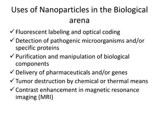
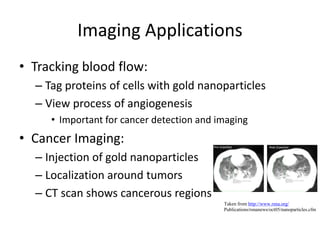
Nanotechnology
Introduction
• Richard Feynman was the first scientist to suggest
that devices and material could someday be
fabricated to atomic specifications in 1959.
• A nanometer is one thousandth of a micrometer
= 10-9 meters.
• Nano comes from the Greek word nanos which
means a little old man or dwarf.
• Pico comes from Spanish where it means a small
quantity.
• Nanotechnology is the science of building
electronic circuits from single atoms and
molecules as they (single atoms and molecules)
are manipulated.
• It is molecular biology viewed from the
perspective of materials science and described in
novel terminologies
• Single molecules or nanostructures are
assembled following specific instructions.
• Its main objectives are to use biological
components to achieve nanoscale tasks
Size Comparisons
Prefixes and sizes
Prefixes and sizes
Visualization at the Nanoscale
• In order to manipulate matter on an atomic scale,
we need to see individual atoms and molecules
• The development of the scanning probe
microscope opened up the field of nanotech.
• It measures some property, such as electric
resistance, magnetism, temperature, or light
absorption, with a tip positioned extremely close
to the sample
• The microscope raster-scans the probe while
measuring the property of interest
• Data are displayed as a raster image similar to
that on a television screen.
• No lenses are used, so the size of the probe
rather than diffraction limits their resolution.
• Some of the microscopes can be used to alter
samples as well as visualize them
Principal of raster scanning
The probe moves to and
fro across the target
region and scans only in
one direction “scan”
In the reverse direction,
the movement is more
rapidly and no contact is
made “flyback”
Scanning Tunneling Microscope (STM)
• Measures electric resistance by sending electrons
through the sample
• Based on the principle that when a metal tip
comes to a conducting surface, electrons can
tunnel from one to the other in either direction
• From a biological perspective, the weakness of
STM is that it requires a conducting surface.
• The atomic force microscope has the advantage
of not needing conductive material and has
therefore been more widely applied in biology.
Atomic Force Microscope
• Invented in 1985 by Gerd Binning, Calvin Quate,
and Christof Gerber
• Often used for visualization at the nanoscale
• Operates by measuring force and not by using a
stream of particles such as photons or elements
• Uses a sharp probe that moves over the surface
of the sample and which bends in response to the
movements between the tip and the sample
• The movement of the probe performs a raster
scan and the resulting topographical image is
displayed on screen.
• It is possible to visualize polymeric biological
molecules such as DNA and cellulose and even
to see the individual monomers and, at high
resolution, even the atoms of which they are
composed of.
Nanoparticles
• Nanotech began with advances in viewing and
measuring the incredibly small substances and then
moved to building structures at the nanoscale.
• Nanoparticles are of submicron scale. Usually spherical
but may be rods, plates, or other shapes.
• Composed of:
 Central functional layer – displays some useful optical
or magnetic behavior
 A protective layer – shields the functional layer from
chemical damage by air, water, or cell components and
conversely shields the cell from any toxic properties of
the chemicals composing the functional layer
 Outer layer – allows biocampatibility (either
hydrophilic or hydrophobic)
Nanoparticles
Assembly of nanocrystals by
microorganisms
• Bacteria may modify metallic elements by
oxidation or reduction
• E.g. certain species of the bacterium
pseudomonas that live in metal-contaminated
areas and the fungus verticillium can both
generate silver nanocrystals.
• It has been shown recently that when E. coli is
exposed to cadmium chloride and sodium sulfide,
it precipitates cadmium sulfide as particles in the
2-5 nm size range.
• In other words, bacteria “biosynthesizes”
semiconductor nanocrystals
• More sophisticated is the use of phage display
to select peptides capable of organizing
semiconductor nanowires
Nanotubes
• Carbon nanotubes are cylinders of pure carbon
with diameters of 1 to 50 nm
• Formed by rolling a single sheet of graphite into a
cylinder
• May be metallic conductor or semiconductors
depending on the diameter and torsion
• Hollow nanotubes may be fabricated to carry a
variety of biologically useful side chains
• Creation of devices by combining biological
molecules with nanotubes is still in its infancy,
but progress in this area is still likely to be rapid
Ion Channel Nanosensors
• Nanoscale ion channels are somewhat more
complex than nanotubes and nanowires
• Designed to allow movement of ions under only
certain conditions
• The ion flow generates an electrical current that
is detected, amplified, and displayed by
appropriate electronic apparatus
• They can be used as biosensors by attaching a
binding site for the target molecule at the entry
to the channel
• The simplest arrangement results in the
channel being open in the absence of the
target molecule and shut when it is detected.
• Currently they are being developed using
modified biological components
• They may be used to detect a variety of target
molecules
Nanoengineering of DNA
• Objective is to make structures using DNA merely
as structural elements, rather than to manipulate
genetic information
• 3D frameworks may be built from DNA whose
sequence is designed to generate branched
structures.
• Such DNA structures may be used as nanoscale
scarfolds for metallic nanowires and circuits.
• In addition, DNA has been proposed as a
framework for nanomachines. Proof of concept
prototypes have been constructed.
Controlled Denaturation of DNA by
Gold Nanoparticles
• Nanoparticles of about 1.4 nm and containing
fewer than 100 atoms of gold are attached to
dsDNA.
• When the structure is exposed to radio waves,
the gold acts as an antenna. It absorbs energy
and heats the DNA molecule to which it is
attached.
• Surrounding molecules are unaffected by the
heat.
• The heat is dissipated in less than 50 picoseconds
and therefore the DNA can be rapidly switched
between ds and ss states by turning the magnetic
field on and off.
Controlled Change of Protein Shape by
DNA
• Shape of proteins can be changed artificially by
mechanical force
• This has been demonstrated by attaching a
single-stranded 60-base segment of DNA
between the poles of a protein.
• Attaching the DNA requires chemical “handles”
which are engineered into the target protein by
replacing AA at appropriate positions with
cycsteine.
• The reactive SH group is then used to chemically
attach the DNA.
• The addition of a complementary strand
generates tension as it binds and creates a
double helix
• Potential applications are a long way in the
future
Uses of Nanoparticles in the Biological
arena
Fluorescent labeling and optical coding
Detection of pathogenic microorganisms and/or
specific proteins
Purification and manipulation of biological
components
Delivery of pharmaceuticals and/or genes
Tumor destruction by chemical or thermal means
Contrast enhancement in magnetic resonance
imaging (MRI)
Medical Applications of Nanoparticles
• New breakthroughs in medicine
– Advanced biomedical research tools
– Labels to experiments
– Study of DNA and its component genes
– Diagnostic tests
– In bone implants etc…
Drug Delivery Methods
• Systems that deliver drugs to specific sites
• Sample Methods:
– Smart Drugs
– Nanocomposite hydrogel systems
– Magnetic Nanoparticles
Drug Delivery
• Smart drugs
– Attack specific antigens
– Immunotoxins that are protein in nature
– Consist of an antibody part and toxic part
Drug Delivery
• Nanocomposite hydrogel systems
– Thermo therapeutic process
– Releases drugs that are encapsulated on heating
– Gold nanoshells/nanoparticles can be used
– Ideal wavelengths of light are infra red i.e 800-
1200nm
Drug Delivery
• Magnetic Nanoparticles
– Drugs are bound to magnetic nanoparticles
– Carry drugs to malignant sites with magnetic fields
– Release the drugs by enzymatic activity
Disease Detection
• Cancer/Virus Detection
– Carbon Nanotubes
– Gold nanoparticles & Nanodots
– Nanowires
• Gene Detection
– Silicon nanowires
Picture taken from
http://mednews.wustl.edu/tips/page/normal/5036.html
Cancer/Virus Detection
• Carbon Nanotubes:
– Covered with monoclonal antibodies
– Antibodies for growth factor receptor commonly found in cancer cells
– Current increases measured
• Silicon Nanowires
– Similar in use to nanotubes
– Antibodies attached to wire
– Current changes measured
– Can be applied to cancer cells and viruses
Taken from http://www.news.harvard.edu/gazette/
2004/10.07/01-nanovirus.html
Cancer/Virus Detection
• Gold Nanoparticles & Nanodots
– Similar application
– Antibodies attached to nanoparticles
– Nanoparticle antibodies bind to cancer cells
– Colors reflected when light hits particles
– Shapes and sizes affect color
Taken from http://www.gatech.edu/news-room
/release.php?id=561
Gene Detection
• Silicon nanowire:
– Can detect specific genes
– Nucleic acids attached to nanowires
– Specific sequences can be created
– Sensor capable of differentiating mutated and
nonmutated genes
– PCR not needed -> detection time lowered
Imaging Techniques
• Conventional Techniques:
– X-ray, MRI, Fluoroscopy
– CAT scan
• Limitations
– Limited detail
– Difficult to track movement
Taken from: http://www.besttreatments.co.uk/btuk
/images/lung_cancer_xray.jpg
Imaging Applications
• Molecular Tracking:
– Use Quantum Dots as labels
• Dots attached to molecules before injection
– Fluoroscopy used to track movement
• Colors from dots seen and imaged
Imaging Applications
• Tracking blood flow:
– Tag proteins of cells with gold nanoparticles
– View process of angiogenesis
• Important for cancer detection and imaging
• Cancer Imaging:
– Injection of gold nanoparticles
– Localization around tumors
– CT scan shows cancerous regions
Taken from http://www.rsna.org/
Publications/rsnanews/oct05/nanoparticles.cfm
Possible Concerns
• Negative biological side-effects:
– Toxicity of quantum nanodots
– Effects on living organisms not well known
• Gold nanoparticles safer:
– Biologically inert
– Won’t interact with other chemicals

Medical Biotechnology lecture 7.pptx

  • 1.
  • 2.
    Introduction • Richard Feynmanwas the first scientist to suggest that devices and material could someday be fabricated to atomic specifications in 1959. • A nanometer is one thousandth of a micrometer = 10-9 meters. • Nano comes from the Greek word nanos which means a little old man or dwarf. • Pico comes from Spanish where it means a small quantity.
  • 3.
    • Nanotechnology isthe science of building electronic circuits from single atoms and molecules as they (single atoms and molecules) are manipulated. • It is molecular biology viewed from the perspective of materials science and described in novel terminologies • Single molecules or nanostructures are assembled following specific instructions. • Its main objectives are to use biological components to achieve nanoscale tasks
  • 4.
  • 5.
  • 6.
  • 7.
    Visualization at theNanoscale • In order to manipulate matter on an atomic scale, we need to see individual atoms and molecules • The development of the scanning probe microscope opened up the field of nanotech. • It measures some property, such as electric resistance, magnetism, temperature, or light absorption, with a tip positioned extremely close to the sample • The microscope raster-scans the probe while measuring the property of interest
  • 8.
    • Data aredisplayed as a raster image similar to that on a television screen. • No lenses are used, so the size of the probe rather than diffraction limits their resolution. • Some of the microscopes can be used to alter samples as well as visualize them
  • 9.
    Principal of rasterscanning The probe moves to and fro across the target region and scans only in one direction “scan” In the reverse direction, the movement is more rapidly and no contact is made “flyback”
  • 10.
    Scanning Tunneling Microscope(STM) • Measures electric resistance by sending electrons through the sample • Based on the principle that when a metal tip comes to a conducting surface, electrons can tunnel from one to the other in either direction • From a biological perspective, the weakness of STM is that it requires a conducting surface. • The atomic force microscope has the advantage of not needing conductive material and has therefore been more widely applied in biology.
  • 12.
    Atomic Force Microscope •Invented in 1985 by Gerd Binning, Calvin Quate, and Christof Gerber • Often used for visualization at the nanoscale • Operates by measuring force and not by using a stream of particles such as photons or elements • Uses a sharp probe that moves over the surface of the sample and which bends in response to the movements between the tip and the sample • The movement of the probe performs a raster scan and the resulting topographical image is displayed on screen.
  • 13.
    • It ispossible to visualize polymeric biological molecules such as DNA and cellulose and even to see the individual monomers and, at high resolution, even the atoms of which they are composed of.
  • 14.
    Nanoparticles • Nanotech beganwith advances in viewing and measuring the incredibly small substances and then moved to building structures at the nanoscale. • Nanoparticles are of submicron scale. Usually spherical but may be rods, plates, or other shapes. • Composed of:  Central functional layer – displays some useful optical or magnetic behavior  A protective layer – shields the functional layer from chemical damage by air, water, or cell components and conversely shields the cell from any toxic properties of the chemicals composing the functional layer  Outer layer – allows biocampatibility (either hydrophilic or hydrophobic)
  • 15.
  • 16.
    Assembly of nanocrystalsby microorganisms • Bacteria may modify metallic elements by oxidation or reduction • E.g. certain species of the bacterium pseudomonas that live in metal-contaminated areas and the fungus verticillium can both generate silver nanocrystals. • It has been shown recently that when E. coli is exposed to cadmium chloride and sodium sulfide, it precipitates cadmium sulfide as particles in the 2-5 nm size range.
  • 17.
    • In otherwords, bacteria “biosynthesizes” semiconductor nanocrystals • More sophisticated is the use of phage display to select peptides capable of organizing semiconductor nanowires
  • 18.
    Nanotubes • Carbon nanotubesare cylinders of pure carbon with diameters of 1 to 50 nm • Formed by rolling a single sheet of graphite into a cylinder • May be metallic conductor or semiconductors depending on the diameter and torsion • Hollow nanotubes may be fabricated to carry a variety of biologically useful side chains • Creation of devices by combining biological molecules with nanotubes is still in its infancy, but progress in this area is still likely to be rapid
  • 19.
    Ion Channel Nanosensors •Nanoscale ion channels are somewhat more complex than nanotubes and nanowires • Designed to allow movement of ions under only certain conditions • The ion flow generates an electrical current that is detected, amplified, and displayed by appropriate electronic apparatus • They can be used as biosensors by attaching a binding site for the target molecule at the entry to the channel
  • 20.
    • The simplestarrangement results in the channel being open in the absence of the target molecule and shut when it is detected. • Currently they are being developed using modified biological components • They may be used to detect a variety of target molecules
  • 21.
    Nanoengineering of DNA •Objective is to make structures using DNA merely as structural elements, rather than to manipulate genetic information • 3D frameworks may be built from DNA whose sequence is designed to generate branched structures. • Such DNA structures may be used as nanoscale scarfolds for metallic nanowires and circuits. • In addition, DNA has been proposed as a framework for nanomachines. Proof of concept prototypes have been constructed.
  • 22.
    Controlled Denaturation ofDNA by Gold Nanoparticles • Nanoparticles of about 1.4 nm and containing fewer than 100 atoms of gold are attached to dsDNA. • When the structure is exposed to radio waves, the gold acts as an antenna. It absorbs energy and heats the DNA molecule to which it is attached. • Surrounding molecules are unaffected by the heat. • The heat is dissipated in less than 50 picoseconds and therefore the DNA can be rapidly switched between ds and ss states by turning the magnetic field on and off.
  • 24.
    Controlled Change ofProtein Shape by DNA • Shape of proteins can be changed artificially by mechanical force • This has been demonstrated by attaching a single-stranded 60-base segment of DNA between the poles of a protein. • Attaching the DNA requires chemical “handles” which are engineered into the target protein by replacing AA at appropriate positions with cycsteine. • The reactive SH group is then used to chemically attach the DNA.
  • 25.
    • The additionof a complementary strand generates tension as it binds and creates a double helix • Potential applications are a long way in the future
  • 26.
    Uses of Nanoparticlesin the Biological arena Fluorescent labeling and optical coding Detection of pathogenic microorganisms and/or specific proteins Purification and manipulation of biological components Delivery of pharmaceuticals and/or genes Tumor destruction by chemical or thermal means Contrast enhancement in magnetic resonance imaging (MRI)
  • 27.
    Medical Applications ofNanoparticles • New breakthroughs in medicine – Advanced biomedical research tools – Labels to experiments – Study of DNA and its component genes – Diagnostic tests – In bone implants etc…
  • 28.
    Drug Delivery Methods •Systems that deliver drugs to specific sites • Sample Methods: – Smart Drugs – Nanocomposite hydrogel systems – Magnetic Nanoparticles
  • 29.
    Drug Delivery • Smartdrugs – Attack specific antigens – Immunotoxins that are protein in nature – Consist of an antibody part and toxic part
  • 30.
    Drug Delivery • Nanocompositehydrogel systems – Thermo therapeutic process – Releases drugs that are encapsulated on heating – Gold nanoshells/nanoparticles can be used – Ideal wavelengths of light are infra red i.e 800- 1200nm
  • 31.
    Drug Delivery • MagneticNanoparticles – Drugs are bound to magnetic nanoparticles – Carry drugs to malignant sites with magnetic fields – Release the drugs by enzymatic activity
  • 32.
    Disease Detection • Cancer/VirusDetection – Carbon Nanotubes – Gold nanoparticles & Nanodots – Nanowires • Gene Detection – Silicon nanowires Picture taken from http://mednews.wustl.edu/tips/page/normal/5036.html
  • 33.
    Cancer/Virus Detection • CarbonNanotubes: – Covered with monoclonal antibodies – Antibodies for growth factor receptor commonly found in cancer cells – Current increases measured • Silicon Nanowires – Similar in use to nanotubes – Antibodies attached to wire – Current changes measured – Can be applied to cancer cells and viruses Taken from http://www.news.harvard.edu/gazette/ 2004/10.07/01-nanovirus.html
  • 34.
    Cancer/Virus Detection • GoldNanoparticles & Nanodots – Similar application – Antibodies attached to nanoparticles – Nanoparticle antibodies bind to cancer cells – Colors reflected when light hits particles – Shapes and sizes affect color Taken from http://www.gatech.edu/news-room /release.php?id=561
  • 35.
    Gene Detection • Siliconnanowire: – Can detect specific genes – Nucleic acids attached to nanowires – Specific sequences can be created – Sensor capable of differentiating mutated and nonmutated genes – PCR not needed -> detection time lowered
  • 36.
    Imaging Techniques • ConventionalTechniques: – X-ray, MRI, Fluoroscopy – CAT scan • Limitations – Limited detail – Difficult to track movement Taken from: http://www.besttreatments.co.uk/btuk /images/lung_cancer_xray.jpg
  • 37.
    Imaging Applications • MolecularTracking: – Use Quantum Dots as labels • Dots attached to molecules before injection – Fluoroscopy used to track movement • Colors from dots seen and imaged
  • 38.
    Imaging Applications • Trackingblood flow: – Tag proteins of cells with gold nanoparticles – View process of angiogenesis • Important for cancer detection and imaging • Cancer Imaging: – Injection of gold nanoparticles – Localization around tumors – CT scan shows cancerous regions Taken from http://www.rsna.org/ Publications/rsnanews/oct05/nanoparticles.cfm
  • 39.
    Possible Concerns • Negativebiological side-effects: – Toxicity of quantum nanodots – Effects on living organisms not well known • Gold nanoparticles safer: – Biologically inert – Won’t interact with other chemicals