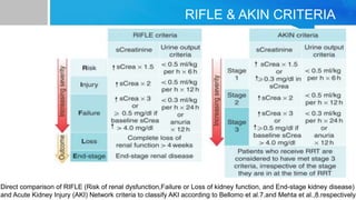
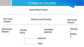
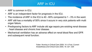
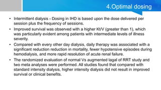
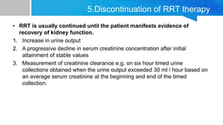
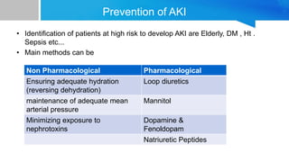
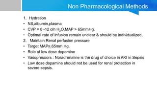
AKI IN ICU
Definition
• Acute kidney injury (AKI) is defined as deterioration of renal function over
hours, days to weeks. The mortality rate of AKI is 50-80% in intensive care
unit (ICU) patients, and has not declined significantly since the initial marked
benefit of acute dialysis therapy.
- ISN (International Society of Nephrology)
• Following long advocacy and persistent work for definition, a system,
RIFLE system was ultimately developed, involving a broad consensus of
experts.
• The acronym RIFLE represents the increasing severity classes Risk, Injury,
and Failure and the two outcome classes Loss and End-Stage Kidney
Disease. The severity grades R–F are defined on the basis of changes in
serum creatinine or urine output, wherein the worst of each criterion is used.
The two outcome criteria, L and E, are defined by the duration of loss of
kidney function.
RIFLE & AKIN CRITERIA
Direct comparison of RIFLE (Risk of renal dysfunction,Failure or Loss of kidney function, and End-stage kidney disease)
and Acute Kidney Injury (AKI) Network criteria to classify AKI according to Bellomo et al.7.and Mehta et al.,8.respectively.
COMMON CAUSES
Acute Renal Failure
Pre renal
Causes
Intrinsic renal Causes Post renal
Causes
Glomerular
disease
Tubular
Injury
Interstitial
nephritis
Vascula
r
Diseas
e
Ischemic
Toxic
Prerenal Causes
• Prerenal (Reduced renal perfusion)
1. Volume Depletion
• Renal loss : diuretics,osmotic diuresis (DKA)
• Extrarenal loss : vomiting,diarrhea,skin losses
• Hemorrhage
• Pancreatitis
2. Hypotension (regardless of cause)
3.Cardiovascular
• Congestive heart failure,reduced myocardial function
• Severe valvular heart disease
4) Hemodynamic (intense internal vasoconstriction)
• Radiographic contrast
• Prostaglandin inhibition
• Cyclosporine and tacrolimus
• ACE inhibitors
5) Hepatorenal syndrome
• (bland urinary sediment,oliguria, low urine sodium,not reversed with volume
repletion,reversible with successful liver transplant).
Intrinsic / Internal renal cause
1)Vascular :
• Renal infarction,renal artery stenosis,renal vein thrombosis
• Malignant hypertension,scleroderma renal crisis
2) Tubular :
• Ischemic prolonged prenatal state,sepsis syndrome
• Nephrotoxic aminoglycosides,cisplatin,myoglobin
3)Glomerular :
• Acute glomerulonephritis
• Good pasture syndrome
4)Interstitium:
• Medications:penicillins,cephalosporins,ciprofloxecin
• Tumor infiltration
Post renal ca
• Prostate hypertrophy,neurogenic bladder
• Intraureteral obstruction - crystals stones,clots,tumor.
• Extraureteral obstruction - tumor ( cervical,prostate),
Retro peritoneal fibrosis.
• Ureteric ligation during pelvic surgery.
Investigation of Acute Renal Failure
• Blood urea nitrogen and serum creatinin are elevated.
• ABG , electrolytes , CBC and serology.
• U/s kidneys (the size of kidney is usually normal)
• Serology : ANA , ANCA,Anti DNA , HBV , HCV, Anti GBM.
• Urine Analysis :
 Differentiates ATN vs AIN vs AGN
Unremarkable in pre and post renal causes
Urine electrolytes and osmolality
• Hansel stain for Eosinophiluria.
ARF in ICU
• ARF is common in ICU.
• ARF is an independent factor for prognosis in the ICU.
• The incidence of ARF in the ICU is 40 - 60% compared to 1 - 3% in the ward.
• ARF still has a mortality of 50% since it occurs in very sick patients with multi
organ failure.
• Predisposing factors to ARF include old age sepsis pre existing renal disease
, heart disease and chronic liver disease.
• Mechanical ventilation has an adverse effect on renal blood flow and GFR
and subsequent renal function.
Faber. Nursing in Critical Care 2009; 14: 4 Foot. Current
Anaesthesia and Critical Care 2005; 16: 321 -329
Classification
• Patients with ICU acquired ARF were classified into following:
A) According to the urine Output:
1.Oliguric (urine volume of < 400 ml/day)
2.Nonoliguric (urine volume of > 400 ml/day)
3.Anuric (urine volume <100 ml/day)
B) According to the cause:
1. Prerenal ARF
2. Ischemic acute tubular necrosis
3.Nephrotoxic ARF
4.Sepsis induced ARF
5.Hepato Renal Syndrome
6.Other causes (e.g. obstructive uropathy,pigment nephropathy)
Prerenal ARF
1.Prerenal ARF
Defined as ARF to renal hypoperfusion with recovery
after correction of hemodynamic disturbances.
2. Ischemic Acute tubular necrosis (IATN)
It is diagnosed when renal function does not improve after correction of
possible prerenal causes.
3.Nephrotoxic ARF :
A)Nephrotoxic Acute Interstitial Nephritis : History of drug ingestion,fever,rash,or
arthritis.
B) Nephrotoxic Acute Tubular Necrosis : was defined as ARF occurring after
administration of drugs known to cause ATN (e.g.,aminoglycosides,amphotericin)
4.Sepsis induced ARF : It is diagnosed if ARF is associated with at least one of
the following three conditions:
A. Documented bacteremia
B. A known focus of infection
C. Immunosuppression with neutropenia
5. Hepatorenal syndrome : It is assigned as the cause of renal failure if the
patient had severe liver failure (e.g. ascites - jaundice,hepatic encephalopathy)
and a urine sodium concentration less than 10 mEq/Litre)
6.Other causes : obstruction was determined to be the cause of renal failure.
RRT in AKI
• The initiation of RRT in patients with AKI prevents uremia and immediate
death from the adverse complications of renal failure.
• It is possible that variations in the timing of initiation,modalities, and/or dosing
may affect clinical outcomes.
• Multiple modalities of RRT are currently available.These include intermittent
hemodialysis (IHD), continuous renal replacement therapies (CRRTs), and
hybrid therapies such as sustained low efficiency dialysis (SLED).
• Despite these varied techniques mortality in patients with ARF remains high
greater than 50% in severely ill patients.
Dialysis in critically ill patients
1. Indications for dialysis
2. Timing of initiation of dialysis
3. Optimal modality
4. Optimal Dosing
5. Discontinuation of therapy.
1. Refractory fluid overload
2. Hyperkalemia (plasma potassium concentration > 6.5 meq/L)
3. Metabolic acidosis (pH less than 7.1)
4. Signs of uremia e.g. pericarditis and decline in mental state
5. Certain alcohol and drug intoxication.
1.Indications for dialysis in AKI
2.Timing of Initiation of Dialysis
• Studies published during the 1960s and 1970s suggested that improved
outcomes were associated with the initiation of hemodialysis when BUN
reached exceeded 150 to 200 mg/dL.
• More recent studies have evaluated the relationship between the timing of
RRT initiation and clinical outcomes.
• Several non randomized studies have reported that improved
outcomes,including survival,are associated with early versus late initiation of
RRT.
• It has been suggested that initiation of RRT dialysis prior to the development
of covert symptoms and signs of renal failure due to AKI improves the
outcome.
3.RRT Modalities
1. Intermittent hemodialysis (IHD)
2. Continuous renal replacement therapy (CRRT)
3. Peritoneal dialysis
Principal of Dialysis
• Dialysis is Passive movement of
solutes across a semi permeable
membrane down concentration
gradient.
• It works on principal for diffusion.
• It is good for small molecules.
• (Ultra) Filtration -
• Convection = solute+fluid removal
across semi permeable membrane
down a pressure gradient (solvent
drag)
• It is better for removal of fluid &
medium size molecule. Faber , Nursing in crital care 2009 ; 14; 4 Foot.
Current Anaesthesia and critical care 2005; 16: 321-329
• Oldest and most common technique.
• Primarily diffusive treatment : blood and dialysate are calculated in counter
current manner.
• Also some fluid removal by ultra filtration due to pressure driving through
circuit.
• Best for removal of small molecules.
• Typically performed 4 hours 3× / wk or daily.
Principal of Dialysis
1.Intermittent Hemodialysis(IHD)
2.Continuous Renal Replacement Therapy (CRRT)
• CRRT strategies are particularly useful in haemodynamically compromised
patients with ARF.
• They allow slow and gentle removal of solutes and fluid avoiding major
intravascular fluid shifts and minimizing electrolyte disturbances,hypotension
and arrhythmia.
• Inflammatory mediators may also be continuously removed by CRRT, so it
may be useful in sepsis syndrome.
Types of CRRT
1. Blood access
• Continuous arteriovenous hemofiltration (CAVH)
• Continuous arteriovenous hemodialysis (CAVHD)
• Continuous arteriovenous hemodiafiltration (CAVHDF)
• Continuous venovenous hemofiltration (CVVH)
• Continuous venovenous hemodialysis (CVVHD)
• Continuous venovenous hemodiafiltration (CVVHDF)
• Slow low efficiency dialysis (SLED)
• Slow continuous ultrafiltration (SCUF)
• Extended daily dialysis (EDD)
2. Peritoneal access
• Continuous equilibrium peritoneal dialysis.
4.Optimal dosing
• Intermittent dialysis - Dosing in IHD is based upon the dose delivered per
session plus the frequency of sessions.
• Improved survival was observed with a higher Kt/V (greater than 1), which
was particularly evident among patients with intermediate levels of illness
severity.
• Compared with every other day dialysis, daily therapy was associated with a
significant reduction reduction in mortality, fewer hypotensive episodes during
hemodialysis, and more rapid resolution of acute renal failure.
• The randomized evaluation of normal Vs augmented lagal of RRT study and
two meta analyses were performed. All studies found that compared with
standard intensity dialysis, higher intensity dialysis did not result in improved
survival or clinical benefits.
5.Discontinuation of RRT therapy
• RRT is usually continued until the patient manifests evidence of
recovery of kidney function.
1. Increase in urine output
2. A progressive decline in serum creatinine concentration after initial
attainment of stable values
3. Measurement of creatinine clearance e.g. on six hour timed urine
collections obtained when the urine output exceeded 30 ml / hour based on
an average serum creatinine at the beginning and end of the timed
collection.
Prevention of AKI
• Identification of patients at high risk to develop AKI are Elderly, DM , Ht .
Sepsis etc...
• Main methods can be
Non Pharmacological Pharmacological
Ensuring adequate hydration
(reversing dehydration)
Loop diuretics
maintenance of adequate mean
arterial pressure
Mannitol
Minimizing exposure to
nephrotoxins
Dopamine &
Fenoldopam
Natriuretic Peptides
Non Pharmacological Methods
1. Hydration
• NS,albumin,plasma
• CVP = 8 -12 cm H2O,MAP > 65mmHg.
• Optimal rate of infusion remain unclear & should be individualized.
2. Maintain Renal perfusion pressure
• Target MAP> 65mm Hg.
• Role of low dose dopamine
• Vasopresoors : Noradrenaline is the drug of choice in AKI in Sepsis
• Low dose dopamine should not be used for renal protection in
severe sepsis.
Pharmacological Methods
1. Mannitol & Loop Diuretics :
• In animal studies the use of mannitol & loop diuretics minimize the degree of
renal injury if given at the time of ischemic injury.
• Loop diuretics increases the active Na transport in the thick ascending loop
decreasing the energy requirement.
Pharmacological Methods
2. ANP & Calcium channel Blockers
• ANP had been tried in experimental models without any
benefit despite their ability to increase renal blood flow and Na
excretion.
• Calcium channel blockers decrease Ca influx to the cells that
lead to cell injury.
• Most human studies were done on established ATN.
• Uncontrolled studies showed that those patients who respond
to diuretics/mannitol may have better outcome but they have
less severe disease.
• Controlled studies failed to show any evidence that low dose
dopamine have any protection in ischemic renal damage.
• There is now clear evidence that ARF is associated with excess
mortality,irrespective of whether the patient requires renal
replacement therapy.
• As no drugs are available to enhance renal recovery once arf
occurs, prevention is the only powerful tool to improve outcome of
AKI.
Take Home Message
References
• References p p p p Miller: Miller's Anesthesia, 7 th ed. 2009 Uchino S,
Kellum JA, Bellomo R, et al: for the Beginning and Ending Supportive
Therapy for the Kidney (BEST Kidney) Investigators.
• Acute renal failure in critically ill patients: A multinational, multicenter study.
JAMA 2005; 294: 813 -818. Bouman CS, Oudemans-Van Straaten HM,
Tijssen JG, Zandstra DF, Kesecioglu J.
• Continuous versus intermittent renal replacement therapy for critically ill
patients with acute kidney injury: a meta-analysis. Crit Care Med 2008, 36:
610 -617. Rabindranath K, Adams J, Macleod AM, Muirhead N
• Intermittent versus continuous renal replacement therapy for acute renal
failure in adults. Cochrane Database Syst Rev 2007, 3: CD 003773.
AKI IN ICU.pptx

AKI IN ICU.pptx

  • 1.
  • 2.
    Definition • Acute kidneyinjury (AKI) is defined as deterioration of renal function over hours, days to weeks. The mortality rate of AKI is 50-80% in intensive care unit (ICU) patients, and has not declined significantly since the initial marked benefit of acute dialysis therapy. - ISN (International Society of Nephrology) • Following long advocacy and persistent work for definition, a system, RIFLE system was ultimately developed, involving a broad consensus of experts. • The acronym RIFLE represents the increasing severity classes Risk, Injury, and Failure and the two outcome classes Loss and End-Stage Kidney Disease. The severity grades R–F are defined on the basis of changes in serum creatinine or urine output, wherein the worst of each criterion is used. The two outcome criteria, L and E, are defined by the duration of loss of kidney function.
  • 3.
    RIFLE & AKINCRITERIA Direct comparison of RIFLE (Risk of renal dysfunction,Failure or Loss of kidney function, and End-stage kidney disease) and Acute Kidney Injury (AKI) Network criteria to classify AKI according to Bellomo et al.7.and Mehta et al.,8.respectively.
  • 4.
    COMMON CAUSES Acute RenalFailure Pre renal Causes Intrinsic renal Causes Post renal Causes Glomerular disease Tubular Injury Interstitial nephritis Vascula r Diseas e Ischemic Toxic
  • 5.
    Prerenal Causes • Prerenal(Reduced renal perfusion) 1. Volume Depletion • Renal loss : diuretics,osmotic diuresis (DKA) • Extrarenal loss : vomiting,diarrhea,skin losses • Hemorrhage • Pancreatitis 2. Hypotension (regardless of cause) 3.Cardiovascular • Congestive heart failure,reduced myocardial function • Severe valvular heart disease
  • 6.
    4) Hemodynamic (intenseinternal vasoconstriction) • Radiographic contrast • Prostaglandin inhibition • Cyclosporine and tacrolimus • ACE inhibitors 5) Hepatorenal syndrome • (bland urinary sediment,oliguria, low urine sodium,not reversed with volume repletion,reversible with successful liver transplant).
  • 7.
    Intrinsic / Internalrenal cause 1)Vascular : • Renal infarction,renal artery stenosis,renal vein thrombosis • Malignant hypertension,scleroderma renal crisis 2) Tubular : • Ischemic prolonged prenatal state,sepsis syndrome • Nephrotoxic aminoglycosides,cisplatin,myoglobin 3)Glomerular : • Acute glomerulonephritis • Good pasture syndrome 4)Interstitium: • Medications:penicillins,cephalosporins,ciprofloxecin • Tumor infiltration
  • 8.
    Post renal ca •Prostate hypertrophy,neurogenic bladder • Intraureteral obstruction - crystals stones,clots,tumor. • Extraureteral obstruction - tumor ( cervical,prostate), Retro peritoneal fibrosis. • Ureteric ligation during pelvic surgery.
  • 9.
    Investigation of AcuteRenal Failure • Blood urea nitrogen and serum creatinin are elevated. • ABG , electrolytes , CBC and serology. • U/s kidneys (the size of kidney is usually normal) • Serology : ANA , ANCA,Anti DNA , HBV , HCV, Anti GBM. • Urine Analysis :  Differentiates ATN vs AIN vs AGN Unremarkable in pre and post renal causes Urine electrolytes and osmolality • Hansel stain for Eosinophiluria.
  • 10.
    ARF in ICU •ARF is common in ICU. • ARF is an independent factor for prognosis in the ICU. • The incidence of ARF in the ICU is 40 - 60% compared to 1 - 3% in the ward. • ARF still has a mortality of 50% since it occurs in very sick patients with multi organ failure. • Predisposing factors to ARF include old age sepsis pre existing renal disease , heart disease and chronic liver disease. • Mechanical ventilation has an adverse effect on renal blood flow and GFR and subsequent renal function. Faber. Nursing in Critical Care 2009; 14: 4 Foot. Current Anaesthesia and Critical Care 2005; 16: 321 -329
  • 11.
    Classification • Patients withICU acquired ARF were classified into following: A) According to the urine Output: 1.Oliguric (urine volume of < 400 ml/day) 2.Nonoliguric (urine volume of > 400 ml/day) 3.Anuric (urine volume <100 ml/day) B) According to the cause: 1. Prerenal ARF 2. Ischemic acute tubular necrosis 3.Nephrotoxic ARF 4.Sepsis induced ARF 5.Hepato Renal Syndrome 6.Other causes (e.g. obstructive uropathy,pigment nephropathy)
  • 12.
    Prerenal ARF 1.Prerenal ARF Definedas ARF to renal hypoperfusion with recovery after correction of hemodynamic disturbances. 2. Ischemic Acute tubular necrosis (IATN) It is diagnosed when renal function does not improve after correction of possible prerenal causes. 3.Nephrotoxic ARF : A)Nephrotoxic Acute Interstitial Nephritis : History of drug ingestion,fever,rash,or arthritis. B) Nephrotoxic Acute Tubular Necrosis : was defined as ARF occurring after administration of drugs known to cause ATN (e.g.,aminoglycosides,amphotericin)
  • 13.
    4.Sepsis induced ARF: It is diagnosed if ARF is associated with at least one of the following three conditions: A. Documented bacteremia B. A known focus of infection C. Immunosuppression with neutropenia 5. Hepatorenal syndrome : It is assigned as the cause of renal failure if the patient had severe liver failure (e.g. ascites - jaundice,hepatic encephalopathy) and a urine sodium concentration less than 10 mEq/Litre) 6.Other causes : obstruction was determined to be the cause of renal failure.
  • 14.
    RRT in AKI •The initiation of RRT in patients with AKI prevents uremia and immediate death from the adverse complications of renal failure. • It is possible that variations in the timing of initiation,modalities, and/or dosing may affect clinical outcomes. • Multiple modalities of RRT are currently available.These include intermittent hemodialysis (IHD), continuous renal replacement therapies (CRRTs), and hybrid therapies such as sustained low efficiency dialysis (SLED). • Despite these varied techniques mortality in patients with ARF remains high greater than 50% in severely ill patients.
  • 15.
    Dialysis in criticallyill patients 1. Indications for dialysis 2. Timing of initiation of dialysis 3. Optimal modality 4. Optimal Dosing 5. Discontinuation of therapy.
  • 16.
    1. Refractory fluidoverload 2. Hyperkalemia (plasma potassium concentration > 6.5 meq/L) 3. Metabolic acidosis (pH less than 7.1) 4. Signs of uremia e.g. pericarditis and decline in mental state 5. Certain alcohol and drug intoxication. 1.Indications for dialysis in AKI
  • 17.
    2.Timing of Initiationof Dialysis • Studies published during the 1960s and 1970s suggested that improved outcomes were associated with the initiation of hemodialysis when BUN reached exceeded 150 to 200 mg/dL. • More recent studies have evaluated the relationship between the timing of RRT initiation and clinical outcomes. • Several non randomized studies have reported that improved outcomes,including survival,are associated with early versus late initiation of RRT. • It has been suggested that initiation of RRT dialysis prior to the development of covert symptoms and signs of renal failure due to AKI improves the outcome.
  • 18.
    3.RRT Modalities 1. Intermittenthemodialysis (IHD) 2. Continuous renal replacement therapy (CRRT) 3. Peritoneal dialysis
  • 19.
    Principal of Dialysis •Dialysis is Passive movement of solutes across a semi permeable membrane down concentration gradient. • It works on principal for diffusion. • It is good for small molecules. • (Ultra) Filtration - • Convection = solute+fluid removal across semi permeable membrane down a pressure gradient (solvent drag) • It is better for removal of fluid & medium size molecule. Faber , Nursing in crital care 2009 ; 14; 4 Foot. Current Anaesthesia and critical care 2005; 16: 321-329
  • 20.
    • Oldest andmost common technique. • Primarily diffusive treatment : blood and dialysate are calculated in counter current manner. • Also some fluid removal by ultra filtration due to pressure driving through circuit. • Best for removal of small molecules. • Typically performed 4 hours 3× / wk or daily. Principal of Dialysis 1.Intermittent Hemodialysis(IHD)
  • 21.
    2.Continuous Renal ReplacementTherapy (CRRT) • CRRT strategies are particularly useful in haemodynamically compromised patients with ARF. • They allow slow and gentle removal of solutes and fluid avoiding major intravascular fluid shifts and minimizing electrolyte disturbances,hypotension and arrhythmia. • Inflammatory mediators may also be continuously removed by CRRT, so it may be useful in sepsis syndrome.
  • 22.
    Types of CRRT 1.Blood access • Continuous arteriovenous hemofiltration (CAVH) • Continuous arteriovenous hemodialysis (CAVHD) • Continuous arteriovenous hemodiafiltration (CAVHDF) • Continuous venovenous hemofiltration (CVVH) • Continuous venovenous hemodialysis (CVVHD) • Continuous venovenous hemodiafiltration (CVVHDF) • Slow low efficiency dialysis (SLED) • Slow continuous ultrafiltration (SCUF) • Extended daily dialysis (EDD) 2. Peritoneal access • Continuous equilibrium peritoneal dialysis.
  • 23.
    4.Optimal dosing • Intermittentdialysis - Dosing in IHD is based upon the dose delivered per session plus the frequency of sessions. • Improved survival was observed with a higher Kt/V (greater than 1), which was particularly evident among patients with intermediate levels of illness severity. • Compared with every other day dialysis, daily therapy was associated with a significant reduction reduction in mortality, fewer hypotensive episodes during hemodialysis, and more rapid resolution of acute renal failure. • The randomized evaluation of normal Vs augmented lagal of RRT study and two meta analyses were performed. All studies found that compared with standard intensity dialysis, higher intensity dialysis did not result in improved survival or clinical benefits.
  • 24.
    5.Discontinuation of RRTtherapy • RRT is usually continued until the patient manifests evidence of recovery of kidney function. 1. Increase in urine output 2. A progressive decline in serum creatinine concentration after initial attainment of stable values 3. Measurement of creatinine clearance e.g. on six hour timed urine collections obtained when the urine output exceeded 30 ml / hour based on an average serum creatinine at the beginning and end of the timed collection.
  • 25.
    Prevention of AKI •Identification of patients at high risk to develop AKI are Elderly, DM , Ht . Sepsis etc... • Main methods can be Non Pharmacological Pharmacological Ensuring adequate hydration (reversing dehydration) Loop diuretics maintenance of adequate mean arterial pressure Mannitol Minimizing exposure to nephrotoxins Dopamine & Fenoldopam Natriuretic Peptides
  • 26.
    Non Pharmacological Methods 1.Hydration • NS,albumin,plasma • CVP = 8 -12 cm H2O,MAP > 65mmHg. • Optimal rate of infusion remain unclear & should be individualized. 2. Maintain Renal perfusion pressure • Target MAP> 65mm Hg. • Role of low dose dopamine • Vasopresoors : Noradrenaline is the drug of choice in AKI in Sepsis • Low dose dopamine should not be used for renal protection in severe sepsis.
  • 27.
    Pharmacological Methods 1. Mannitol& Loop Diuretics : • In animal studies the use of mannitol & loop diuretics minimize the degree of renal injury if given at the time of ischemic injury. • Loop diuretics increases the active Na transport in the thick ascending loop decreasing the energy requirement.
  • 28.
    Pharmacological Methods 2. ANP& Calcium channel Blockers • ANP had been tried in experimental models without any benefit despite their ability to increase renal blood flow and Na excretion. • Calcium channel blockers decrease Ca influx to the cells that lead to cell injury. • Most human studies were done on established ATN. • Uncontrolled studies showed that those patients who respond to diuretics/mannitol may have better outcome but they have less severe disease. • Controlled studies failed to show any evidence that low dose dopamine have any protection in ischemic renal damage.
  • 29.
    • There isnow clear evidence that ARF is associated with excess mortality,irrespective of whether the patient requires renal replacement therapy. • As no drugs are available to enhance renal recovery once arf occurs, prevention is the only powerful tool to improve outcome of AKI. Take Home Message
  • 30.
    References • References pp p p Miller: Miller's Anesthesia, 7 th ed. 2009 Uchino S, Kellum JA, Bellomo R, et al: for the Beginning and Ending Supportive Therapy for the Kidney (BEST Kidney) Investigators. • Acute renal failure in critically ill patients: A multinational, multicenter study. JAMA 2005; 294: 813 -818. Bouman CS, Oudemans-Van Straaten HM, Tijssen JG, Zandstra DF, Kesecioglu J. • Continuous versus intermittent renal replacement therapy for critically ill patients with acute kidney injury: a meta-analysis. Crit Care Med 2008, 36: 610 -617. Rabindranath K, Adams J, Macleod AM, Muirhead N • Intermittent versus continuous renal replacement therapy for acute renal failure in adults. Cochrane Database Syst Rev 2007, 3: CD 003773.

Editor's Notes

  • #10 https://www.semanticscholar.org/paper/Cardiorenal-syndrome.-Ronco-Haapio/afea71a4ea0845018b58214c6c2f643fba38cfca
  • #12 https://www.semanticscholar.org/paper/Cardiorenal-syndrome.-Ronco-Haapio/afea71a4ea0845018b58214c6c2f643fba38cfca