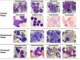
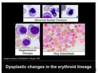
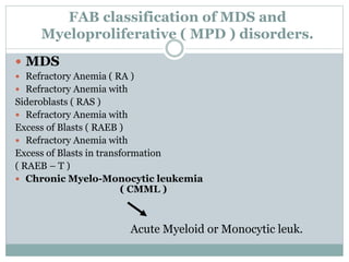
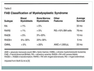
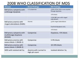
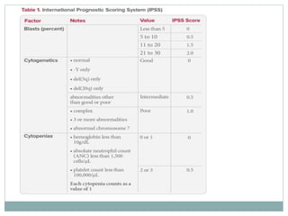
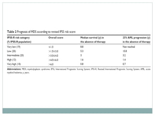
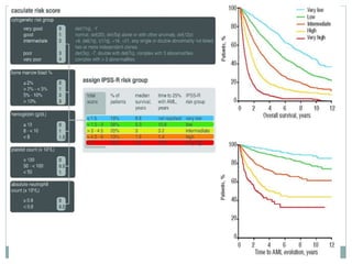
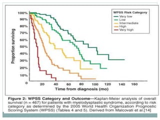
MYELODYSPLASTIC
SYNDROMES
WHAT ARE THEY??
 The term “myelodysplastic syndrome” (MDS)
refers to a group of clonal stem cell disorders
 characterized by maturation defects
 that are associated with ineffective
hematopoiesis
 and a high risk of transformation to AML
 In MDS the bone marrow is partly or wholly replaced
by the clonal progeny of
- a neoplastic multipotent stem cell that retains the
capacity to differentiate but does so in an ineffective
and disordered fashion.
 These abnormal cells stay within the bone marrow
and hence the patients have peripheral blood
cytopenias.
 MDS may be either primary (idiopathic) or
secondary to previous genotoxic drug or radiation
therapy (t-MDS).
-t-MDS usually appears from 2 to 8 years after the
genotoxic exposure.
 All forms of MDS can transform to AML, but
transformation occurs with highest frequency and
most rapidly in t-MDS.
 Although characteristic morphologic
changes are typically seen in the marrow and the
peripheral blood, the diagnosis frequently requires
correlation with other laboratory tests.
 Cytogenetic analysis is particularly helpful, since
certain chromosomal aberrations.
PATHOGENESIS
 The pathogenesis of MDS is poorly understood.
 The important new insights have come from recent
deep sequencing of MDS genomes, which has
identifed a number of recurrently mutated genes.
 These genes can be grouped into three major
functional categories, as follows:
Epigenetic factors. Frequent mutations are seen
involving many of the same epigenetic factors that
are mutated in AML, including factors that regulate
DNA methylation and histone modifcations;
thus, like AML, dysregulation of the epigenome
appears to be important in the genesis of MDS.
 RNA splicing factors:
A subset of tumors has mutations involving
components of the 3’ end of the RNA splicing
machinery. The impact of these mutations on RNA
splicing and other nuclear functions is not yet
known.
 Transcription factors. These mutations affect
transcription factors that are are required for normal
myelopoiesis and may contribute to the deranged
differentiation that characterizes MDS.
 chromosomal abnormalities, including monosomies
5 and 7, deletions of 5q, 7q, and 20 q, and trisomy 8.
 As with aneuploidy in other cancers, it is not known
how these aberrations contribute to MDS
 MYC gene is located on chromosome 8, and trisomy
8 is one of the most common forms of aneuploidy in
a wide range of myeloid tumors.
 Similarly, the region that is commonly lost on
chromosome 5q contains a gene encoding
the ribosomal protein RPS14
 In experimental systems, loss
of one copy of RPS14 produces ineffective
erythropoiesis, one of the hallmarks of MDS.
MORPHOLOGY
 Although the marrow is usually
 hypercellular at diagnosis,
 It is sometimes normocellular
 or, less commonly, hypocellular.
 The most characteristic finding is
disordered (dysplastic) differentiation
affecting the
 erythroid,
 granulocytic,
 monocytic,
 and megakaryocytic lineages to varying
degrees.
 Within the erythroid series, common
abnormalities include
 ring sideroblasts,
 erythroblasts with iron-laden mitochondria
visible as perinuclear granules in Prussian
blue-stained aspirates or biopsies. .
 megaloblastoid maturation and nuclear
budding abnormalities,
 recognized as nuclei with misshapen, often
polyploid, outlines .
 Neutrophils frequently contain decreased
numbers of secondary granules, toxic
granulations, and/or Döhle bodies.
 Pseudo-Pelger-Hüet cells, neutrophils
with only two nuclear lobes, are commonly
observed, and neutrophils are seen
occasionally that completely lack nuclear
segmentation.
 Megakaryocytes with single nuclear lobes or
multiple separate nuclei (pawn ball
megakaryocytes) are also characteristic.
 Myeloid blasts may be increased but make
up less than 20% of the overall
marrow cellularity. The blood often
contains
 pseudo-Pelger-Hüet cells,
 giant platelets,
 macrocytes,
 and poikilocytes,
 accompanied by a relative or absolute
monocytosis. Myeloid blasts usually make
up less than 10% of the leukocytes in
the blood.
Nucleated red cell progenitors with multilobated or multiple
nuclei.
 Ringed sideroblasts,
 erythroid progenitors with iron-laden
mitochondria seen as blue perinuclear
granules (Prussian blue stain).
 Pseudo-Pelger-Hüet cells, neutrophils
with only two nuclear lobes instead of
the normal three to four, are observed at
the top and bottom of this feld.
 Megakaryocytes with multiple nuclei
instead of the normal single multilobated
nucleus.
FAB classification of MDS and
Myeloproliferative ( MPD ) disorders.
 MDS
 Refractory Anemia ( RA )
 Refractory Anemia with
Sideroblasts ( RAS )
 Refractory Anemia with
Excess of Blasts ( RAEB )
 Refractory Anemia with
Excess of Blasts in transformation
( RAEB – T )
 Chronic Myelo-Monocytic leukemia
( CMML )
Acute Myeloid or Monocytic leuk.
MDS – WHO classification
CLINICAL FEATURES
 Primary MDS is predominantly a
disease of older adults; the mean age of onset is 70 years.
 In up to half of the cases, it is discovered incidentally on
routine blood testing.
 When symptomatic, it presents
with weakness, infections, and hemorrhages, all due to
pancytopenia.
 Primary MDS is divided into eight categories based
on morphologic and cytogenetic features in the WHO
 In brief, worse outcomes are predicted by higher
blast counts and more severe cytopenias, as well as
the presence of multiple clonal chromosomal
abnormalities.
PROGNOSIS
SURVIVAL RATE
 The median survival in primary MDS varies from 9
to 29 months, but some individuals in good
prognostic groups may live for 5 years or more.
 Overall, progression to AML occurs in 10% to 40% of
individuals and is usually accompanied by the
appearance of additional cytogenetic abnormalities.
 Patients often succumb to the complications of
thrombocytopenia (bleeding) and neutropenia
(infection).
 The outlook is even grimmer in t-MDS, which has a
median survival of only 4 to 8 months.
 In t-MDS, cytopenias tend to be more severe and
progression to AML is often rapid.
TREATMENT
 Treatment options are fairly limited.
 In younger patients,allogeneic hematopoietic stem
cell transplantation offers hope for reconstitution of
normal hematopoiesis and possible cure.
 Older patients with MDS are treated supportively
with antibiotics and blood product transfusions.
 Thalidomide-like drugs and DNA methylation
inhibitors improve the effectiveness of hematopoiesis
and the peripheral blood counts in a subset of
patients.
THANK YOU

Myelodysplastic syndromes

  • 1.
  • 2.
    WHAT ARE THEY?? The term “myelodysplastic syndrome” (MDS) refers to a group of clonal stem cell disorders  characterized by maturation defects  that are associated with ineffective hematopoiesis  and a high risk of transformation to AML
  • 3.
     In MDSthe bone marrow is partly or wholly replaced by the clonal progeny of - a neoplastic multipotent stem cell that retains the capacity to differentiate but does so in an ineffective and disordered fashion.
  • 4.
     These abnormalcells stay within the bone marrow and hence the patients have peripheral blood cytopenias.
  • 5.
     MDS maybe either primary (idiopathic) or secondary to previous genotoxic drug or radiation therapy (t-MDS). -t-MDS usually appears from 2 to 8 years after the genotoxic exposure.
  • 6.
     All formsof MDS can transform to AML, but transformation occurs with highest frequency and most rapidly in t-MDS.
  • 7.
     Although characteristicmorphologic changes are typically seen in the marrow and the peripheral blood, the diagnosis frequently requires correlation with other laboratory tests.  Cytogenetic analysis is particularly helpful, since certain chromosomal aberrations.
  • 8.
    PATHOGENESIS  The pathogenesisof MDS is poorly understood.  The important new insights have come from recent deep sequencing of MDS genomes, which has identifed a number of recurrently mutated genes.
  • 9.
     These genescan be grouped into three major functional categories, as follows: Epigenetic factors. Frequent mutations are seen involving many of the same epigenetic factors that are mutated in AML, including factors that regulate DNA methylation and histone modifcations; thus, like AML, dysregulation of the epigenome appears to be important in the genesis of MDS.
  • 10.
     RNA splicingfactors: A subset of tumors has mutations involving components of the 3’ end of the RNA splicing machinery. The impact of these mutations on RNA splicing and other nuclear functions is not yet known.
  • 11.
     Transcription factors.These mutations affect transcription factors that are are required for normal myelopoiesis and may contribute to the deranged differentiation that characterizes MDS.
  • 12.
     chromosomal abnormalities,including monosomies 5 and 7, deletions of 5q, 7q, and 20 q, and trisomy 8.  As with aneuploidy in other cancers, it is not known how these aberrations contribute to MDS
  • 13.
     MYC geneis located on chromosome 8, and trisomy 8 is one of the most common forms of aneuploidy in a wide range of myeloid tumors.  Similarly, the region that is commonly lost on chromosome 5q contains a gene encoding the ribosomal protein RPS14
  • 14.
     In experimentalsystems, loss of one copy of RPS14 produces ineffective erythropoiesis, one of the hallmarks of MDS.
  • 15.
    MORPHOLOGY  Although themarrow is usually  hypercellular at diagnosis,  It is sometimes normocellular  or, less commonly, hypocellular.
  • 16.
     The mostcharacteristic finding is disordered (dysplastic) differentiation affecting the  erythroid,  granulocytic,  monocytic,  and megakaryocytic lineages to varying degrees.
  • 17.
     Within theerythroid series, common abnormalities include  ring sideroblasts,  erythroblasts with iron-laden mitochondria visible as perinuclear granules in Prussian blue-stained aspirates or biopsies. .
  • 18.
     megaloblastoid maturationand nuclear budding abnormalities,  recognized as nuclei with misshapen, often polyploid, outlines .
  • 19.
     Neutrophils frequentlycontain decreased numbers of secondary granules, toxic granulations, and/or Döhle bodies.  Pseudo-Pelger-Hüet cells, neutrophils with only two nuclear lobes, are commonly observed, and neutrophils are seen occasionally that completely lack nuclear segmentation.
  • 20.
     Megakaryocytes withsingle nuclear lobes or multiple separate nuclei (pawn ball megakaryocytes) are also characteristic.
  • 23.
     Myeloid blastsmay be increased but make up less than 20% of the overall marrow cellularity. The blood often contains  pseudo-Pelger-Hüet cells,  giant platelets,  macrocytes,  and poikilocytes,  accompanied by a relative or absolute monocytosis. Myeloid blasts usually make up less than 10% of the leukocytes in the blood.
  • 24.
    Nucleated red cellprogenitors with multilobated or multiple nuclei.
  • 26.
     Ringed sideroblasts, erythroid progenitors with iron-laden mitochondria seen as blue perinuclear granules (Prussian blue stain).
  • 28.
     Pseudo-Pelger-Hüet cells,neutrophils with only two nuclear lobes instead of the normal three to four, are observed at the top and bottom of this feld.
  • 30.
     Megakaryocytes withmultiple nuclei instead of the normal single multilobated nucleus.
  • 31.
    FAB classification ofMDS and Myeloproliferative ( MPD ) disorders.  MDS  Refractory Anemia ( RA )  Refractory Anemia with Sideroblasts ( RAS )  Refractory Anemia with Excess of Blasts ( RAEB )  Refractory Anemia with Excess of Blasts in transformation ( RAEB – T )  Chronic Myelo-Monocytic leukemia ( CMML ) Acute Myeloid or Monocytic leuk.
  • 33.
    MDS – WHOclassification
  • 35.
    CLINICAL FEATURES  PrimaryMDS is predominantly a disease of older adults; the mean age of onset is 70 years.  In up to half of the cases, it is discovered incidentally on routine blood testing.  When symptomatic, it presents with weakness, infections, and hemorrhages, all due to pancytopenia.  Primary MDS is divided into eight categories based on morphologic and cytogenetic features in the WHO
  • 36.
     In brief,worse outcomes are predicted by higher blast counts and more severe cytopenias, as well as the presence of multiple clonal chromosomal abnormalities.
  • 37.
  • 43.
    SURVIVAL RATE  Themedian survival in primary MDS varies from 9 to 29 months, but some individuals in good prognostic groups may live for 5 years or more.
  • 44.
     Overall, progressionto AML occurs in 10% to 40% of individuals and is usually accompanied by the appearance of additional cytogenetic abnormalities.
  • 45.
     Patients oftensuccumb to the complications of thrombocytopenia (bleeding) and neutropenia (infection).  The outlook is even grimmer in t-MDS, which has a median survival of only 4 to 8 months.  In t-MDS, cytopenias tend to be more severe and progression to AML is often rapid.
  • 46.
    TREATMENT  Treatment optionsare fairly limited.  In younger patients,allogeneic hematopoietic stem cell transplantation offers hope for reconstitution of normal hematopoiesis and possible cure.  Older patients with MDS are treated supportively with antibiotics and blood product transfusions.
  • 47.
     Thalidomide-like drugsand DNA methylation inhibitors improve the effectiveness of hematopoiesis and the peripheral blood counts in a subset of patients.
  • 48.