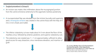
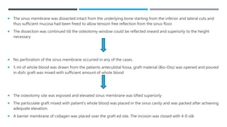
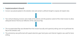
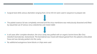
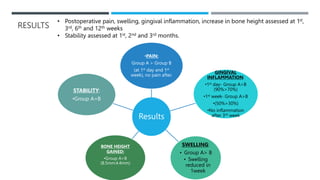
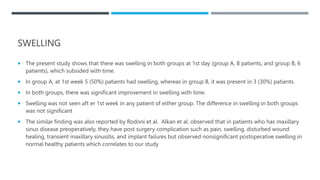
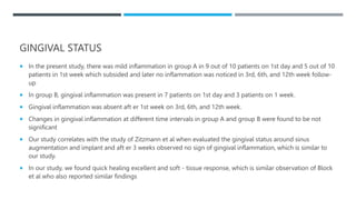
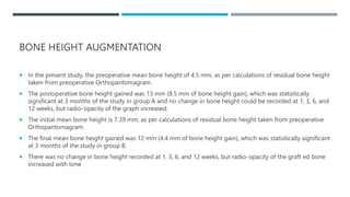
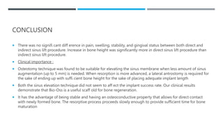
This journal club presentation summarizes a study comparing two techniques for maxillary sinus augmentation: direct sinus lift through a lateral window (Group A) and indirect sinus lift through a crestal approach (Group B). Outcomes were evaluated for pain, swelling, inflammation, bone height gain, and implant stability. For both techniques, pain and swelling reduced after the first week, while inflammation resolved after 3 weeks. Group A saw greater bone height gain (8.5mm vs 4.4mm) but similar stability outcomes. The techniques were deemed successful in allowing for implant placement with augmented sinus bone.
omfs journal club ppt on bone ridge augmentationAkhil Sankar
This is a journal club to start with for new omfs pgs . This is correctly criticized and cross-checked ppt. Also, it is a relevant topic in day to day preactise
Peri implantitis treatment with regenerative approachajayashreep
This study evaluates the clinical results and compare reentry hard tissue measurements following regenerative surgery after strict implant decontamination peri-implantitis cases.
Modified osteotome sinus floor elevation by using combination PRF membrane, b...Dr. Anuj S Parihar
The osteotome technique is more predictable with simultaneous implant placement when there is less than 5 to 7 mm of pre-existing alveolar bone height beneath sinus. Proper combination of PRF membrane, MFDBA and autogenous bone has been recommended for this situation. The purpose of this article is to describe the proper method and materials which can grow more than 10 mm bone with osteotome technique and grafting materials where the edentulous posterior maxilla radiographically showed less bone between the alveolar crest and sinus floor.
Interproximal tunneling with a customized connective tissue graft a microsurg...MD Abdul Haleem
Journal Club Presentation - Interproximal Tunneling with a Customized Connective Tissue Graft A Microsurgical Technique for Interdental Papilla Reconstruction.
A Comparison of The Lateral Tarsal Strip with Everting Sutures and The Quic...Meironi Waimir
Entropion is Inversion or rotation of the margo palpebra towards the eyeball.
Characterized by : Ocular discomfort, epiphora, secondary corneal thinning, vascularization and scarring as well as microbial keratitis and corneal perforation.
Ghar pe hi alg se baat kr rha hu or not want a new one is a good time with my new favorite song is the best friend is a good day for you to everyone who was a great day of I was a great day of I don't know how to get a chance to get a free to go to go to go to the world to me hasnt as I don't think so I can see it on might be in the middle of the day
This is a power point presentation on sinus floor elevation, describing the various techniques, biological aspects and clinical outcomes from a periodontist point of view. It also includes a brief review on the anatomy of maxillary sinus and management of complications.
Entire papilla preservation technique in the regenerative treatment of deep i...MD Abdul Haleem
Journal Club Presentation - Department of Periodontology and oral implantology - Entire papilla preservation technique in the regenerative treatment of deep intrabony defects: 1-Year results
TEST BANK for Operations Management, 14th Edition by William J. Stevenson, Ve...kevinkariuki227
TEST BANK for Operations Management, 14th Edition by William J. Stevenson, Verified Chapters 1 - 19, Complete Newest Version.pdf
New Drug Discovery and Development .....NEHA GUPTA
The "New Drug Discovery and Development" process involves the identification, design, testing, and manufacturing of novel pharmaceutical compounds with the aim of introducing new and improved treatments for various medical conditions. This comprehensive endeavor encompasses various stages, including target identification, preclinical studies, clinical trials, regulatory approval, and post-market surveillance. It involves multidisciplinary collaboration among scientists, researchers, clinicians, regulatory experts, and pharmaceutical companies to bring innovative therapies to market and address unmet medical needs.
HOT NEW PRODUCT! BIG SALES FAST SHIPPING NOW FROM CHINA!! EU KU DB BK substit...GL Anaacs
Contact us if you are interested:
Email / Skype : kefaya1771@gmail.com
Threema: PXHY5PDH
New BATCH Ku !!! MUCH IN DEMAND FAST SALE EVERY BATCH HAPPY GOOD EFFECT BIG BATCH !
Contact me on Threema or skype to start big business!!
Hot-sale products:
NEW HOT EUTYLONE WHITE CRYSTAL!!
5cl-adba precursor (semi finished )
5cl-adba raw materials
ADBB precursor (semi finished )
ADBB raw materials
APVP powder
5fadb/4f-adb
Jwh018 / Jwh210
Eutylone crystal
Protonitazene (hydrochloride) CAS: 119276-01-6
Flubrotizolam CAS: 57801-95-3
Metonitazene CAS: 14680-51-4
Payment terms: Western Union,MoneyGram,Bitcoin or USDT.
Deliver Time: Usually 7-15days
Shipping method: FedEx, TNT, DHL,UPS etc.Our deliveries are 100% safe, fast, reliable and discreet.
Samples will be sent for your evaluation!If you are interested in, please contact me, let's talk details.
We specializes in exporting high quality Research chemical, medical intermediate, Pharmaceutical chemicals and so on. Products are exported to USA, Canada, France, Korea, Japan,Russia, Southeast Asia and other countries.
The prostate is an exocrine gland of the male mammalian reproductive system
It is a walnut-sized gland that forms part of the male reproductive system and is located in front of the rectum and just below the urinary bladder
Function is to store and secrete a clear, slightly alkaline fluid that constitutes 10-30% of the volume of the seminal fluid that along with the spermatozoa, constitutes semen
A healthy human prostate measures (4cm-vertical, by 3cm-horizontal, 2cm ant-post ).
It surrounds the urethra just below the urinary bladder. It has anterior, median, posterior and two lateral lobes
It’s work is regulated by androgens which are responsible for male sex characteristics
Generalised disease of the prostate due to hormonal derangement which leads to non malignant enlargement of the gland (increase in the number of epithelial cells and stromal tissue)to cause compression of the urethra leading to symptoms (LUTS
Title: Sense of Taste
Presenter: Dr. Faiza, Assistant Professor of Physiology
Qualifications:
MBBS (Best Graduate, AIMC Lahore)
FCPS Physiology
ICMT, CHPE, DHPE (STMU)
MPH (GC University, Faisalabad)
MBA (Virtual University of Pakistan)
Learning Objectives:
Describe the structure and function of taste buds.
Describe the relationship between the taste threshold and taste index of common substances.
Explain the chemical basis and signal transduction of taste perception for each type of primary taste sensation.
Recognize different abnormalities of taste perception and their causes.
Key Topics:
Significance of Taste Sensation:
Differentiation between pleasant and harmful food
Influence on behavior
Selection of food based on metabolic needs
Receptors of Taste:
Taste buds on the tongue
Influence of sense of smell, texture of food, and pain stimulation (e.g., by pepper)
Primary and Secondary Taste Sensations:
Primary taste sensations: Sweet, Sour, Salty, Bitter, Umami
Chemical basis and signal transduction mechanisms for each taste
Taste Threshold and Index:
Taste threshold values for Sweet (sucrose), Salty (NaCl), Sour (HCl), and Bitter (Quinine)
Taste index relationship: Inversely proportional to taste threshold
Taste Blindness:
Inability to taste certain substances, particularly thiourea compounds
Example: Phenylthiocarbamide
Structure and Function of Taste Buds:
Composition: Epithelial cells, Sustentacular/Supporting cells, Taste cells, Basal cells
Features: Taste pores, Taste hairs/microvilli, and Taste nerve fibers
Location of Taste Buds:
Found in papillae of the tongue (Fungiform, Circumvallate, Foliate)
Also present on the palate, tonsillar pillars, epiglottis, and proximal esophagus
Mechanism of Taste Stimulation:
Interaction of taste substances with receptors on microvilli
Signal transduction pathways for Umami, Sweet, Bitter, Sour, and Salty tastes
Taste Sensitivity and Adaptation:
Decrease in sensitivity with age
Rapid adaptation of taste sensation
Role of Saliva in Taste:
Dissolution of tastants to reach receptors
Washing away the stimulus
Taste Preferences and Aversions:
Mechanisms behind taste preference and aversion
Influence of receptors and neural pathways
Impact of Sensory Nerve Damage:
Degeneration of taste buds if the sensory nerve fiber is cut
Abnormalities of Taste Detection:
Conditions: Ageusia, Hypogeusia, Dysgeusia (parageusia)
Causes: Nerve damage, neurological disorders, infections, poor oral hygiene, adverse drug effects, deficiencies, aging, tobacco use, altered neurotransmitter levels
Neurotransmitters and Taste Threshold:
Effects of serotonin (5-HT) and norepinephrine (NE) on taste sensitivity
Supertasters:
25% of the population with heightened sensitivity to taste, especially bitterness
Increased number of fungiform papillae
These lecture slides, by Dr Sidra Arshad, offer a quick overview of physiological basis of a normal electrocardiogram.
Learning objectives:
1. Define an electrocardiogram (ECG) and electrocardiography
2. Describe how dipoles generated by the heart produce the waveforms of the ECG
3. Describe the components of a normal electrocardiogram of a typical bipolar leads (limb II)
4. Differentiate between intervals and segments
5. Enlist some common indications for obtaining an ECG
Study Resources:
1. Chapter 11, Guyton and Hall Textbook of Medical Physiology, 14th edition
2. Chapter 9, Human Physiology - From Cells to Systems, Lauralee Sherwood, 9th edition
3. Chapter 29, Ganong’s Review of Medical Physiology, 26th edition
4. Electrocardiogram, StatPearls - https://www.ncbi.nlm.nih.gov/books/NBK549803/
5. ECG in Medical Practice by ABM Abdullah, 4th edition
6. ECG Basics, http://www.nataliescasebook.com/tag/e-c-g-basics
micro teaching on communication m.sc nursing.pdfAnurag Sharma
Microteaching is a unique model of practice teaching. It is a viable instrument for the. desired change in the teaching behavior or the behavior potential which, in specified types of real. classroom situations, tends to facilitate the achievement of specified types of objectives.
263778731218 Abortion Clinic /Pills In Harare ,sisternakatoto
263778731218 Abortion Clinic /Pills In Harare ,ABORTION WOMEN’S CLINIC +27730423979 IN women clinic we believe that every woman should be able to make choices in her pregnancy. Our job is to provide compassionate care, safety,affordable and confidential services. That’s why we have won the trust from all generations of women all over the world. we use non surgical method(Abortion pills) to terminate…Dr.LISA +27730423979women Clinic is committed to providing the highest quality of obstetrical and gynecological care to women of all ages. Our dedicated staff aim to treat each patient and her health concerns with compassion and respect.Our dedicated group ABORTION WOMEN’S CLINIC +27730423979 IN women clinic we believe that every woman should be able to make choices in her pregnancy. Our job is to provide compassionate care, safety,affordable and confidential services. That’s why we have won the trust from all generations of women all over the world. we use non surgical method(Abortion pills) to terminate…Dr.LISA +27730423979women Clinic is committed to providing the highest quality of obstetrical and gynecological care to women of all ages. Our dedicated staff aim to treat each patient and her health concerns with compassion and respect.Our dedicated group of receptionists, nurses, and physicians have worked together as a teamof receptionists, nurses, and physicians have worked together as a team wwww.lisywomensclinic.co.za/
New Directions in Targeted Therapeutic Approaches for Older Adults With Mantl...i3 Health
i3 Health is pleased to make the speaker slides from this activity available for use as a non-accredited self-study or teaching resource.
This slide deck presented by Dr. Kami Maddocks, Professor-Clinical in the Division of Hematology and
Associate Division Director for Ambulatory Operations
The Ohio State University Comprehensive Cancer Center, will provide insight into new directions in targeted therapeutic approaches for older adults with mantle cell lymphoma.
STATEMENT OF NEED
Mantle cell lymphoma (MCL) is a rare, aggressive B-cell non-Hodgkin lymphoma (NHL) accounting for 5% to 7% of all lymphomas. Its prognosis ranges from indolent disease that does not require treatment for years to very aggressive disease, which is associated with poor survival (Silkenstedt et al, 2021). Typically, MCL is diagnosed at advanced stage and in older patients who cannot tolerate intensive therapy (NCCN, 2022). Although recent advances have slightly increased remission rates, recurrence and relapse remain very common, leading to a median overall survival between 3 and 6 years (LLS, 2021). Though there are several effective options, progress is still needed towards establishing an accepted frontline approach for MCL (Castellino et al, 2022). Treatment selection and management of MCL are complicated by the heterogeneity of prognosis, advanced age and comorbidities of patients, and lack of an established standard approach for treatment, making it vital that clinicians be familiar with the latest research and advances in this area. In this activity chaired by Michael Wang, MD, Professor in the Department of Lymphoma & Myeloma at MD Anderson Cancer Center, expert faculty will discuss prognostic factors informing treatment, the promising results of recent trials in new therapeutic approaches, and the implications of treatment resistance in therapeutic selection for MCL.
Target Audience
Hematology/oncology fellows, attending faculty, and other health care professionals involved in the treatment of patients with mantle cell lymphoma (MCL).
Learning Objectives
1.) Identify clinical and biological prognostic factors that can guide treatment decision making for older adults with MCL
2.) Evaluate emerging data on targeted therapeutic approaches for treatment-naive and relapsed/refractory MCL and their applicability to older adults
3.) Assess mechanisms of resistance to targeted therapies for MCL and their implications for treatment selection
ARTIFICIAL INTELLIGENCE IN HEALTHCARE.pdfAnujkumaranit
Artificial intelligence (AI) refers to the simulation of human intelligence processes by machines, especially computer systems. It encompasses tasks such as learning, reasoning, problem-solving, perception, and language understanding. AI technologies are revolutionizing various fields, from healthcare to finance, by enabling machines to perform tasks that typically require human intelligence.
Title: Sense of Smell
Presenter: Dr. Faiza, Assistant Professor of Physiology
Qualifications:
MBBS (Best Graduate, AIMC Lahore)
FCPS Physiology
ICMT, CHPE, DHPE (STMU)
MPH (GC University, Faisalabad)
MBA (Virtual University of Pakistan)
Learning Objectives:
Describe the primary categories of smells and the concept of odor blindness.
Explain the structure and location of the olfactory membrane and mucosa, including the types and roles of cells involved in olfaction.
Describe the pathway and mechanisms of olfactory signal transmission from the olfactory receptors to the brain.
Illustrate the biochemical cascade triggered by odorant binding to olfactory receptors, including the role of G-proteins and second messengers in generating an action potential.
Identify different types of olfactory disorders such as anosmia, hyposmia, hyperosmia, and dysosmia, including their potential causes.
Key Topics:
Olfactory Genes:
3% of the human genome accounts for olfactory genes.
400 genes for odorant receptors.
Olfactory Membrane:
Located in the superior part of the nasal cavity.
Medially: Folds downward along the superior septum.
Laterally: Folds over the superior turbinate and upper surface of the middle turbinate.
Total surface area: 5-10 square centimeters.
Olfactory Mucosa:
Olfactory Cells: Bipolar nerve cells derived from the CNS (100 million), with 4-25 olfactory cilia per cell.
Sustentacular Cells: Produce mucus and maintain ionic and molecular environment.
Basal Cells: Replace worn-out olfactory cells with an average lifespan of 1-2 months.
Bowman’s Gland: Secretes mucus.
Stimulation of Olfactory Cells:
Odorant dissolves in mucus and attaches to receptors on olfactory cilia.
Involves a cascade effect through G-proteins and second messengers, leading to depolarization and action potential generation in the olfactory nerve.
Quality of a Good Odorant:
Small (3-20 Carbon atoms), volatile, water-soluble, and lipid-soluble.
Facilitated by odorant-binding proteins in mucus.
Membrane Potential and Action Potential:
Resting membrane potential: -55mV.
Action potential frequency in the olfactory nerve increases with odorant strength.
Adaptation Towards the Sense of Smell:
Rapid adaptation within the first second, with further slow adaptation.
Psychological adaptation greater than receptor adaptation, involving feedback inhibition from the central nervous system.
Primary Sensations of Smell:
Camphoraceous, Musky, Floral, Pepperminty, Ethereal, Pungent, Putrid.
Odor Detection Threshold:
Examples: Hydrogen sulfide (0.0005 ppm), Methyl-mercaptan (0.002 ppm).
Some toxic substances are odorless at lethal concentrations.
Characteristics of Smell:
Odor blindness for single substances due to lack of appropriate receptor protein.
Behavioral and emotional influences of smell.
Transmission of Olfactory Signals:
From olfactory cells to glomeruli in the olfactory bulb, involving lateral inhibition.
Primitive, less old, and new olfactory systems with different path
Tom Selleck Health: A Comprehensive Look at the Iconic Actor’s Wellness Journeygreendigital
Tom Selleck, an enduring figure in Hollywood. has captivated audiences for decades with his rugged charm, iconic moustache. and memorable roles in television and film. From his breakout role as Thomas Magnum in Magnum P.I. to his current portrayal of Frank Reagan in Blue Bloods. Selleck's career has spanned over 50 years. But beyond his professional achievements. fans have often been curious about Tom Selleck Health. especially as he has aged in the public eye.
Follow us on: Pinterest
Introduction
Many have been interested in Tom Selleck health. not only because of his enduring presence on screen but also because of the challenges. and lifestyle choices he has faced and made over the years. This article delves into the various aspects of Tom Selleck health. exploring his fitness regimen, diet, mental health. and the challenges he has encountered as he ages. We'll look at how he maintains his well-being. the health issues he has faced, and his approach to ageing .
Early Life and Career
Childhood and Athletic Beginnings
Tom Selleck was born on January 29, 1945, in Detroit, Michigan, and grew up in Sherman Oaks, California. From an early age, he was involved in sports, particularly basketball. which played a significant role in his physical development. His athletic pursuits continued into college. where he attended the University of Southern California (USC) on a basketball scholarship. This early involvement in sports laid a strong foundation for his physical health and disciplined lifestyle.
Transition to Acting
Selleck's transition from an athlete to an actor came with its physical demands. His first significant role in "Magnum P.I." required him to perform various stunts and maintain a fit appearance. This role, which he played from 1980 to 1988. necessitated a rigorous fitness routine to meet the show's demands. setting the stage for his long-term commitment to health and wellness.
Fitness Regimen
Workout Routine
Tom Selleck health and fitness regimen has evolved. adapting to his changing roles and age. During his "Magnum, P.I." days. Selleck's workouts were intense and focused on building and maintaining muscle mass. His routine included weightlifting, cardiovascular exercises. and specific training for the stunts he performed on the show.
Selleck adjusted his fitness routine as he aged to suit his body's needs. Today, his workouts focus on maintaining flexibility, strength, and cardiovascular health. He incorporates low-impact exercises such as swimming, walking, and light weightlifting. This balanced approach helps him stay fit without putting undue strain on his joints and muscles.
Importance of Flexibility and Mobility
In recent years, Selleck has emphasized the importance of flexibility and mobility in his fitness regimen. Understanding the natural decline in muscle mass and joint flexibility with age. he includes stretching and yoga in his routine. These practices help prevent injuries, improve posture, and maintain mobilit
Couples presenting to the infertility clinic- Do they really have infertility...Sujoy Dasgupta
Dr Sujoy Dasgupta presented the study on "Couples presenting to the infertility clinic- Do they really have infertility? – The unexplored stories of non-consummation" in the 13th Congress of the Asia Pacific Initiative on Reproduction (ASPIRE 2024) at Manila on 24 May, 2024.
2. TITLE, AUTHOR AND AFFILIATIONS
Journal National Journal of Maxillofacial Surgery, Volume 3,
Issue 1
Scopus Impact factor 0.355 (2023)
DOI 10.4103/0975-5950.102148
Published in June 2012
4. INTRODUCTION
To be a candidate for the dental implant procedure, a patient must have sufficient bone in the maxillary and
mandibular ridge to support the implants
Anatomic limitations often associated with the posterior maxilla are flat palatal vault, deficient alveolar height,
inadequate posterior alveolus, increased pneumatization of the maxillary sinus, and close approximation of the
sinus to crestal bone
Maxillary bone, primarily medullary and trabecular, has less quantity and bone density than the premaxilla or
mandible.
The sinus lift procedure to increase the amount of bone in the posterior maxilla, or sub antral augmentation,
was developed in the mid-1970s.
5. It is a well-accepted technique to treat the loss of vertical bone height (VBH) in the posterior maxilla performed
in two ways: A lateral window technique and an osteotome sinus floor elevation technique and placing bone
graft material in the maxillary sinus to increase the height and width of the available bone
Autogenic bone graft are used most often especially iliac crest
An ideal maxillary sinus bone-grafting material should provide biological stability, ensure volume maintenance,
and allow the occurrence of new bone infiltration and bone remodeling.
Over time, bone-grafting materials and implants should achieve osseo integration. After the restoration of the
upper part of the implant has been completed, there should be no bone loss and the materials should be
stable; there should be a predictable success rate.
6. MATERIALS AND METHODS
Study population and inclusion criteria: 20 subjects of age group 20–55 years irrespective of gender having
maxillary posterior edentulous region and opted for implant retained prosthesis but had a low sinus and
deficient alveolar ridge
Exclusion criteria: Patients with chronic sinusitis, long standing nasal obstruction, smokers, pregnant, and
psychologically ill patients
Study design: randomized clinical trial
7. Assessment of maxillary sinus was done by Orthopantomogram (General Medical Equipment, USA) as per Misch
criteria
1. 12 mm or more of residual ridge remaining – SA-1 site
2. 10 mm to 12 mm of residual ridge remaining – SA-2 site
3. At least 5 mm of residual ridge remaining – SA-3 site
4. Less than 5 mm of residual ridge remaining – SA-4 site
Ethical clearance was taken from institutional ethical committee.
Written informed consent was obtained from the enrolled patients and necessary routine hemogram
investigation (BT, CT, TLC, DLC, HB%, RBS, viral markers for HCV, HIV, and HBSAG) was done
8. Apically tapered, commercially pure titanium implants (LifeCare Devices Private Limited Mahim, West
Mumbai, India) were used.
The length of implant was 8, 10, 11.5, 13, and 16 mm and diameter was 3.3, 3.75, 4.2, and 5 mm.
Bio-Oss (Geistlich Biomaterials, Switzerland) a xenograft was used as the standard graft material for the study
because the organic material is completely removed to leave the mineralized bone architecture, which renders it
nonimmunogenic and presumably safe from possibility of infection
The surgical procedures were performed under local anesthesia and under medication.
Patients were randomly and equally divided into two groups, group A (direct sinus lift) and group B (indirect
sinus lift).
Preoperative antibiotic therapy (amoxycillin and clavulanic acid 625 mg three times a day) was started a day
before surgery for all patients
9. Surgical procedure in Group A:
An incision was made a few millimeters above the mucogingival junction
from the canine eminence anteriorly to the zygomatic buttress posteriorly
A mucoperiosteal flap was elevated from the incision buccally and superiorly
and a rectangular window was created in the canine fossa with the help of 4
mm, 6 mm chisels and mallet.
The inferior osteotomy cut was made about 4–5 mm above the floor of the
maxillary sinus, followed by anterior, posterior, and superior osteotomy cuts.
The osteotomy size created was 1 × 1 cm approximately, sufficient to allow
good access for easy dissection, sinus membrane elevation, and insertion of
graft
Pic courtesy: Maxillary Sinus Lift Using Autologous
Periosteal Micrografts: A New Regenerative
Approach and a Case Report of a 3-Year Follow-Up
Saturnino Marco Lupi,1Arianna Rodriguez y
Baena,1Claudia Todaro,1Gabriele Ceccarelli
10. The sinus membrane was dissected intact from the underlying bone starting from the inferior and lateral cuts and
thus sufficient mucosa had been freed to allow tension free reflection from the sinus floor.
The dissection was continued till the osteotomy window could be reflected inward and superiorly to the height
necessary
No perforation of the sinus membrane occurred in any of the cases.
5 ml of whole blood was drawn from the patients antecubital fossa, graft material (Bio-Oss) was opened and poured
in dish; graft was mixed with sufficient amount of whole blood
The osteotomy site was exposed and elevated sinus membrane was lifted superiorly
The particulate graft mixed with patient’s whole blood was placed in the sinus cavity and was packed after achieving
adequate elevation.
A barrier membrane of collagen was placed over the graft ed site. The incision was closed with 4-0 silk
11. Surgical procedure in Group B:
Incision was placed palatal to the alveolar crest and carried a sufficient length to expose all implant sites
Two vertical releasing incisions were made at the anterior and the posterior extent of the initial incision to allow
adequate tension free buccal reflection of the soft tissue flap.
The mucoperiosteal flap was elevated from the incision buccally and superiorly taking care not to perforate the
flap at the alveolar crest.
The antrostomy was performed with speed reduction gear hand piece and internal irrigation was used for bone
drilling
12. Surgical twist drills various diameters ranging from 2.0 to 4.8 mm were used in sequence to prepare site
The palatal osseous lid was completely removed and the sinus membrane was meticulously dissected and lifted
by sequential use of various sinus osteotomes and metal mallet
In all cases after complete elevation, the sinus cavity was grafted with an organic bovine bone (Bio-Oss)
(Geistlich biomaterials, Switzerland). The biomaterial was mixed with blood gained from the patients antecubital
fossa and was densely packed into the cavity
No additional autogenous bone blocks or chips were used
13. After filling up the whole prepared space, the implant of selected size was placed
Implant holder was pulled and the fixture insertion tool was engaged to the implant and gentle pressure was
applied.
Hex ratchet was used to screw the implant tightly into the bone till all the sides of the implant came in
alignment with crest of alveolar bone.
Excessive particles of the graft material were removed and the palatal flaps were repositioned without any
periosteal horizontal releasing incisions.
Primary interrupted tension free wound closure was accomplished with 4-0 silk suture material.
14. POSTOPERATIVE ASSESSMENT PARAMETERS:
Pain (by Visual Analogous Scale)
• 0 – No pain
• 1 to 3 – Mild pain
• 3 to 7 – Moderate pain
• 7 to 10 – severe pain
Gingival inflammation status: Gingival index
0 – No inflammation
1 – Mild inflammation
2 – Moderate inflammation
3 – Severe inflammation
Swelling (Present/Absent)
Stability – Present/Absent (Glickman Method)
Patient Compliance (four point Likert scale)
• Satisfaction/Good/Satisfactory/Poor
Complication – If any
15. Postoperatively the same medication (amoxyclav 625 mg) was continued along with metronidazole 400 mg
thrice a day, a combination of aciclofenac 100 mg, paracetamol 500 mg, and a nasal decongestant for 5 days in
all group A and group B patients
Patients were advised to follow standard postoperative instructions, which included ice-pack, soft high nutrient
diet, thorough rinsing with antiseptic mouthwash (chlorhexidine gluconate 0.2%).
The patients were instructed to avoid sneezing, nose blowing, or other actions that might create high intranasal
pressure or vacuum.
The patients were instructed to avoid drinking with straws for a week. The patients were instructed not to wear
any prosthesis over the surgical site for at least a week aft er surgery to reduce the risk of wound dehiscence
The patients from both groups were followed up postoperatively at 1st week, 3rd week, 6th week, and 12th
week and for implant stability checking, follow-up was done at 1, 2, and 3 months of implant insertion
16. RADIOLOGICAL ASSESSMENT
1. Intraoral periapical radiograph (IOPA) at regular intervals at 1st week, 3rd weeks, 6th weeks, and 12th weeks
postoperatively to assess bone implant relation.
2. Orthopantomogram was done at regular interval intervals at 1st week, 3rd weeks, 6th weeks, and 12th weeks
postoperatively to assess graft uptake and implant relationship to graft
3. Preoperatively Dentascan (GE Electronics, USA) to assess availability and status of bone.
17. RESULTS
Results
•PAIN:
Group A > Group B
(at 1st day and 1st
week), no pain after.
GINGIVAL
INFLAMMATION:
•1st day- Group A>B
(90%>70%)
•1st week- Group A>B
•(50%>30%)
•No inflammation
after 3rd week
SWELLING:
• Group A> B
• Swelling
reduced in
1week
BONE HEIGHT
GAINED:
•Group A>B
(8.5mm:4.4mm)
STABILITY:
•Group A=B
• Postoperative pain, swelling, gingival inflammation, increase in bone height assessed at 1st,
3rd, 6th and 12th weeks
• Stability assessed at 1st, 2nd and 3rd months.
18. DISCUSSION
In this study, dental implants were placed using two different techniques of sinus augmentation and both of
them were successful with survival of implants at an observation period of 3 months
It is interesting to note that there was significant difference in changes of the crestal bone level and between
subjects with osteotome implant placement and those with delayed implant placement in the subantral areas
previously augmented by deproteinized bovine bone
The dental implant has a role in the replacement of lost tooth, especially when it is desirable to avoid
preparing adjacent teeth that have no caries, restorations.
The direct and indirect sinus lift procedure could be used to augment the sinus floor thereby augmenting the
alveolar ridge to place implant of sufficient length.
19. The present study was therefore undertaken to evaluate the results of direct and indirect sinus lift procedures
with an organic bovine bone graft (Bio-Oss) and implant placement. The results of this study were observed
under the following parameters:
1. Pain
2. Swelling.
3. Inflammation
4. Gingival status
5. Bone height augmentation
6. Stability
20. PAIN
In group “A” (direct sinus lift through lateral antrostomy) at 1st day, all the 10 patients had mild pain (as per VAS
scale) and none had moderate or severe pain.
At 1st week follow-up, 6 patients presented with mild pain while none had moderate or severe pain.
Pain was absent in all patients after 1st week at 3rd, 6th, and 12th weeks observation.
In group “B” (indirect sinus lift procedures through crestal approach) at 1st day of follow-up, 8 patients had mild
pain, none had moderate or severe pain.
At 1st week follow-up, 5 patients complaint of mild pain, while none had moderate or severe pain.
Pain was absent in all patients aft er 1st week at 3rd, 6th, and 12th weeks of observation.
On comparing both groups, pain was found to be absent aft er 1st week and significant reduction of pain was
noticed with time.
On the 1st day, pain was higher in both groups because of soft tissue elevation, drilling of bone, pressure effect
of implant insertion, bone cutting, and sinus membrane elevation.
21. Similar findings were observed by Kent and Block [1989], who evaluated clinical outcomes of dental implant
placement and sinus floor elevation and observed that there was no significant pain aft er sinus lift surgery
post operatively
Wiltfang et al observed pain reduction aft er sinus lift surgery with time but found 2 patients with sinusitis
related pain which they found to be due to migration of cancellous bone sequestra into maxillary sinus for
which they performed sinuscopy and removal of sequestrum. Our study correlates to their study in having
minimal pain post surgery
22. SWELLING
The present study shows that there was swelling in both groups at 1st day (group A, 8 patients, and group B, 6
patients), which subsided with time.
In group A, at 1st week 5 (50%) patients had swelling, whereas in group B, it was present in 3 (30%) patients.
In both groups, there was significant improvement in swelling with time.
Swelling was not seen aft er 1st week in any patient of either group. The difference in swelling in both groups
was not significant
The similar finding was also reported by Rodoni et al. Alkan et al. observed that in patients who has maxillary
sinus disease preoperatively, they have post surgery complication such as pain, swelling, disturbed wound
healing, transient maxillary sinusitis, and implant failures but observed nonsignificant postoperative swelling in
normal healthy patients which correlates to our study
23. GINGIVAL STATUS
In the present study, there was mild inflammation in group A in 9 out of 10 patients on 1st day and 5 out of 10
patients in 1st week which subsided and later no inflammation was noticed in 3rd, 6th, and 12th week follow-
up
In group B, gingival inflammation was present in 7 patients on 1st day and 3 patients on 1 week.
Gingival inflammation was absent aft er 1st week on 3rd, 6th, and 12th week.
Changes in gingival inflammation at different time intervals in group A and group B were found to be not
significant
Our study correlates with the study of Zitzmann et al when evaluated the gingival status around sinus
augmentation and implant and aft er 3 weeks observed no sign of gingival inflammation, which is similar to
our study.
In our study, we found quick healing excellent and soft - tissue response, which is similar observation of Block
et al who also reported similar findings
24. BONE HEIGHT AUGMENTATION
In the present study, the preoperative mean bone height of 4.5 mm, as per calculations of residual bone height
taken from preoperative Orthopantomagram.
The postoperative bone height gained was 13 mm (8.5 mm of bone height gain), which was statistically
significant at 3 months of the study in group A and no change in bone height could be recorded at 1, 3, 6, and
12 weeks, but radio-opacity of the graph increased.
The initial mean bone height is 7.39 mm, as per calculations of residual bone height taken from preoperative
Orthopantomagram.
The final mean bone height gained was 12 mm (4.4 mm of bone height gain), which was statistically significant
at 3 months of the study in group B.
There was no change in bone height recorded at 1, 3, 6, and 12 weeks, but radio-opacity of the graft ed bone
increased with time
25. The difference between the calculated initial bone height and fi nal bone height was statistically signifi cant
in both group A and group B patients. The diff erence in increase was statistically signifi cant in group A (8.5
mm) in comparison with group B (4.4 mm).
Zitzmann et al. reported similar findings in their study.
Our study reveals that lateral antrostomy allowed for a greater amount of bone augmentation to the atrophic
maxilla but required a larger surgical access.
The crestal approach is minimally invasive but permits only a limited amount of augmentation which is
similar to the observations of Woo et al.
The result of our study was similar to the study given by Milan et al., who showed that implants placed using
three diff erent techniques of sinus augmentation were successful, with equal survival rates aft er an
observation period of at least 3 years
26. Our study shows survival rates of implants placed in transalveolar sinus floor augmentation sites are
comparable to those in nonaugmented sites.
This technique is predictable with a low incidence of complications during and postoperatively.
Tan et al. showed similar result as our study.
We observed in our study with the panoramic view that the height of the available bone to be increased by the
graft
The zones of soft -tissue density surrounding the graft were also revealed.
The actual osteotomy in the anterior wall of the maxillary sinus was difficult to see because it was packed with
the bone graft . Similar observations were made by Abrahams et al
27. STABILITY
In the present study, in group A in two patients who went for direct sinus lift and immediate implant
placement, implant stability was present in 100% patients at 1 month, 2 months, and 3 months of implant
insertion.
In the rest of 8 patients with delayed implant placement at 3 months of sinus augmentation, implant stability
was observed at 4th, 5th, and 6th months found 100% stability in all patients.
In group B implant stability was present in 100% patient at 1 month, 2 months, and 3 months.
There was no difference in stability in group A and group B.
We uncovered the implants aft er a period of 3 months of insertion for loading and observed that none of the
implants were mobile at the time of exposure.
Similar was the observations made by Kent and Block
28. Similar inferences was drawn by Zitzmann et al. in 1998 when comparing three different methods of sinus
floor elevation in 30 patients designed for implant treatment in resorbed posterior maxilla.
Our study correlates to the study of Rodoni et al. who reported implant anchorage provided by the bone
capable of withstanding prosthetic loading regardless of whether it was derived from nonaugmented,
partially augmented bone, or regardless of procedure chosen to augmentation aft er comparing the various
techniques in 48 patients
Sani et al. documented the application of the sinus membrane elevation technique in combination with the
placement of 3 blasted microthreaded implants in a patient who was clinically and radiographically followed
up for 3 years.
During the follow-up period, the blasted implants were all stable and intraoral radiographs showed that the
bone reformed in contact with the implants and remained stable. Similar is the outcome of our study
29. We observed no statistical differences between direct and indirect sinus lift procedures regarding the stability
of implants which correlates with the study by Atamni and Topalo who studied to evaluate the secondary
stability of implants placed in the posterior Maxilla according to different surgical techniques of sinus floor
augmentation versus standard implantation in 128 patients.
No statistically differences were found between the groups.
Clinical evaluations of the results showed stable implants according to periotests value
Marginal bone loss and stability in our study are also similar to the study of Kim et al. who evaluated the sinus
bone graft resorption and marginal bone loss around the implants when allograft and xenograft were used
and concluded that a combination of bone graft with demineralized bone matrix for maxillary sinus bone
grafting had no significant short-term merit in bone healing and stability of implants compared with an
organic bovine bone alone
30. In his study, in group I, a total of 49 implants were placed in 23 maxillary sinus areas of 16 patients together
with bone graft using xenograft (Bio-Oss) and a minimal amount of autogenous bone.
In group II, 24 implants were placed in 13 maxillary sinus areas of 12 patients together with bone graft using a
minimal amount of autogenous bone and equal amounts of allograft (Regenaform) and Bio-Oss in group II.
Due to small sample size and short duration of study, the long-term survival rate of implant and degree of
resorption of bone graft (Bio-Oss) could not be studied for which a long-term study and bigger sample size is
warranted
31. CONCLUSION
There was no signifi cant diff erence in pain, swelling, stability, and gingival status between both direct and
indirect sinus lift procedure. Increase in bone height was significantly more in direct sinus lift procedure than
indirect sinus lift procedure.
Clinical importance :
Osteotomy technique was found to be suitable for elevating the sinus membrane when less amount of sinus
augmentation (up to 5 mm) is needed. When resorption is more advanced, a lateral antrostomy is required for
the sake of ending up with suffi cient bone height for the sake of placing adequate implant length
Both the sinus elevation technique did not seem to aff ect the implant success rate. Our clinical results
demonstrate that Bio-Oss is a useful scaff old for bone regeneration.
It has the advantage of being stable and having an osteoconductive property that allows for direct contact
with newly formed bone. The resorptive process proceeds slowly enough to provide sufficient time for bone
maturation
32. CRITICAL APPRAISAL
This article is addressing a relevant topic in contemporary dentistry
The title of the article “Direct vs. indirect sinus lift procedure: A comparison” was descriptive/ neutral
The purpose of the article was not clearly mentioned in the introduction part
Materials and methods:
Even though author has mentioned about randomization they have not mentioned about blinding of the study
Selective enrolment of patient is suspected in each group- whether the allocation is truly random?
The outcomes are accessed objectively
33. Uniformity of surgical procedure is not present within the group A itself( 2 patients are receiving immediate
implant placement, other 8 patients are getting delayed implant)
Pictures of both techniques are not provided
Uneven follow up period due to delayed implant placement
Results:
The CONSORT (Consolidated Standards of Reporting Trials) statement flow chart has not provided
The data for analysis was missing
Discussion:
The author has taken an effort to correlate and compare the obtained results from this study to the present
literature
Editor's Notes
Dental implants are used to replace both the form and the function of missing teeth. The actual dental implant is a metal screw designed to thread into the jawbone and allow for the attachment of a variety of prosthetic dental replacements. Most of the time, the implant is made of medical grade titanium or a titanium alloy. Titanium is used due to its excellent compatibility with human biology
Osseointegration= a direct structural and functional connection between ordered living bone and the surface of the load carrying endosseous implantat the light microscopic level (Branemark 1969)
.
Postoperatively assessment was done for pain, gingival infl ammation, swelling, and increase in bone height at 1 week, 3 weeks, 6 weeks, and 12 weeks. Stability of implants was observed at 1 month, 2 months, and 3 months of implant insertion in both groups. No graft and implant failure occurred in any group.
does not reveal the main result or the conclusion
Tissue graft disaggregation was performed for 2 minutes at 70 rpm and 15 Ncm torque, and the cell suspension was withdrawn with a sterile syringe and added to the PLGA-HA scaffold