Download as PDF, PPTX


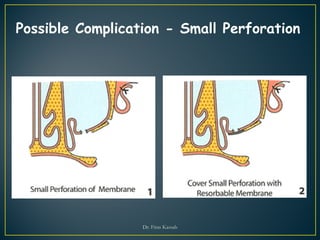


The document provides information on the anatomy and surgical procedures related to maxillary sinus augmentation. It discusses the maxillary sinus anatomy, blood supply, drainage pathways, and membrane characteristics. It then describes various sinus lift surgical techniques categorized based on residual bone height (SA-1 to SA-4). For each category, it details the surgical steps, grafting materials used, healing timelines and risks. It also outlines post-operative care instructions and possible complications along with their management.


![maxillarysinus-170705134531 [Autosaved].pptx](https://cdn.slidesharecdn.com/ss_thumbnails/maxillarysinus-170705134531autosaved-240606120847-09cc00a0-thumbnail.jpg?width=640&height=640&fit=bounds)

![MAXILLARY SINUS AND ITS SURGICAL ANATOMY (2) (1) [Autosaved].ppt](https://cdn.slidesharecdn.com/ss_thumbnails/maxillarysinusanditssurgicalanatomy21autosaved-240927151609-5597be7b-thumbnail.jpg?width=640&height=640&fit=bounds)


