Downloaded 252 times


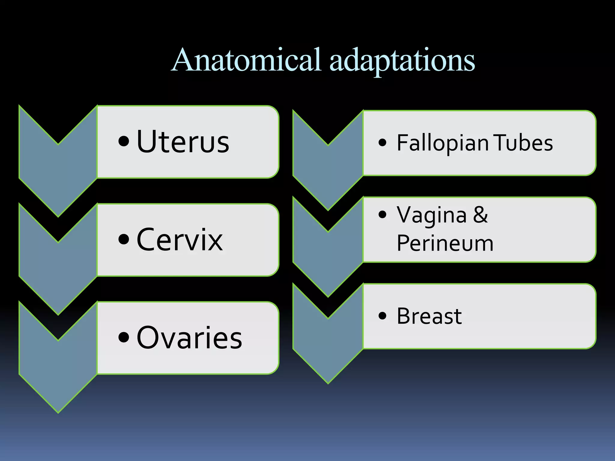


This document discusses the extensive physiological and anatomical changes that occur during normal human pregnancy. It provides details on adaptations in multiple organ systems to support the growth and development of the fetus. The main changes include increased blood volume and cardiac output, anatomical changes to the uterus and cervix, hormonal changes involving hCG and estrogen, and metabolic adaptations to provide optimal nutrition for the fetus. All major body systems are impacted in ways that precisely meet the needs of pregnancy.


