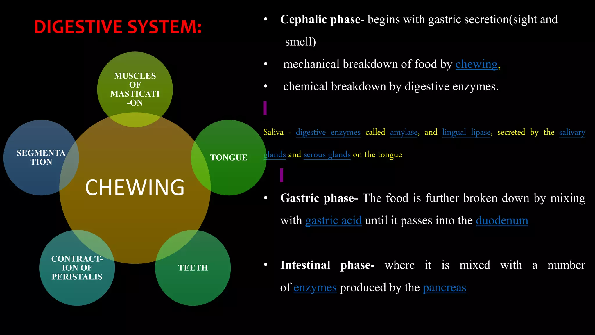
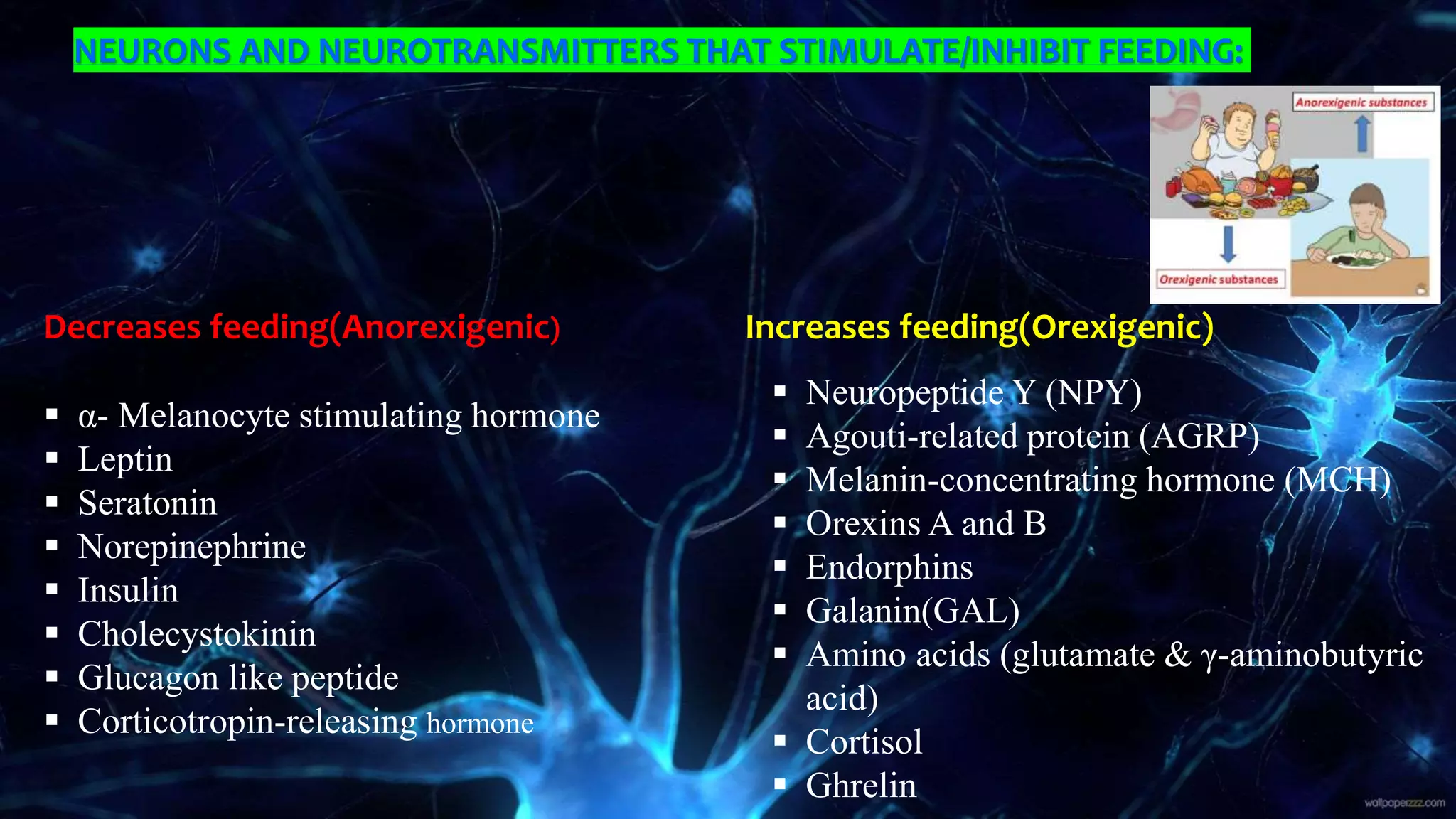
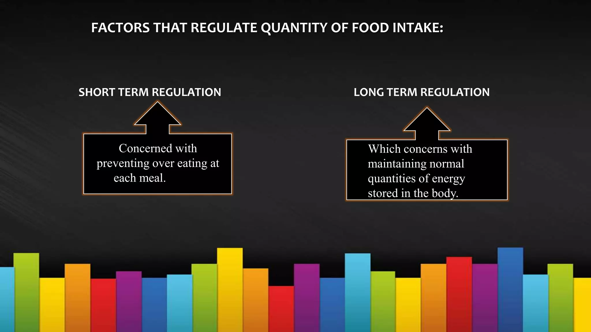
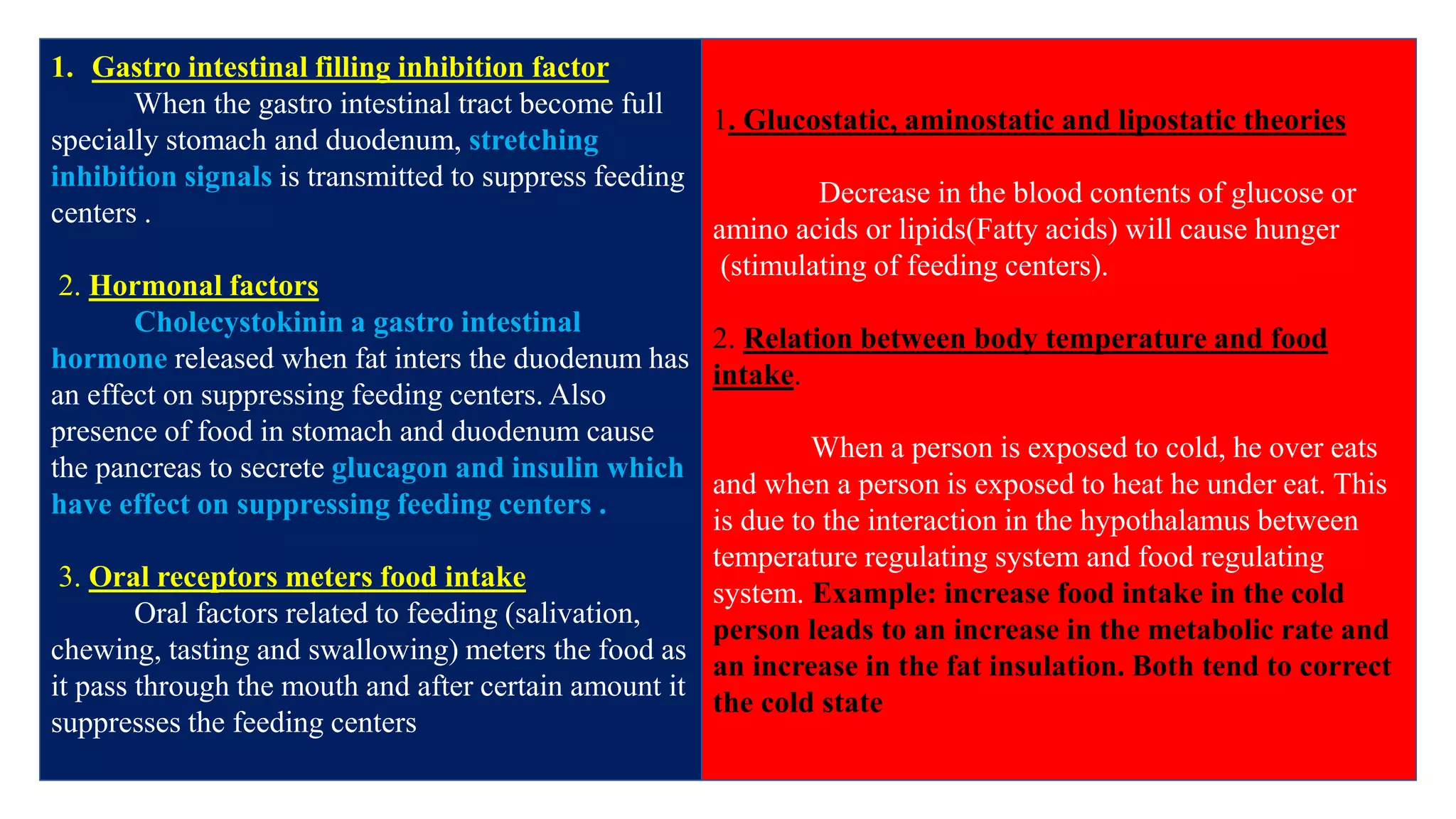
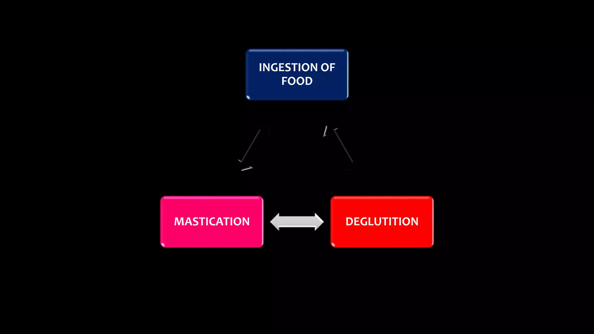
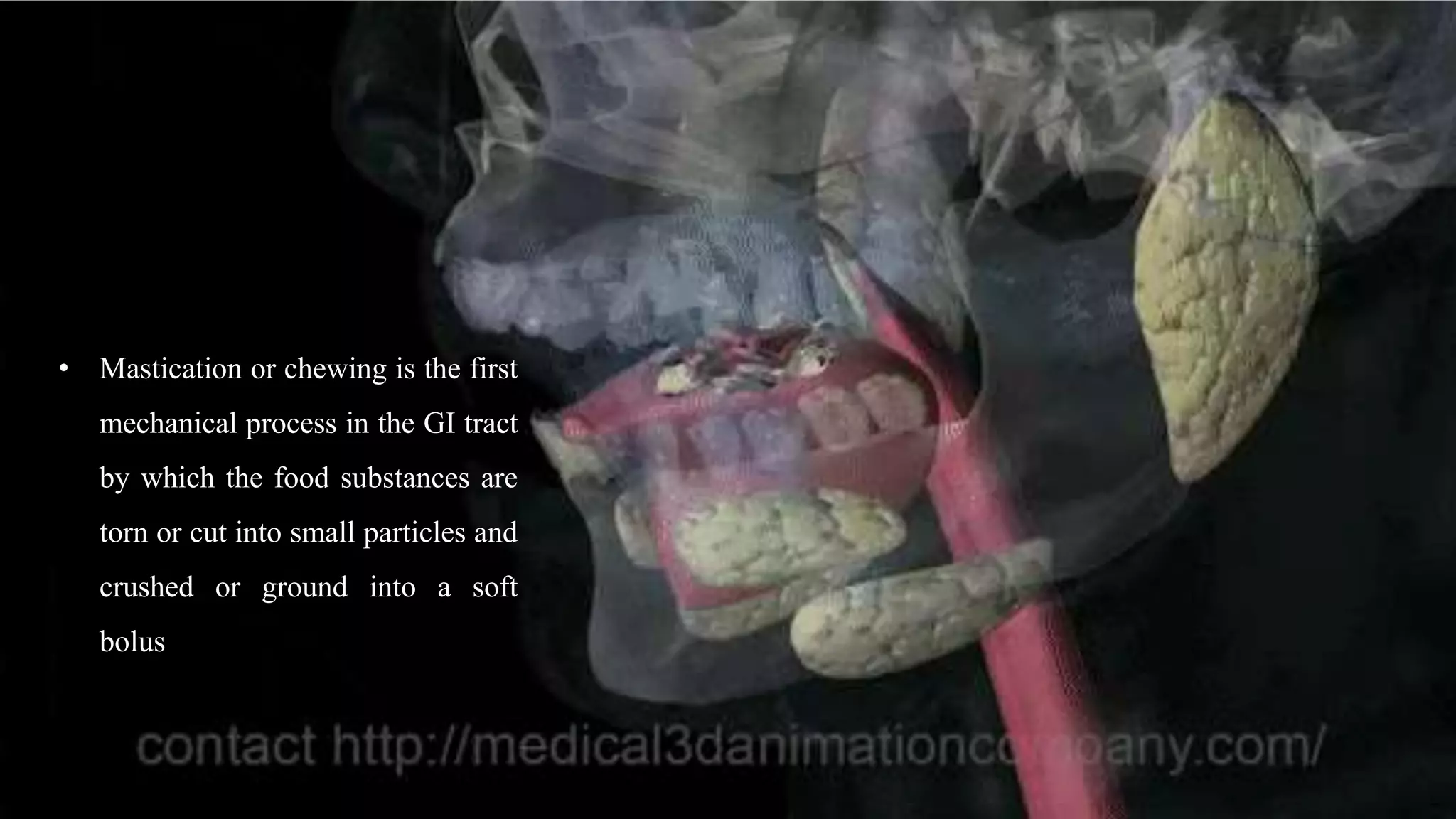
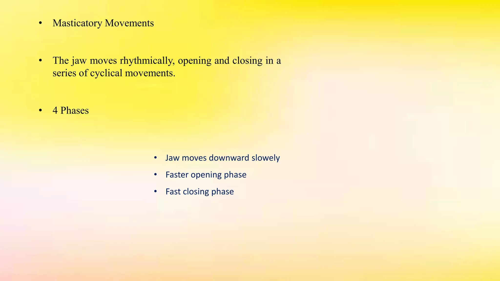
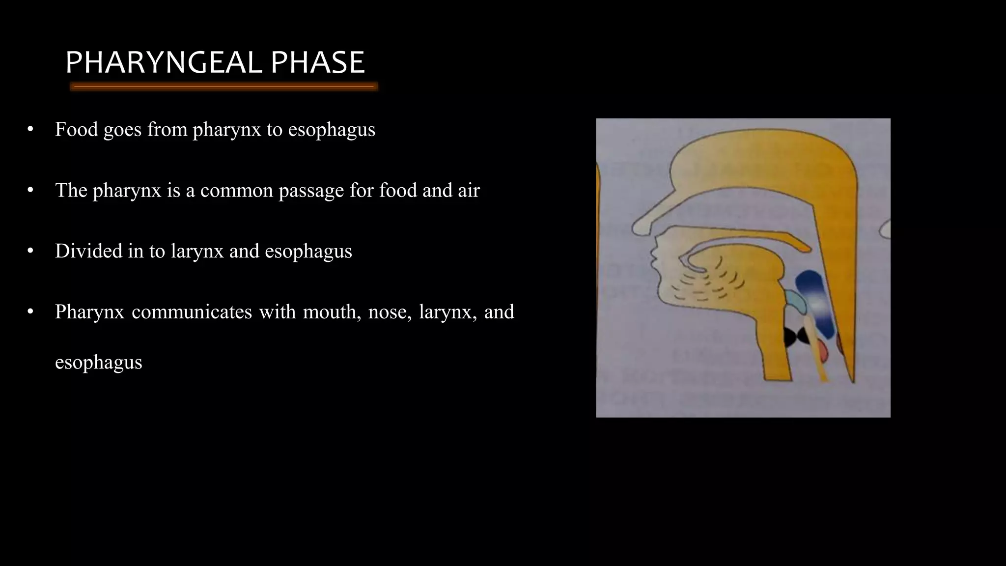
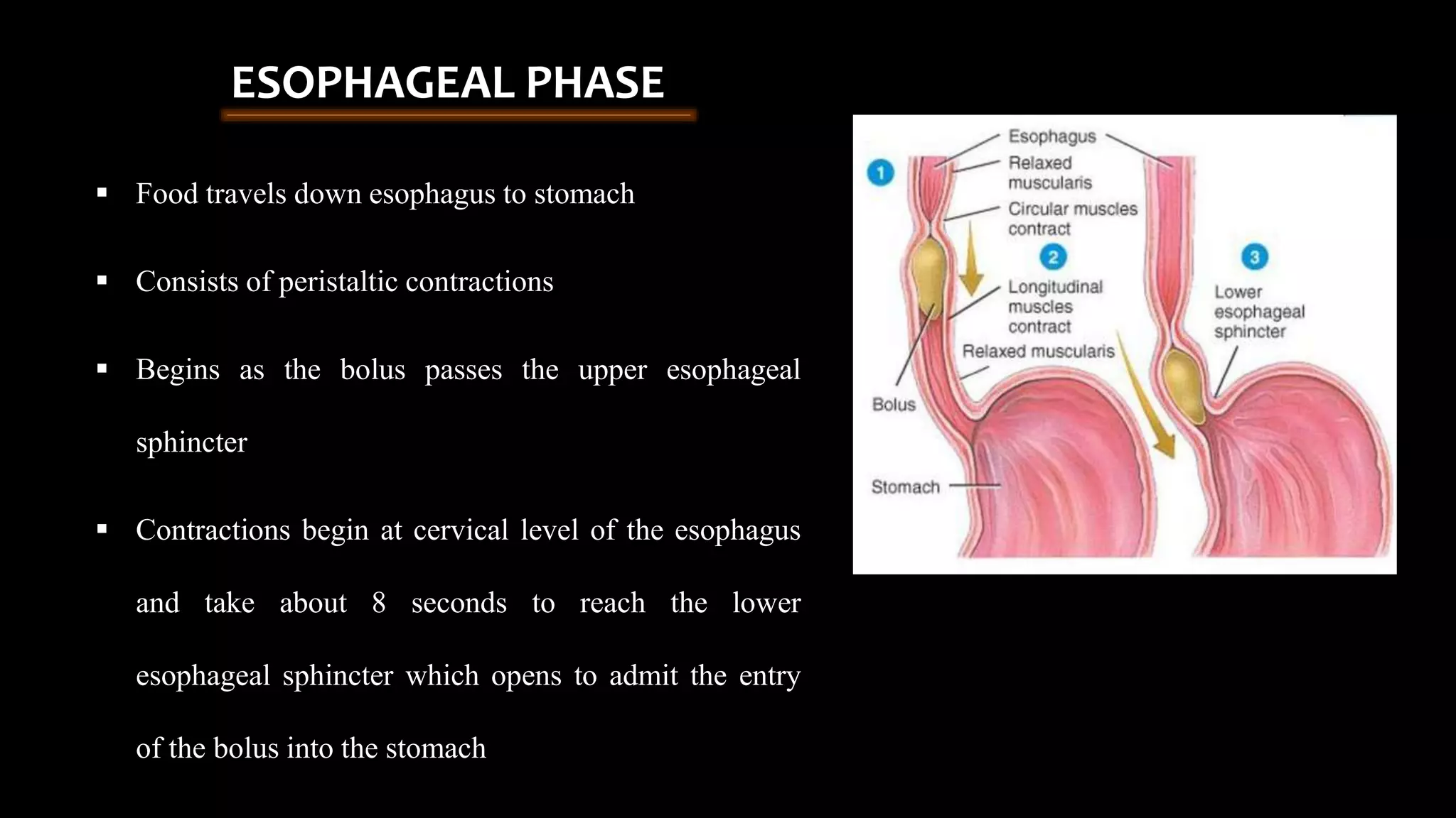
GOOD MORNING!!!
MASTICATION AND DEGLUTITION
Presented by
Dr.keerthanasri. MDS
(Public health dentistry)
CONTENTS
• INTRODUCTION
• DIGESTIVE SYSTEM
• FOOD INTAKE
• NEURONS AND NEUROTRANSMITTERS
• INGESTION OF FOOD
• MASTICATION
• DEGLUTITION
• APPLIED PHYSIOLOGY
• CONCLUSION
INTRODUCTION
• Cephalic phase- begins with gastric secretion(sight and
smell)
• mechanical breakdown of food by chewing,
• chemical breakdown by digestive enzymes.
Saliva - digestive enzymes called amylase, and lingual lipase, secreted by the salivary
glands and serous glands on the tongue
• Gastric phase- The food is further broken down by mixing
with gastric acid until it passes into the duodenum
• Intestinal phase- where it is mixed with a number
of enzymes produced by the pancreas
DIGESTIVE SYSTEM:
CHEWING
MUSCLES
OF
MASTICATI
-ON
TONGUE
TEETH
CONTRACT-
ION OF
PERISTALIS
SEGMENTA
TION
• Peristalsis is the rhythmic contraction of muscles that
begins in the esophagus and continues along the wall of
the stomach and the rest of the gastrointestinal tract.
• This initially results in the production of chyme which
when fully broken down in the small intestine is
absorbed as chyle into the lymphatic system. Most of the
digestion of food takes place in the small intestine. Water
and some minerals are reabsorbed back into the blood in
the colon of the large intestine. The waste products of
digestion (feces) are defecated from the anus via
the rectum.
INGESTION OF FOOD
HUNGER APPETITE
Amount of food that a person
ingests is determined by intrinsic
desire for food called hunger.
The Type of food that a
person preferentially seeks is
determined by appetite
HYPOTHALAMUS
HUNGER CENTER SATIETY
• The lateral nuclei of the hypothalamus serve
as a feeding center.
• The stimulation of this area causes a person
to eat varociously (hyperphagia).
• Destruction of the lateral hypothalamus
causes lack of desire for food and
progressive inanition (marked weight loss,
muscle weakness & decreased metabolism)
• If the quest for food is successful , the feeling
of satiety occurs.
• The ventromedial nuclei of the hypothalamus
serve as the satiety center.
• Electrical stimulation of this region can cause
complete satiety, and even in the presence of
appetizing food, the person refuses to eat
(aphagia).
• Destruction of the ventromedial nuclei causes
voracious and continued
• The paraventricular, dorsomedial and arcuate nuclei
of the hypothalamus play a major role in regulating
food intake.
• Lesion of Paraventricular nuclei- excessive eating.
• Lesion of Dorsomedial nuclei- depress eating
behaviour.
• Arcuate nuclei- regulate food intake and energy
expenditure.
NEURONS AND NEUROTRANSMITTERS THAT STIMULATE/INHIBIT FEEDING:
Decreases feeding(Anorexigenic) Increases feeding(Orexigenic)
 α- Melanocyte stimulating hormone
 Leptin
 Seratonin
 Norepinephrine
 Insulin
 Cholecystokinin
 Glucagon like peptide
 Corticotropin-releasing hormone
 Neuropeptide Y (NPY)
 Agouti-related protein (AGRP)
 Melanin-concentrating hormone (MCH)
 Orexins A and B
 Endorphins
 Galanin(GAL)
 Amino acids (glutamate & γ-aminobutyric
acid)
 Cortisol
 Ghrelin
FACTORS THAT REGULATE QUANTITY OF FOOD INTAKE:
SHORT TERM REGULATION LONG TERM REGULATION
Concerned with
preventing over eating at
each meal.
Which concerns with
maintaining normal
quantities of energy
stored in the body.
1. Gastro intestinal filling inhibition factor
When the gastro intestinal tract become full
specially stomach and duodenum, stretching
inhibition signals is transmitted to suppress feeding
centers .
2. Hormonal factors
Cholecystokinin a gastro intestinal
hormone released when fat inters the duodenum has
an effect on suppressing feeding centers. Also
presence of food in stomach and duodenum cause
the pancreas to secrete glucagon and insulin which
have effect on suppressing feeding centers .
3. Oral receptors meters food intake
Oral factors related to feeding (salivation,
chewing, tasting and swallowing) meters the food as
it pass through the mouth and after certain amount it
suppresses the feeding centers
1. Glucostatic, aminostatic and lipostatic theories
Decrease in the blood contents of glucose or
amino acids or lipids(Fatty acids) will cause hunger
(stimulating of feeding centers).
2. Relation between body temperature and food
intake.
When a person is exposed to cold, he over eats
and when a person is exposed to heat he under eat. This
is due to the interaction in the hypothalamus between
temperature regulating system and food regulating
system. Example: increase food intake in the cold
person leads to an increase in the metabolic rate and
an increase in the fat insulation. Both tend to correct
the cold state
INGESTION OF
FOOD
DEGLUTITIONMASTICATION
MASTICATION
• Mastication or chewing is the first
mechanical process in the GI tract
by which the food substances are
torn or cut into small particles and
crushed or ground into a soft
bolus
Significances of mastication:
• Breakdown of food stuffs into smaller particles
• Mixing of saliva with food substances thoroughly
• Lubrication and moistening of dry food by saliva so that,
the bolus can be easily swallowed
• Appreciation of taste of the food
FUNCTIONS
OF TEETH
CUTTING
TEARING
GRINDING
FORCE
• 55 Pounds on incisors
• 200 pounds on molars
Presence of bolus in the mouth
Reflux inhibition of muscles of mastication
Lower jaw to drops
Raises the jaw to cause closure of the teeth
Inhibits jaw muscles
CHEWING REFLUX
Initiates a stretch reflux of jaw muscles (Rebound contraction)
Repeated again And again
Masticatory moments
• Chewing stroke
• Tooth contact
• Forces of mastication
• Role of soft tissue
• Role of saliva
• Muscles activity
• Masticatory Movements
• The jaw moves rhythmically, opening and closing in a
series of cyclical movements.
• 4 Phases
• Jaw moves downward slowely
• Faster opening phase
• Fast closing phase
Tongue -Positioning of the food between occlusal
surfaces of teeth.
Tongue moves forward and
expand beneath the food
The hyoid bone and the
tongue retract, forms a
trough. Moves the food
to post oral cavity
Food is moved
backward below soft
palate by squeezing
effect of tongue.
SLOW OPENING PHASE FAST OPENING AND CLOSING PHASE
CHEWING STROKE
• Rhythmic
• Cyclic movement
• CPG (Central pattern generator) located
in brain stem
Opening movement
Closing movement
Crushing phase
Grinding phase
Interconnected neural circuits form a neural oscillatory network that is capable of
generating the pattern of masticatory movements. Other pattern generators
responsible for respiration ad swallowing
• During opening phase there is initial
rotation of the mandible for the first
20-27mm of interincisal distance.
• Thereafter there is translatory or
bodily shift of the mandible anteriorly
and in downward direction.
Crushing phase
• starts when the mandible starts closing.
• As the mandible closes the bolus of food is
trapped.
Grinding phase
• bolus gets trapped between the cusps and is
grinded.
• Bolus is trapped by the buccinator bucally and
by the tongue lingually
Opening movement Closing movement
TOOTH CONTACT
• Gliding contact
• Centric contact
 Average length of time for tooth contact during mastication is 194
msec.
 Occlusal condition influences the entire chewing stroke .
 Tall cusps and deep fossae promote a predominantly vertical chewing
stroke .
 Flattened or worn teeth encourage a broader chewing stroke area.
ROLE OF SOFT TISSUES
Maneuvering the food within the
oral cavity
Initiate the breaking of food
Pushes food to occlusal surface
of teeth
Control intake
Seal off the cavity
◦ During opening phase repositions the crushed food
◦ After eating the tongue sweeps the teeth to remove any
food residue that has been trapped in the oral cavity
MUSCLES ACTIVITY
 Contraction of muscles control the jaw during mastication.
 The pattern of activity is determined by a factors such as :
◦ Type of food,
◦ Degree of food breakdown,
 The general pattern of muscle activity during chewing cycle.
◦ Closing muscles are inactive during jaw opening.
◦ Activity of the jaw closing muscles increases slowly as the teeth begin
to interdigitate or as soon as food is encountered between the teeth.
◦ The jaw closing muscles are more active then the jaw opening muscles.
JAW CLOSING MUSCLESJAW OPENING MUSCLES
 Lateral pterygoid
 Masseter
 Temporalis
 Medial pterygoid
MUSCLES OF MASTICATION
CONTROL OF MUSCLES OF MASTICATION
• Action of mastication is mostly a reflux process
• Voluntarily
• Center of mastication –Medulla and Cerebral cortex
• 5th cranial (Trigeminal nerve)
CONTROL OF MASTICATION
• Cyclical movements generated and controlled at the level of
brainstem.
• Complex interactions between several motor nuclei and
sensory input from oral cavity, terminating primarily in the
trigeminal sensory and mesencephalic nuclei.
Cortical output neurons
Central Pattern
Generator
Motor nuclei
Sensory input terminating in trigeminal nucleus
Final pattern of mastication is interaction at the brain stem and
peripheral sensory input
MASTICATION
BREAKIKNG
OF
INDIGESTIBLE
MEMBRANE
SRUFACE
AREA OF
DIGESTIVE
TRACT
PREVENT
EXCORIATION
OF GIT
EASY
PASSAGE OF
FOOD
IMPORTANCE OF MASTICATION
DEGLUTITION
Swallowing of food is known as deglutition
1)Esophageal
phase
1)Pharynge
al phase
1)Oral phase
 Textbook of sembulingam(4th edition)
Chewed food goes from mouth to pharynx
 voluntary
 collection of chewed food into a bolus by tongue
 The bolus is placed on the posterodorsal of the tongue.-
Preparatory phase
 Anterior part of the tongue is retracted and depressed
 The posterior part of the tongue is elevated and retracted
against hard palate
 The forceful contraction of tongue against the palate produces
a positive pressure in the posterior part of hard palate
ORAL PHASE
PHARYNGEAL PHASE
• Food goes from pharynx to esophagus
• The pharynx is a common passage for food and air
• Divided in to larynx and esophagus
• Pharynx communicates with mouth, nose, larynx, and
esophagus
The bolus from the pharynx can enter in to 4 paths
• Back into mouth
Tongue against the soft palate
Intraoral pressure
• Upwards into nasopharynx
Elevation of soft palate
• Forwards into larynx
Forward upward moment of the larynx
Backward movement of the epiglottis
Temporary arrest of breathing (apnea)
• downward into esophagus (factors)
Upward movement of the larynx
The upper 3 to 4cm of esophagus relax (Upper esophageal sphincter)
Peristalic contraction in the pharynx due to contraction of muscles
 Food travels down esophagus to stomach
 Consists of peristaltic contractions
 Begins as the bolus passes the upper esophageal
sphincter
 Contractions begin at cervical level of the esophagus
and take about 8 seconds to reach the lower
esophageal sphincter which opens to admit the entry
of the bolus into the stomach
ESOPHAGEAL PHASE
PRESSURE CHANGES
 Food is moved by a positive-pressure wave.
 Developed by the piston type action of the tongue and peristaltic muscle contractions
 At the start of a swallowing sequence the pressure in the mouth and pharynx is close to
atmospheric pressure but at upper gastro esophageal sphincter it is 5-10 mm Hg
pressure.
 Similarly at the lower gastro esophageal sphincter it is 30 mm Hg pressure.
CONTROL OF SWALLOWING
 Preparatory and oral phases are under voluntary
control
 The pharyngeal and esophageal phases are
involuntary
Organization of the swallowing motor sequence depends
on the activity of brain stem neurons that belong to a
functionally defined swallowing center
• Neural control of swallowing
• Two areas of brain are involved
• Cerebral cortex
• Brain stem
Neural control (initiation)
• Certain trigger areas like fauces / mucosa of posterior
pharyngeal wall
• Afferent nerve is the glossopharyngeal nerve
• Nucleus tractus solitarius & spinal nucleus of
trigeminal nerve play a vital role
• Efferents involve several cranial nerve nuclei which
include nucleus ambiguous (muscles of palate,
pharynx and larynx), hypoglossal nucleus supplying
the muscles of the tongue, motor nuclei of trigeminal
and facial nerves supplying the muscles of face, jaws
and lips
FOOD IN THE MOUTH
SOMATOSENSORY
RECEPTORS
SWALLOWING
CENTER IN THE
MEDULLA
STRAITED MUCLE OF
PHARYNX AND UPPER
THIRD OF
ESOPHAGUS
SWALLOWINGG
AFFERENT
GLOSSOPHARYNGEAL NERVE
VAGUS NERVE
TYPES OF SWALLOWING
Swallowing can be classified according to how the mandible is braced:
Somatic (Adult) swallow
Visceral (Infantile) swallow
• mandible is stabilized by tongue pushing forwards
between the arches and against the anterior part of
the palate
• uses the teeth to stabilize the mandible
INFANTILE SWALLOW
• A type of swallowing pattern seen in infants.
• In it tongue is being thrusted between their gum pads to obtain
the seal during swallowing.
• This type of swallowing changes to mature swallowing when
central incisors erupt.
• When this type of swallowing is retained even after teething,
then it is referred to as “retained infantile swallow”
• It is the major cause of malocclusion.
Suckling reflex
 Fetus is capable of suckling and swallowing amniotic fluid in utero
indicating development of motor activities before birth.
 Fetal swallowing takes in episodes 1 to 9 mins at around 20 times/day
 Volume of amniotic fluid swallowed is similar to volume of milk intake in
the neonatal period.
Rooting reflex
 Response of infant to tactile stimulation of the cheek or lips; causes infant
to turn toward the stimulus and open his mouth.
APPLIED PHYSIOLOGY
DYSPHAGIA
Dysphagia means difficulty in swallowing
It may be a sensation that suggests difficulty in the passage of solids or liquids
from the mouth to the stomach
Dysphagia is classified into two major types:
Oropharyngeal dysphagia
Esophageal dysphagia
ORAL
DYSPHAGIA
Tonsillitis
Peri
tonsillar
abscess
Stomatitis
Tongue
cancer
Paralysis
of soft
palate
Bell's
palsy
Xerostomia
LUMEN • Impacted foreign
body
WALL
• Pharyngitis
• Paterson-Kelly
syndrome
• Pharyngeal spasms
• Malignant neoplasm
OUTSIDE
THE
WALL
• Retropharyngeal
abscess
• Lymphadenopathy of
cervical lymph nodes
• Thyroid malignancy
PHARYNGEAL
DYSPHAGIA
LUMEN
• Impacted foreign
body
WALL
• esophageal atresia
• Crohn's disease
• Candida esophagitis
•
OUTSIDE
THE
WALL
• Retrosternal goitre
• Malignancy
• Thyroid swelling
• Zenker's diverticulum
• Mediastinal growth
• Dysphagia lusoria
•
ESOPHAGIAL
DYSPHAGIA
ACHALASA CARDIA
• Neurodegenerative disorder
• Failure of lower esophageal sphincter
to relax during swallowing
• Dilation of esophagus
Dysphagia
Weight loss
Chest pain
cough
 Textbook of sembulingam(4th edition)
STRUCTURAL
ABNORMALLITIES
Cleft lip and palate
• It hampers control for sucking
• Decrease the oral suction
• Nasal regurgitation
Functional Abnormalities
• Drooling- reduced closing pressure of the lip
• Premature leakage of bolus into pharynx- Weak
contraction of tongue and soft palate
• Tongue Dysfunction- Impaired mastication, bolus
formation and bolus transport.
• Loss of teeth
• Xerostomia
• Impaired opening of UES- Weakness of ant.
suprahyoid muscles
• Zenker diverticulum is a diverticulum of the hypopharynx that occurs at a
weak spot in the muscular well.
• The bolus can enter in the diverticulum and be regurgitated to the pharynx,
Which may result in coughing or aspiration
GASTROESOPHAGEAL REFLUX DISEASE
• Regurgitation of acidic gastric content into esophagus
• It flows pharynx to mouth
• Weakness or incompetence of lower esophageal sphincter
Heart
burn
Esophagi
tis
Dysphagia
Cough and
change of
voice
Esophageal ulcer
or cancer
 Textbook of sembulingam(4th edition)
CONCLUSION
• Food being the basic needs for proper nutrition and ill free health
• Mastication as a basic and initial function for fulfilling the basic need, proper
knowledge on this essential in ruling out the root cause for many disease
REFRENCES
 Essentials of Oral physiology-Robert M.Bradely
 Textbook of medical physiology- Guyton (10th edition)
 Textbook of sembulingam(4th edition) pg no. 243-246
• Lau C. Development of Suck and Swallow Mechanisms in
Infants. Ann Nutr Metab. 2015;66 Suppl 5(0 5):7–14.
doi:10.1159/000381361
• Matsuo K, Palmer JB. Anatomy and physiology of feeding and
swallowing: normal and abnormal. Phys Med Rehabil Clin N Am.
2008;19(4):691–vii. doi:10.1016/j.pmr.2008.06.001
THANK YOU!!!

Mastication and deglutition

  • 1.
  • 2.
    MASTICATION AND DEGLUTITION Presentedby Dr.keerthanasri. MDS (Public health dentistry)
  • 3.
    CONTENTS • INTRODUCTION • DIGESTIVESYSTEM • FOOD INTAKE • NEURONS AND NEUROTRANSMITTERS • INGESTION OF FOOD • MASTICATION • DEGLUTITION • APPLIED PHYSIOLOGY • CONCLUSION
  • 4.
  • 5.
    • Cephalic phase-begins with gastric secretion(sight and smell) • mechanical breakdown of food by chewing, • chemical breakdown by digestive enzymes. Saliva - digestive enzymes called amylase, and lingual lipase, secreted by the salivary glands and serous glands on the tongue • Gastric phase- The food is further broken down by mixing with gastric acid until it passes into the duodenum • Intestinal phase- where it is mixed with a number of enzymes produced by the pancreas DIGESTIVE SYSTEM: CHEWING MUSCLES OF MASTICATI -ON TONGUE TEETH CONTRACT- ION OF PERISTALIS SEGMENTA TION
  • 6.
    • Peristalsis isthe rhythmic contraction of muscles that begins in the esophagus and continues along the wall of the stomach and the rest of the gastrointestinal tract. • This initially results in the production of chyme which when fully broken down in the small intestine is absorbed as chyle into the lymphatic system. Most of the digestion of food takes place in the small intestine. Water and some minerals are reabsorbed back into the blood in the colon of the large intestine. The waste products of digestion (feces) are defecated from the anus via the rectum.
  • 7.
    INGESTION OF FOOD HUNGERAPPETITE Amount of food that a person ingests is determined by intrinsic desire for food called hunger. The Type of food that a person preferentially seeks is determined by appetite
  • 8.
    HYPOTHALAMUS HUNGER CENTER SATIETY •The lateral nuclei of the hypothalamus serve as a feeding center. • The stimulation of this area causes a person to eat varociously (hyperphagia). • Destruction of the lateral hypothalamus causes lack of desire for food and progressive inanition (marked weight loss, muscle weakness & decreased metabolism) • If the quest for food is successful , the feeling of satiety occurs. • The ventromedial nuclei of the hypothalamus serve as the satiety center. • Electrical stimulation of this region can cause complete satiety, and even in the presence of appetizing food, the person refuses to eat (aphagia). • Destruction of the ventromedial nuclei causes voracious and continued
  • 9.
    • The paraventricular,dorsomedial and arcuate nuclei of the hypothalamus play a major role in regulating food intake. • Lesion of Paraventricular nuclei- excessive eating. • Lesion of Dorsomedial nuclei- depress eating behaviour. • Arcuate nuclei- regulate food intake and energy expenditure.
  • 10.
    NEURONS AND NEUROTRANSMITTERSTHAT STIMULATE/INHIBIT FEEDING: Decreases feeding(Anorexigenic) Increases feeding(Orexigenic)  α- Melanocyte stimulating hormone  Leptin  Seratonin  Norepinephrine  Insulin  Cholecystokinin  Glucagon like peptide  Corticotropin-releasing hormone  Neuropeptide Y (NPY)  Agouti-related protein (AGRP)  Melanin-concentrating hormone (MCH)  Orexins A and B  Endorphins  Galanin(GAL)  Amino acids (glutamate & γ-aminobutyric acid)  Cortisol  Ghrelin
  • 11.
    FACTORS THAT REGULATEQUANTITY OF FOOD INTAKE: SHORT TERM REGULATION LONG TERM REGULATION Concerned with preventing over eating at each meal. Which concerns with maintaining normal quantities of energy stored in the body.
  • 12.
    1. Gastro intestinalfilling inhibition factor When the gastro intestinal tract become full specially stomach and duodenum, stretching inhibition signals is transmitted to suppress feeding centers . 2. Hormonal factors Cholecystokinin a gastro intestinal hormone released when fat inters the duodenum has an effect on suppressing feeding centers. Also presence of food in stomach and duodenum cause the pancreas to secrete glucagon and insulin which have effect on suppressing feeding centers . 3. Oral receptors meters food intake Oral factors related to feeding (salivation, chewing, tasting and swallowing) meters the food as it pass through the mouth and after certain amount it suppresses the feeding centers 1. Glucostatic, aminostatic and lipostatic theories Decrease in the blood contents of glucose or amino acids or lipids(Fatty acids) will cause hunger (stimulating of feeding centers). 2. Relation between body temperature and food intake. When a person is exposed to cold, he over eats and when a person is exposed to heat he under eat. This is due to the interaction in the hypothalamus between temperature regulating system and food regulating system. Example: increase food intake in the cold person leads to an increase in the metabolic rate and an increase in the fat insulation. Both tend to correct the cold state
  • 13.
  • 14.
  • 15.
    • Mastication orchewing is the first mechanical process in the GI tract by which the food substances are torn or cut into small particles and crushed or ground into a soft bolus
  • 16.
    Significances of mastication: •Breakdown of food stuffs into smaller particles • Mixing of saliva with food substances thoroughly • Lubrication and moistening of dry food by saliva so that, the bolus can be easily swallowed • Appreciation of taste of the food
  • 17.
    FUNCTIONS OF TEETH CUTTING TEARING GRINDING FORCE • 55Pounds on incisors • 200 pounds on molars
  • 18.
    Presence of bolusin the mouth Reflux inhibition of muscles of mastication Lower jaw to drops Raises the jaw to cause closure of the teeth Inhibits jaw muscles CHEWING REFLUX Initiates a stretch reflux of jaw muscles (Rebound contraction) Repeated again And again
  • 19.
    Masticatory moments • Chewingstroke • Tooth contact • Forces of mastication • Role of soft tissue • Role of saliva • Muscles activity
  • 20.
    • Masticatory Movements •The jaw moves rhythmically, opening and closing in a series of cyclical movements. • 4 Phases • Jaw moves downward slowely • Faster opening phase • Fast closing phase
  • 21.
    Tongue -Positioning ofthe food between occlusal surfaces of teeth. Tongue moves forward and expand beneath the food The hyoid bone and the tongue retract, forms a trough. Moves the food to post oral cavity Food is moved backward below soft palate by squeezing effect of tongue. SLOW OPENING PHASE FAST OPENING AND CLOSING PHASE
  • 22.
    CHEWING STROKE • Rhythmic •Cyclic movement • CPG (Central pattern generator) located in brain stem Opening movement Closing movement Crushing phase Grinding phase Interconnected neural circuits form a neural oscillatory network that is capable of generating the pattern of masticatory movements. Other pattern generators responsible for respiration ad swallowing
  • 23.
    • During openingphase there is initial rotation of the mandible for the first 20-27mm of interincisal distance. • Thereafter there is translatory or bodily shift of the mandible anteriorly and in downward direction. Crushing phase • starts when the mandible starts closing. • As the mandible closes the bolus of food is trapped. Grinding phase • bolus gets trapped between the cusps and is grinded. • Bolus is trapped by the buccinator bucally and by the tongue lingually Opening movement Closing movement
  • 24.
    TOOTH CONTACT • Glidingcontact • Centric contact  Average length of time for tooth contact during mastication is 194 msec.  Occlusal condition influences the entire chewing stroke .  Tall cusps and deep fossae promote a predominantly vertical chewing stroke .  Flattened or worn teeth encourage a broader chewing stroke area.
  • 25.
    ROLE OF SOFTTISSUES Maneuvering the food within the oral cavity Initiate the breaking of food Pushes food to occlusal surface of teeth Control intake Seal off the cavity ◦ During opening phase repositions the crushed food ◦ After eating the tongue sweeps the teeth to remove any food residue that has been trapped in the oral cavity
  • 26.
    MUSCLES ACTIVITY  Contractionof muscles control the jaw during mastication.  The pattern of activity is determined by a factors such as : ◦ Type of food, ◦ Degree of food breakdown,  The general pattern of muscle activity during chewing cycle. ◦ Closing muscles are inactive during jaw opening. ◦ Activity of the jaw closing muscles increases slowly as the teeth begin to interdigitate or as soon as food is encountered between the teeth. ◦ The jaw closing muscles are more active then the jaw opening muscles.
  • 27.
    JAW CLOSING MUSCLESJAWOPENING MUSCLES  Lateral pterygoid  Masseter  Temporalis  Medial pterygoid
  • 28.
  • 29.
    CONTROL OF MUSCLESOF MASTICATION • Action of mastication is mostly a reflux process • Voluntarily • Center of mastication –Medulla and Cerebral cortex • 5th cranial (Trigeminal nerve)
  • 30.
    CONTROL OF MASTICATION •Cyclical movements generated and controlled at the level of brainstem. • Complex interactions between several motor nuclei and sensory input from oral cavity, terminating primarily in the trigeminal sensory and mesencephalic nuclei.
  • 31.
    Cortical output neurons CentralPattern Generator Motor nuclei Sensory input terminating in trigeminal nucleus Final pattern of mastication is interaction at the brain stem and peripheral sensory input
  • 32.
  • 33.
  • 34.
    Swallowing of foodis known as deglutition 1)Esophageal phase 1)Pharynge al phase 1)Oral phase  Textbook of sembulingam(4th edition)
  • 35.
    Chewed food goesfrom mouth to pharynx  voluntary  collection of chewed food into a bolus by tongue  The bolus is placed on the posterodorsal of the tongue.- Preparatory phase  Anterior part of the tongue is retracted and depressed  The posterior part of the tongue is elevated and retracted against hard palate  The forceful contraction of tongue against the palate produces a positive pressure in the posterior part of hard palate ORAL PHASE
  • 36.
    PHARYNGEAL PHASE • Foodgoes from pharynx to esophagus • The pharynx is a common passage for food and air • Divided in to larynx and esophagus • Pharynx communicates with mouth, nose, larynx, and esophagus
  • 37.
    The bolus fromthe pharynx can enter in to 4 paths • Back into mouth Tongue against the soft palate Intraoral pressure • Upwards into nasopharynx Elevation of soft palate • Forwards into larynx Forward upward moment of the larynx Backward movement of the epiglottis Temporary arrest of breathing (apnea) • downward into esophagus (factors) Upward movement of the larynx The upper 3 to 4cm of esophagus relax (Upper esophageal sphincter) Peristalic contraction in the pharynx due to contraction of muscles
  • 38.
     Food travelsdown esophagus to stomach  Consists of peristaltic contractions  Begins as the bolus passes the upper esophageal sphincter  Contractions begin at cervical level of the esophagus and take about 8 seconds to reach the lower esophageal sphincter which opens to admit the entry of the bolus into the stomach ESOPHAGEAL PHASE
  • 39.
    PRESSURE CHANGES  Foodis moved by a positive-pressure wave.  Developed by the piston type action of the tongue and peristaltic muscle contractions  At the start of a swallowing sequence the pressure in the mouth and pharynx is close to atmospheric pressure but at upper gastro esophageal sphincter it is 5-10 mm Hg pressure.  Similarly at the lower gastro esophageal sphincter it is 30 mm Hg pressure.
  • 40.
    CONTROL OF SWALLOWING Preparatory and oral phases are under voluntary control  The pharyngeal and esophageal phases are involuntary Organization of the swallowing motor sequence depends on the activity of brain stem neurons that belong to a functionally defined swallowing center
  • 41.
    • Neural controlof swallowing • Two areas of brain are involved • Cerebral cortex • Brain stem
  • 42.
    Neural control (initiation) •Certain trigger areas like fauces / mucosa of posterior pharyngeal wall • Afferent nerve is the glossopharyngeal nerve • Nucleus tractus solitarius & spinal nucleus of trigeminal nerve play a vital role • Efferents involve several cranial nerve nuclei which include nucleus ambiguous (muscles of palate, pharynx and larynx), hypoglossal nucleus supplying the muscles of the tongue, motor nuclei of trigeminal and facial nerves supplying the muscles of face, jaws and lips
  • 43.
    FOOD IN THEMOUTH SOMATOSENSORY RECEPTORS SWALLOWING CENTER IN THE MEDULLA STRAITED MUCLE OF PHARYNX AND UPPER THIRD OF ESOPHAGUS SWALLOWINGG AFFERENT GLOSSOPHARYNGEAL NERVE VAGUS NERVE
  • 44.
    TYPES OF SWALLOWING Swallowingcan be classified according to how the mandible is braced: Somatic (Adult) swallow Visceral (Infantile) swallow • mandible is stabilized by tongue pushing forwards between the arches and against the anterior part of the palate • uses the teeth to stabilize the mandible
  • 45.
    INFANTILE SWALLOW • Atype of swallowing pattern seen in infants. • In it tongue is being thrusted between their gum pads to obtain the seal during swallowing. • This type of swallowing changes to mature swallowing when central incisors erupt. • When this type of swallowing is retained even after teething, then it is referred to as “retained infantile swallow” • It is the major cause of malocclusion.
  • 46.
    Suckling reflex  Fetusis capable of suckling and swallowing amniotic fluid in utero indicating development of motor activities before birth.  Fetal swallowing takes in episodes 1 to 9 mins at around 20 times/day  Volume of amniotic fluid swallowed is similar to volume of milk intake in the neonatal period. Rooting reflex  Response of infant to tactile stimulation of the cheek or lips; causes infant to turn toward the stimulus and open his mouth.
  • 47.
  • 48.
    DYSPHAGIA Dysphagia means difficultyin swallowing It may be a sensation that suggests difficulty in the passage of solids or liquids from the mouth to the stomach Dysphagia is classified into two major types: Oropharyngeal dysphagia Esophageal dysphagia
  • 49.
  • 50.
    LUMEN • Impactedforeign body WALL • Pharyngitis • Paterson-Kelly syndrome • Pharyngeal spasms • Malignant neoplasm OUTSIDE THE WALL • Retropharyngeal abscess • Lymphadenopathy of cervical lymph nodes • Thyroid malignancy PHARYNGEAL DYSPHAGIA
  • 51.
    LUMEN • Impacted foreign body WALL •esophageal atresia • Crohn's disease • Candida esophagitis • OUTSIDE THE WALL • Retrosternal goitre • Malignancy • Thyroid swelling • Zenker's diverticulum • Mediastinal growth • Dysphagia lusoria • ESOPHAGIAL DYSPHAGIA
  • 52.
    ACHALASA CARDIA • Neurodegenerativedisorder • Failure of lower esophageal sphincter to relax during swallowing • Dilation of esophagus Dysphagia Weight loss Chest pain cough  Textbook of sembulingam(4th edition)
  • 53.
    STRUCTURAL ABNORMALLITIES Cleft lip andpalate • It hampers control for sucking • Decrease the oral suction • Nasal regurgitation
  • 54.
    Functional Abnormalities • Drooling-reduced closing pressure of the lip • Premature leakage of bolus into pharynx- Weak contraction of tongue and soft palate • Tongue Dysfunction- Impaired mastication, bolus formation and bolus transport. • Loss of teeth • Xerostomia • Impaired opening of UES- Weakness of ant. suprahyoid muscles
  • 55.
    • Zenker diverticulumis a diverticulum of the hypopharynx that occurs at a weak spot in the muscular well. • The bolus can enter in the diverticulum and be regurgitated to the pharynx, Which may result in coughing or aspiration
  • 56.
    GASTROESOPHAGEAL REFLUX DISEASE •Regurgitation of acidic gastric content into esophagus • It flows pharynx to mouth • Weakness or incompetence of lower esophageal sphincter Heart burn Esophagi tis Dysphagia Cough and change of voice Esophageal ulcer or cancer  Textbook of sembulingam(4th edition)
  • 57.
  • 58.
    • Food beingthe basic needs for proper nutrition and ill free health • Mastication as a basic and initial function for fulfilling the basic need, proper knowledge on this essential in ruling out the root cause for many disease
  • 59.
    REFRENCES  Essentials ofOral physiology-Robert M.Bradely  Textbook of medical physiology- Guyton (10th edition)  Textbook of sembulingam(4th edition) pg no. 243-246 • Lau C. Development of Suck and Swallow Mechanisms in Infants. Ann Nutr Metab. 2015;66 Suppl 5(0 5):7–14. doi:10.1159/000381361 • Matsuo K, Palmer JB. Anatomy and physiology of feeding and swallowing: normal and abnormal. Phys Med Rehabil Clin N Am. 2008;19(4):691–vii. doi:10.1016/j.pmr.2008.06.001
  • 60.