
Recommended
More Related Content
Similar to 13_Digestion in oral cavity and stomach.pptx
Similar to 13_Digestion in oral cavity and stomach.pptx (20)
More from GauravPrakashGaurav
Recently uploaded
Recently uploaded (20)
13_Digestion in oral cavity and stomach.pptx
- 2. Digestion is complex physiological process, initial stage of exchange of substances and energy in the organism. During the digestive process the food loses its specific features and is transformed (broken down) into simple components: proteins amino acids; carbohydrates glucose; lipids (fats) free fatty acids and cholesterol.
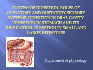
- 3. Digestive system is made up of gastrointestinal tract (GI tract) or alimentary canal and accessory organs, which help in the process of digestion and absorption. GI tract is formed by two types of organs: 1. Primary; 2. Accessory.
- 4. Maintaining of homeostasis. Maintaining required level of metabolism and energy exchange in organism. Excretory function – excreting some end products with excrements (feces). Hormonal function – producing some hormones and biologically active substances. Significant role in process of erythropoesis. Takes part in the process of adaptation of organism.
- 5. Mechanical processing of the food – formation of smaller pieces (fragmentation), mixing and propelling the food throughout the gastro- intestinal tract (GI tract). Term motility refers to the contraction of GI walls. Secretion of digestive juices – the enzymes in them dissolve (broke down) proteins, carbohydrates and fats. Absorption – the process of transport of the end products of digestion, water, salts and vitamins through the epithelial mucous membrane of GI tract into blood and lymph.
- 6. The food is ingested via mouth that is the initial part of digestive system. The oral part of digestive system has attitude to the intake of food, analysing of its features, preparing of food for chemical digestion and propelling it to the esophagus.
- 7. Mechanical processing of the food (done by chewing and swallowing acts). Partial chemical processing of the food. Partial absorption of the food. Protective function – it is a primary barrier against the infectious agents that come with food. Trophic (nutritive) function. Sensory – approbation, sensation of food properties.
- 8. Chewing (mastication) is a physiological act during which the food is being fragmented, moistened by saliva, mixed and partially chemically processed. As a result of chewing there is a formation of digestive bolus. In the act of chewing such organs take part: upper and lower jaws with teeth, chewing and mimic muscles, mucosa of oral cavity, tongue, soft palate, salivary glands. Normally chewing is an inborn reflex (!).
- 9. It provides complex coordination of conditioned and unconditioned digestive motor reflexes that contribute to the time of presence of food in oral cavity. It provides primary mechanical processing of the food. The more the food is chewed the more will be effective digestion of it in the lower parts of digestive system. Chewing act influences on secretory functions of digestive system (particularly on secretion of gastric and pancreatic juices).
- 10. Food irritates the receptors of oral cavity afferent impulses travel via trigeminal nerve (V cranial), glosso-pharyngeal nerve (IX cranial), superior laryngeal nerve (part of vagus) and chorda tympani (part of facial nerve, VII cranial) impulses arrive to the sensory nuclei of medulla oblongata (nucleus tractus solitarii, nucleus of trigeminal nerve) than information is travelling to the cortex where it is being analysed (good or bad food) efferent fibers go via trigeminal, facial and hypoglossus nerves to the chewing and mimic muscles, that provide the chewing act.
- 11. Is a physiological reflex act during which the digestive bolus is propelled from the oral cavity into the esophagus. Receptors for this act are located in the root of tongue, soft palate and posterior wall of pharynx. Swallowing act consists of 3 phases: Oral phase – food is moistened by saliva, masticated and moved to the posterior part of oral cavity (in general voluntary phase). Pharyngeal phase – starts when digestive bolus comes behind the palatinal arc. Complex process carried by IX and X cranial nerves. Nasopharynx and larynx close and the bolus moves into esophagus (unvoluntary). Esophageal phase – propelling of bolus via the tube of esophagus (unvoluntary).
- 12. Though the beginning of swallowing is a voluntary act, later it becomes involuntary and is carried out by a reflex action called deglutition reflex. It occurs during the pharyngeal and esophageal stages. Stimulus When the bolus enters the oropharyngeal region, the receptors present in this region are stimulated. Afferent Fibers Afferent impulses from the oropharyngeal receptors pass via the glossopharyngeal nerve fibers to the deglutition center.
- 13. Center Deglutition center is at the floor of the fourth ventricle in medulla oblongata of brain. Efferent Fibers Impulses from deglutition center travel through glossopharyngeal and vagus nerves (parasympathetic motor fibers) and reach soft palate, pharynx and esophagus. The glossopharyngeal nerve is concerned with pharyngeal stage of swallowing. The vagus nerve is concerned with esophageal stage. Response The reflex causes upward movement of soft palate, to close nasopharynx and downward movement of epiglotis, to close respiratory passage (larynx), so that bolus enters the esophagus. Now the peristalsis occurs in esophagus, pushing the bolus into stomach.
- 15. Various oral glands produce liquid that is called saliva. It is essential for digestion (!) Functions of saliva include: Significant role in providing of organism with the information about properties of food. Reception of food is possible only when food is moistened by saliva (!). Providing initial chemical processing of the food by enzymes in it. Taking part in the formation of digestive bolus. Protective function – contains immunoglobulin A, lysosyme etc. Trophic action – supplies nutrients for teeth (ions of Calcium, Phosphorus) Has a role in speaking.
- 16. Salivary glands are divided into: 1) major; 2) minor (buccal, lingual) There are 3 pairs of major salivary glands: 1) parotid; 2) submandibular; 3) sublingual. Classification of salivary glands according to their secretion: 1) Serose (protein) glands – produce liquid saliva without mucine, but rich in enzymes (parotid glands). 2) Mucous glands – produce viscous saliva rich in mucous (sublingual glands). 3) Mixed glands – combine the features of serose and mucous glands (submandibular glands)
- 17. Normally humans produce up to 2 litres of saliva per day (approximate rates are: 0.05 ml/min during sleep, 0.5 ml/min at rest, 5 ml/min maximally). pH of saliva is 5,8-7,3. The quantity of produced saliva depends from: 1) level of “food dryness”; 2) level of food fragmentation; 3) chemical composition of food.
- 19. Regulation of salivation divided on nervous and humoral. Nervous regulation is presented by conditional and unconditional reflexes.
- 20. Starts from irritation of receptors in oral cavity. Afferent fibers go via lingual, glossopharyngeal and vagus nerves to center of salivation which is located in medulla oblongata. Efferent fibers divided on sympathetic and parasympathetic. Sympathetic fibers inhibit secretion of saliva (small quantity of thick saliva). Parasympathetic fibers (chorda tympani) stimulate of secretion of saliva (high quantity of watery saliva).
- 21. Starts from irritation of receptors of vision, hearing, and olfaction. Impulses go to specific areas of cortex then to medulla oblongata. Parasympathetic division of A.N.S. secretion of watery saliva lacking enzymes; sympathetic division of A.N.S. secretion of thick saliva rich in enzymes. Humoral regulation (very small role): adrenalin (-), thyroxine (-), acetylcholine (+), sex hormones (-).
- 22. Is a depot (storage) for food. Providing further mechanical processing of food. Providing further chemical processing of food. Protective – gastric acidity (due to presence of hydrochloric acid (HCl)) and lysozyme kill microbes that come with food. It is involved in hemopoesis – gastric mucosa releases so called intrinsic factor of Castle (gastromucoprotein), that in organism combines with the vitamin B12 (extrinsic factor of Castle). Only in such combined state vitamin B12 that is essential for hemopoesis could be absorbed from the intestines into the organism.
- 23. Chief cells – produce pepsinogen that is an inactive form of the proteolytic enzyme pepsin (located mainly in fundus and body of stomach). Parietal cells – produce HCl and intrinsic factor (located mainly in fundus and body of stomach). Enterochomaffin - like cell - produce histamine (stimulates acid).
- 24. Accessory cells – produce gastric mucus. It protects gastric mucosa from digestive action of HCl acid and gastric enzymes (almost totally located in cardiac area of the stomach and also antral/pyloric area). G-cells – endocrine cells that produce hormone gastrin, which influences on motility and secretion of gastrointestinal tract. In order to perform its action gastrin should be firstly released into bloodstream and then with blood it returns back to GI tract and exert the action (located mostly in antral and pyloric areas of stomach). D-cells-produce somatostatin (inhibits acid).
- 25. The fluid secreted by the gastric glands is called gastric juice. Normally 1.5 – 2 litres of gastric juice are produced per day. The quantity of produced gastric juice depends from: 1) time elapsed since the intake of food; 2) chemical composition of food. Juice that is released during the state of fasting is called basal gastric secretion (approximate rate is 5- 15 ml/h). Composition of gastric juice: 99.5 % water; 0.5 % organic and non-organic waste (electrolytes + HCl). One of the most important non-organic compound of gastric juice is hydrochloride acid (HCl).
- 26. Catalyses the transformation of inactive pepsinogen into active pepsin. Provides optimal pH for the action of enzymes, particularly pepsin (normal pH range 1.5 – 2.5). Makes denaturation of proteins (they begin to lose their structure) and it becomes more easier for pepsin to break down proteins. Protective function – killing of microbes, that enter stomach. Stimulates the production of pancreatic juice. Regulates motor function of pylorus. Low acidity caused by the deficiency in HCl production violates the normal passage of chyme (partially digested food in stomach) from the stomach to duodenum as the sphincter stays partially relaxed.
- 27. Proteolytic (Protein-degrading enzymes) - the major function is to break down the proteins into smaller peptides: a) Pepsin b) Gastriksin c) Parapepsin c) Chymosin (babies to 1 year old have this enzyme) Amylolytic (starch-converting enzyme) – gastric amylase, enzyme that digests carbohydrates (starch, glycogen) into oligosaccharides (has low significance in stomach). Lipolytic - enzyme that digests lipids: a) gastric lipase - is mostly important in lipid digestion of infants (!) (triglycerides -----diglyceride); b) gastric phospholipase.
- 28. The digestive actions of the stomach reduce food particles to a solution known as chyme, which contains molecular fragments of proteins and polysaccharides and droplets of fat. Gastric mucus (mucine) lubricates stomach (0,5-1mm). This called barrier of Cholendona. The role of gastric mucus: Protective function from mechanical and chemical damages; Neutralization of hydrochloric acid; Adsorption active enzymes; Provides absorption of vit. B12.
- 29. There are 3 phases of gastric secretion: Cephalic (up to 30% of total value) Gastric (60 %) Intestinal (10 %)
- 30. Cephalic phase of secretion starts even before the food enters the stomach. The main triggers of gastric secretion in this phase are: 1) the look of food; 2) the smell of food; 3) the taste of food when it is already being processed in the oral cavity. Neurogenic signals that cause the cephalic phase of gastric secretion originate in the cerebral cortex and in the appetite centres of the amygdala and hypothalamus. Efferent impulses from these centres go to stomach via vagus nerve and cause secretion of gastric juice.
- 31. Starts when food arrives to the stomach. The main triggers of gastric secretion in this phase are: 1) distension (stretching of stomach wall); 2) chemical composition of food. Stretching of stomach walls activates mechanoreceptors that leads to release of acetylcholine (Ach) both via local secretion and via release from vagus nerves. Released Ach increases the secretion of gastric juice by: 1) direct stimulation of gastric mucosal glands; 2) stimulation of release of hormone gastrin from G-cells of gastric mucosa. Gastrin is a powerful stimulant of secretion of HCl and also of gastric enzymes and mucus. Peptides and amino acids have direct effect of stimulation of G- cells to produce gastrin. Gastrin secretion is inhibited when pH of gastric juice decreases (!).
- 32. Intestinal phase starts when chyme enters to duodenum. The main trigger of gastric secretion in this phase is small amount of gastrin released by the duodenal mucosa. Secretin - a hormone that is released by duodenal mucosa has effect of suppression of gastric acid secretion.
- 33. Parasympathetic nervous system (vagus nerves) has strong secretory influence on gastric secretion. Sympathetic nervous system inhibits gastric secretion.
- 39. Into duodenal lumen pancreatic juice, bile and juice of duodenal glands are excreted. pH of duodenal juice 4-8 and quantity – approximately 0,5 l. Pancreatic juice is produced by pancreas in quantity 1,5-2 liters per day. pH of this juice is 8,0-8,5. Alkaline reaction is provided by presence of high concentration of bicarbonates. Role of alkaline reaction: neutralization of acid chyme which is coming from stomach and support of optimal pH for activity of pancreatic enzymes.
- 41. Proteolytic enzymes: trypsin, chymotrypsin, carboxypeptidase, elastase, ribonuclease and deoxyribonuclease. These enzymes are in an inactive form. Their activation takes place in intestinal lumen. Pancreas releases trypsinogen. Conversion of trypsinogen to trypsin is provided by the enzyme enterokinase that is produced by duodenal glands. Then trypsin acts on the inactive precursors (chymotrypsinogen, procarboxypeptidase, and proelastase) to produce chymotrypsin, carboxypeptidase, elastase. Proteolytic enzymes digest proteins into dipeptides and oligopeptides.
- 42. Amylolytic enzymes: pancreatic amylase. Amylolytic enzymes digest carbohydrates into monosaccharides. Lipolytic enzymes: pancreatic lipase, phospholipase A. Lipolytic enzymes digest emulsified lipids to monoglycerides and free fatty acids.
- 43. Distinguish 3 phases: cephalic (nervous), gastric (nervous and humoral) and intestinal (humoral). The cephalic phase is represented by unconditioned and conditioned food reflexes. The major efferent nerve is vagus nerve. Gastric phase is represented most of all by humoral regulation. The main hormon in this phase is gastrin which increases pancreatic secretion.
- 44. Intestinal phase is represented by secretin and cholecystokinin. In response to acidic chyme (pH lower than 4,0) enteroendocrine cells in the small intestinal mucosa liberate secretin into the blood. Functions of secretin: stimulates pancreatic (high concentration of bicarbonate ions and lower level of enzymes) and bile secretions and inhibits gastric secretion (HCl). In response to fatty acids and amino acids enteroendocrine cells in the small intestinal mucosa liberate cholecystokinin into the blood. Functions of cholecystokinin: stimulates pancreatic secretion rich in enzymes and increase bile secretion and formation.
- 45. Liver produces bile. Each day, hepatocytes secrete 0,5-1 liter of bile. It has a pH 7,6-8,6. Mechanisms of bile secretion and formation: - active production of bile acids; - filtration of water and ions from blood to bile; - reabsorption of water from bile in gallbladder.
- 46. Bile is secreted by hepatocytes. The initial bile contains large quantity of bile acids, bile pigments, cholesterol, lecithin and fatty acids. From hepatocytes, bile passes through small ducts and hepatic ducts and reaches the common hepatic duct. From common hepatic duct, bile is diverted either directly into the intestine or into the gallbladder. Sodium, bicarbonate and water are added to bile when it passes through the ducts. These substances are secreted by the epithelial cells of the ducts. Addition of sodium, bicarbonate and water increases the total quantity of it.
- 47. 97.6% of water and 2.4% of solids which consists of bile acids, bile pigments (bilirubin, biliverdin), cholesterol, mucine, free fatty acids, vitamins A, E, D and K, inorganic components. Most of the bile from liver enters the gallbladder, where it is stored. It is released from gallbladder into the intestine whenever it is required.
- 48. Indexes Duodenal (А) Bladder (В) Liver (С) Color Light-yellow Duck-brown Gold-yellow Volume, ml 15-20 30-60 Determined of zone and time Density g/сm3 1,008-1,012 1,028-1,032 1,008-1,012 рН 7,0-7,5 6,5-7,5 7,5-8,5 Bilirubin mlmol/l 0,5-1,0 1,7-3,4 0,5-1,0 Bile acids g/l 4-5 18-22 4-5 Cholesterol mlmol/l 1,3-2,8 5,2-15,6 1,3-2,8
- 49. increases action of pancreatic enzymes, takes part in digestive processes by emulsification of fats, provides absorption of fatty acids and vitamins A, D, E and K, increases motility of GIT, bactericidal and bacteriostatic actions.
- 50. Bile secretion is a continuous process, the amount is less during fasting. It starts increasing after meals and continues for three hours. Secretion of bile from liver and release of bile from the gallbladder are influenced by some chemical factors, which are categorized into three groups: 1. Choleretics (іubstances which increase the secretion of bile from liver). 2. Cholagogue (an agent which increases the release of bile into the intestine by contracting of gallbladder). 3. Hydrocholeretic agents (a substance which causes the secretion of bile from liver, with large amount of water and less amount of solids).
- 51. Digestion in small intestine In small intestine 3 processes take place: Final stage of mechanical processing of food Final stage of chemical processing of food – hydrolysis (break down of nutrients) Absorption of products of hydrolysis Function of small intestine includes: - Secretory function - Protective (barrier) - Motor function - Absorptive function
- 52. These all mentioned factors are due to: Chyme is more than 6 hr in intestine Glands of small intestine produces a lot of enzymes Mucous membrane of small intestine makes very large surface area for digestion and absorption Intensive blood supply of small intestine.
- 53. Intestinal juice (SUCCUS ENTERICUS) consists of two parts: liquid ( 99.5%) and solid (0.5%). Liquid part consists of water. Functions of liquid part: moisten and lubricate chyme, neutralize chyme (pH of intestinal juice is 7,2- 8,6). Solid part consists of mucus particles where approximately 60-70% of enzymes are located and ions.
- 54. enterokinase - specific activator of pancreatic trypsinogen proteases – digest proteins into amino acids lipases – digest emulsified lipids to finishing products amylolytic enzymes – carbohydrates – digest carbohydrates to monosaccharides.
- 55. In small intestine distinguish two processes of hydrolysis of nutrients: 1. luminal digestion – it is in a lumen of small intestine (20-50% of nutrients); 2. surface (membranous) digestion is performed on surface of mucus membrane of small intestine (50-80%).
- 56. Secretion of succus entericus is regulated by both nervous and hormonal mechanisms. NERVOUS REGULATION Stimulation of parasympathetic nerves causes vasodilatation and increases the secretion of succus entericus. Stimulation of sympathetic nerves causes vasoconstriction and decreases the secretion of succus entericus. But, the role of these nerves in the regulation of intestinal secretion in physiological conditions is uncertain. However, the local nervous reflexes play an important role in increasing the secretion of intestinal juice. When chyme enters the small intestine, the mucosa is stimulated by tactile stimuli or irritation. It causes the development of local nervous reflexes, which stimulate the glands of intestine. HUMORAL REGULATION When chyme enters the small intestine, intestinal mucosa secretes enterocrinine, secretin and cholecystokinin, which promote the secretion of succus entericus by stimulation the intestinal glands.
- 57. Functions of large intestine: Major function is depot of food which contains in this part of GIT absorption of water and water-soluble substances takes part in formation of feces secretory function (this juice contains some enzymes and has pH 8-9)
- 59. The large intestine houses over 700 species of bacteria (microbes )that perform a variety of functions. Role of large intestine microbes (90-95% are anaerobes): protective (this normal flora protects organism from pathogenic infection by production of IgE) production of vitamins K, B2, B6, B12 and other takes part in digestion of carbohydrates inactivation of digestive juices
- 60. Absorption is a process of transfer of substances (nutrients) across the intestinal epithelium into the blood and lymph. Absorption provides organism by plastic and energetic materials. It depends on the structure of mucus, level of digestion of food and composition of nutrients.
- 61. Most nutrients are not absorbed in an oral cavity. Water, alcohol, medicaments (validol, nitroglycerine) and poisons (cyanides) are absorbed in the oral cavity. Some carbohydrates, water, medicaments and alcohol are absorbed in the stomach. All nutrients (end products of disintegration of proteins, carbohydrates and fats) are absorbed in a small intestine.
- 62. There are micro- and macromolecules transport. Transport by phagocytosis and pinocytosis - endocytosis. Endocytosis- intracellular digestion. Some substances may be transported through intercellular spaces - persorption. This mechanism explains the falling into the internal environment a small number of proteins (antibodies, allergens, enzymes) other substances (dyes) and bacteria.
- 63. Passive transport: diffusion (for the gradient of concentration), osmosis ( for osmotic gradient), and filtration (for electrochemical gradient). Water, cations, anions, salts, fatty acids, water- soluble, lipide-soluble vitamins , fructose and folice acid are absorbed by passive transport Active transport (against to the gradient of concentration, osmotic and electrochemical gradients). By help of protein and energy. Active transport need energy of ATP. Amino acids, monosaccharides are absorbed by active transport.
- 64. Motility of the oral cavity is presented by mastication and swallowing. Motility of the stomach: 1. Receptive relaxation; 2. Peristaltic waves. Motility of small intestine is presented by tonic contraction (tension of smooth muscles), peristaltic contraction (provides move of chyme) and rhythmic segmentation (provides mix of chyme). Regulation of motility of small intestine is provided by myogenic, nervous and hormonal mechanisms. Myogenic regulation is provided by pace-makers. The 1st pacemaker is located in the bile duct (ductus choledochus) in the duodenum and provides motility of upper part of small intestine. The 2-nd pace-maker is located in ileocecal corner and provides motility of lower part of small intestine. The nervous regulation is provided by metasympathetic nervous system.