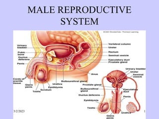
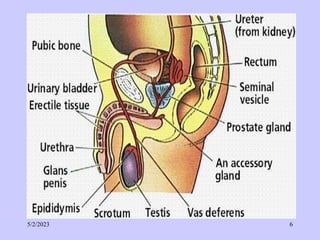
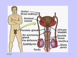
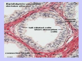
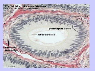
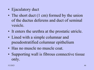
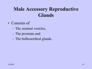
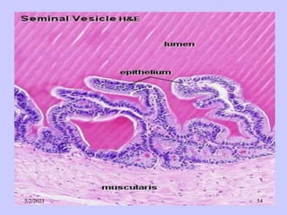
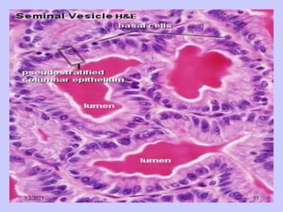
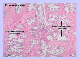
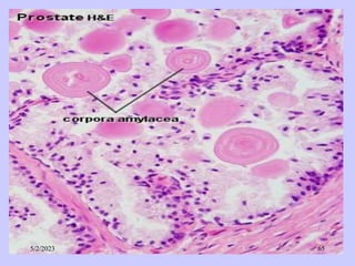
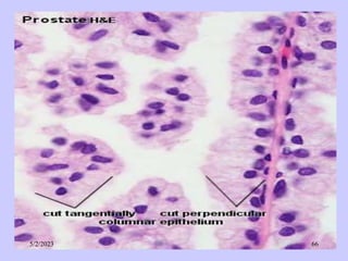
The document provides details about the male reproductive system. It describes the testes, duct system, and accessory sex glands. The testes produce sperm and hormones. Sperm develop through spermatogenesis within the seminiferous tubules of the testes. The duct system includes the rete testis, efferent ducts, epididymis, vas deferens, and ejaculatory duct. The accessory sex glands are the seminal vesicles, prostate gland, and bulbourethral glands. The seminal vesicles secrete fluids that provide energy for sperm.
The testis and duct system consists of highly coiled seminiferous tubules in the testis that produce sperm through spermatogenesis. A system of ducts made up of the rete testis, epididymis, vas deferens, and ejaculatory duct collects and transports sperm. The seminal vesicles and prostate gland secrete fluids that nourish and transport sperm via the penis during ejaculation. Within the seminiferous tubules, Sertoli cells nurture germ cells and Leydig cells produce testosterone.
HISTO. MALE&FEMALE GENITALS FINAL- DR G. TOWO.pptxPharmTecM
The male genital system is composed of the testes, genital ducts, accessory glands, and penis. The testes produce sperm and hormones. The genital ducts and accessory glands secrete fluids that transport sperm and provide nutrients. Sperm and secretions make up semen. In the testes, sperm are produced in seminiferous tubules through spermatogenesis and spermiogenesis. Sertoli cells support this process. Sperm then travel through rete testis, epididymis, and ductus deferens to the urethra, aided by contractions. Accessory glands including seminal vesicles and prostate add secretions to semen. The penis contains erectile
The male reproductive system consists of testes that produce sperm through spermatogenesis in seminiferous tubules. Sperm exit the testes and travel through the epididymis, vas deferens, and urethra. Accessory glands including the seminal vesicles and prostate gland secrete fluids that combine with sperm to form semen. The testes are located in the scrotum to maintain a temperature slightly lower than body temperature, which is vital for sperm production.
The document discusses the male reproductive system and spermatogenesis. It describes the major structures of the male reproductive system including the testes, duct system, and accessory glands. It then focuses on the microscopic structure and function of the seminiferous tubules where spermatogenesis occurs. Spermatogenesis is the process by which spermatogonia differentiate and develop into spermatozoa through spermatocytogenesis, meiosis, and spermiogenesis within the seminiferous tubules of the testes. Sertoli and germ cells are also described in detail for their roles in supporting spermatogenesis.
Anatomy and physiology of male reproductive systemPallavi Lokhande
The organs of the male reproductive system include the testes, a system of ducts (including the epididymis, ductus deferens, ejaculatory ducts, and urethra), accessory sex glands (seminal vesicles, prostate, and bulbourethral glands), and several supporting Structures, including the scrotum and the penis.
and knowledge of this system can help in knowing abnormality in it as well as can be used as study material
The document provides details on the histology of male reproductive organs. It describes the structure and function of the testes, seminiferous tubules, interstitial tissue, spermatogenesis, epididymis, ductus deferens, seminal vesicles, prostate gland, and bulbourethral glands. The testes contain seminiferous tubules where spermatogenesis occurs to produce sperm, and interstitial tissue containing Leydig cells that secrete testosterone. The intratesticular ducts and excretory ducts then transport the mature sperm to the urethra for ejaculation. The accessory glands secrete fluids that mix with sperm to form semen.
This document provides an overview of the histology and histophysiology of the male reproductive system. It describes the key structures and functions of the testes, seminiferous tubules, spermatogenic cells, Leydig cells, and spermatozoa. The testes contain seminiferous tubules that produce spermatozoa through spermatogenesis. Sertoli cells nurture developing spermatogenic cells and form the blood-testis barrier. Leydig cells respond to LH to synthesize testosterone, which supports spermatogenesis. Upon maturation, spermatids are transformed into motile spermatozoa through spermiogenesis.
The document summarizes the histology of the urinary system. It describes the structures and cell types found in the kidney, nephron, ureter, bladder, and urethra. In the kidney, nephrons contain glomeruli and Bowman's capsules for blood filtration. The ureter transports urine from the kidney to the bladder via smooth muscle layers. The bladder stores urine in transitional epithelium before exiting the body through the urethra, lined with stratified squamous epithelium in males and females. Clinical correlates of urinary structures include incontinence and overactive bladder.
The document summarizes the histology of the male reproductive system. It describes the key structures including the testis, seminiferous tubules, Sertoli and Leydig cells involved in spermatogenesis. It then discusses the male duct system including the rete testis, efferent ductules, epididymis, vas deferens, and accessory glands like the seminal vesicles and prostate gland. Diagrams are provided to illustrate the microscopic anatomy of each structure.
The urinary system consists of the kidneys, ureters, urinary bladder, and urethra, with the kidneys being the main organs that filter waste from the blood to produce urine. The kidneys contain nephrons that filter the blood and regulate water and electrolyte levels through selective reabsorption and secretion in the tubules. Urine is then transported from the kidneys through the ureters to the bladder for storage and then exits through the urethra.
The seminiferous tubules contain the seminiferous epithelium, which lines the inside of each tubule. The epithelium contains Sertoli cells and germ cells at various stages of development. Sertoli cells provide support and nourishment to developing sperm cells. Leydig cells located between tubules produce testosterone in response to LH. During spermatogenesis, Sertoli cells phagocytose excess cytoplasm from maturing sperm cells. Fully developed sperm pass through the tubuli recti and rete testis into the epididymis.
Histology of Testis by Dr Mohammad Manzoor MashwaniMohammad Manzoor
The document summarizes the histology of the testis. It describes the size and functions of the testis. It details the layers that make up the testis (tunica vaginalis, tunica albuginea, mediastinum testis, etc). It provides an in-depth explanation of the seminiferous tubules, spermatogenesis, and the key cell types involved like spermatogonia, sertoli cells, and developing sperm cells. It outlines the process by which sperm cells mature and are released from the testis.
The male reproductive system functions to produce sperm and sex hormones. It includes the testes, which produce sperm and testosterone, and a series of ducts that transport sperm. During sexual arousal, blood flow increases in the penis, causing erection. During orgasm, contractions propel semen, containing sperm and fluids, through the urethra and out of the body. Sperm develop through spermatogenesis and spermiogenesis in the testes and mature in the epididymis. The female reproductive system must receive the sperm for fertilization to occur.
The reproductive systems of male and female Ascaris lubricoides worms are described. The male system includes a single coiled testis, vas deferens, seminal vesicle, ejaculatory duct, and two penial spicules. The female system includes two long coiled ovaries, oviducts, uteri which store fertilized eggs, and a vagina. Gametes are formed through a process called telogony, where gametogonia bud off from the gonads and differentiate into gametocytes as they move through zones of proliferation, growth, and maturation.
This document provides an overview of the male reproductive system including both external and internal structures. It begins with describing the external genitalia including the penis and scrotum. It then discusses the internal genitalia such as the testes, epididymis, and vas deferens. Next, it covers the accessory glands including the prostate, seminal vesicles, and bulbourethral glands. It concludes with an explanation of sperm production through spermatogenesis within the seminiferous tubules of the testes. The entire male reproductive system works together to produce, mature, and ejaculate sperm for reproduction.
This document provides an overview of epithelial tissue, including its derivation, structure, functions, and classification. It discusses the key features of epithelium such as polarity, junctions, and basement membrane. Epithelium is classified based on the number of cell layers (simple vs stratified) and cell shape (squamous, cuboidal, columnar). Examples of simple, stratified, and glandular epithelium are provided. The mechanisms of secretion and types of secretory products in glands are also summarized.
The kidney is bean-shaped and located retroperitoneally. It contains nephrons which filter blood to form urine. Each nephron contains a glomerulus for blood filtration and a tubule for urine transport. The kidneys also contain collecting ducts which drain urine from nephrons into the renal pelvis. Urine exits each kidney via the ureter and is stored in the bladder before exiting the body through the urethra.
- The male reproductive system includes the penis, testes, scrotum, prostate gland, and other structures. The testes produce sperm and hormones within the scrotum, which provides a cooler environment for sperm production. The prostate gland secretes fluids that protect and nourish sperm.
- The testes and epididymis work together to produce, mature, and store sperm. The epididymis is a coiled tube that receives sperm from the testes and stores it until ejaculation. The testes have seminiferous tubules where sperm are produced with help from Sertoli cells.
- The scrotum houses the testes and epididymis and contains muscles that help
Epithelial tissue lines body surfaces and forms glands. It has several key functions, including protection, absorption, and secretion. Epithelial cells are closely packed and polarized. They communicate through junctions and microstructures. There are several types of epithelia defined by their cell shape, number of layers, and location in the body. Epithelial tissues contain secretory cells that produce molecules for exocrine and endocrine glands.
The male reproductive system includes the testes, which produce sperm and hormones. Additional structures like the vas deferens, seminal vesicles, prostate and penis work together in ejaculation. The testes contain seminiferous tubules where spermatogenesis occurs to produce sperm. During ejaculation, seminal fluid containing sperm and secretions from the seminal vesicles and prostate mix to form semen. The penis contains erectile tissue that fills with blood to cause an erection, enabling delivery of semen during intercourse.
The document summarizes the development of the urogenital system. It describes how the urinary and genital systems develop from a common intermediate mesoderm and cloaca. The three kidney systems - pronephros, mesonephros, and metanephros - develop sequentially. The metanephros forms the permanent kidney. The urinary bladder, urethra, and genital structures like the uterus, vagina, and external genitalia also develop from the intermediate mesoderm and urogenital sinus. The kidneys ascend to their final position in the lumbar region during development.
The urinary system- BPH 1.2 A, B & C 2023- DR.TOWO.pptxPharmTecM
The urinary system consists of the kidneys, ureters, urinary bladder, and urethra. The kidneys filter waste from the blood to produce urine via nephrons containing glomeruli and tubules. The urine travels from the kidneys through the ureters to the bladder, then exits through the urethra. In addition to waste excretion, the kidneys regulate water balance and electrolyte levels in the body.
This document provides an overview of the histology of the urinary system, including the kidneys, ureters, urinary bladder, and urethra. It describes the microscopic anatomy of each organ in detail, highlighting key structures like the nephron (the functional unit of the kidney), renal corpuscles, proximal and distal tubules, loops of Henle, collecting ducts, and the transitional epithelium lining the ureters, bladder, and urethra. The document also discusses specialized structures in the kidney like the juxtaglomerular apparatus and its role in regulating blood pressure and the renin-angiotensin system.
The cytoskeleton is a network of protein filaments that extends throughout the cytoplasm. It provides structure and organization to the cell, determining shape and positioning organelles. The three main types of filaments are actin filaments, intermediate filaments, and microtubules. Actin filaments are the thinnest filaments and form structures like filopodia, lamellipodia, and stress fibers. Microtubules are hollow cylinders composed of tubulin dimers and originate from the centrosome. They are involved in processes like cell division, organelle transport, and motility. Cilia and flagella project from the cell surface and use microtubule motors for movement.
EWOCS-I: The catalog of X-ray sources in Westerlund 1 from the Extended Weste...Sérgio Sacani
Context. With a mass exceeding several 104 M⊙ and a rich and dense population of massive stars, supermassive young star clusters
represent the most massive star-forming environment that is dominated by the feedback from massive stars and gravitational interactions
among stars.
Aims. In this paper we present the Extended Westerlund 1 and 2 Open Clusters Survey (EWOCS) project, which aims to investigate
the influence of the starburst environment on the formation of stars and planets, and on the evolution of both low and high mass stars.
The primary targets of this project are Westerlund 1 and 2, the closest supermassive star clusters to the Sun.
Methods. The project is based primarily on recent observations conducted with the Chandra and JWST observatories. Specifically,
the Chandra survey of Westerlund 1 consists of 36 new ACIS-I observations, nearly co-pointed, for a total exposure time of 1 Msec.
Additionally, we included 8 archival Chandra/ACIS-S observations. This paper presents the resulting catalog of X-ray sources within
and around Westerlund 1. Sources were detected by combining various existing methods, and photon extraction and source validation
were carried out using the ACIS-Extract software.
Results. The EWOCS X-ray catalog comprises 5963 validated sources out of the 9420 initially provided to ACIS-Extract, reaching a
photon flux threshold of approximately 2 × 10⁻⁸ photons cm−2
s
−1
. The X-ray sources exhibit a highly concentrated spatial distribution,
with 1075 sources located within the central 1 arcmin. We have successfully detected X-ray emissions from 126 out of the 166 known
massive stars of the cluster, and we have collected over 71 000 photons from the magnetar CXO J164710.20-455217.
Or: Beyond linear.
Abstract: Equivariant neural networks are neural networks that incorporate symmetries. The nonlinear activation functions in these networks result in interesting nonlinear equivariant maps between simple representations, and motivate the key player of this talk: piecewise linear representation theory.
Disclaimer: No one is perfect, so please mind that there might be mistakes and typos.
dtubbenhauer@gmail.com
Corrected slides: dtubbenhauer.com/talks.html
More Related Content
Similar to MALE REPRODUCTIVE SYSTEM PRESENTATION.ppt
The document summarizes the histology of the male reproductive system. It describes the key structures including the testis, seminiferous tubules, Sertoli and Leydig cells involved in spermatogenesis. It then discusses the male duct system including the rete testis, efferent ductules, epididymis, vas deferens, and accessory glands like the seminal vesicles and prostate gland. Diagrams are provided to illustrate the microscopic anatomy of each structure.
The urinary system consists of the kidneys, ureters, urinary bladder, and urethra, with the kidneys being the main organs that filter waste from the blood to produce urine. The kidneys contain nephrons that filter the blood and regulate water and electrolyte levels through selective reabsorption and secretion in the tubules. Urine is then transported from the kidneys through the ureters to the bladder for storage and then exits through the urethra.
The seminiferous tubules contain the seminiferous epithelium, which lines the inside of each tubule. The epithelium contains Sertoli cells and germ cells at various stages of development. Sertoli cells provide support and nourishment to developing sperm cells. Leydig cells located between tubules produce testosterone in response to LH. During spermatogenesis, Sertoli cells phagocytose excess cytoplasm from maturing sperm cells. Fully developed sperm pass through the tubuli recti and rete testis into the epididymis.
Histology of Testis by Dr Mohammad Manzoor MashwaniMohammad Manzoor
The document summarizes the histology of the testis. It describes the size and functions of the testis. It details the layers that make up the testis (tunica vaginalis, tunica albuginea, mediastinum testis, etc). It provides an in-depth explanation of the seminiferous tubules, spermatogenesis, and the key cell types involved like spermatogonia, sertoli cells, and developing sperm cells. It outlines the process by which sperm cells mature and are released from the testis.
The male reproductive system functions to produce sperm and sex hormones. It includes the testes, which produce sperm and testosterone, and a series of ducts that transport sperm. During sexual arousal, blood flow increases in the penis, causing erection. During orgasm, contractions propel semen, containing sperm and fluids, through the urethra and out of the body. Sperm develop through spermatogenesis and spermiogenesis in the testes and mature in the epididymis. The female reproductive system must receive the sperm for fertilization to occur.
The reproductive systems of male and female Ascaris lubricoides worms are described. The male system includes a single coiled testis, vas deferens, seminal vesicle, ejaculatory duct, and two penial spicules. The female system includes two long coiled ovaries, oviducts, uteri which store fertilized eggs, and a vagina. Gametes are formed through a process called telogony, where gametogonia bud off from the gonads and differentiate into gametocytes as they move through zones of proliferation, growth, and maturation.
This document provides an overview of the male reproductive system including both external and internal structures. It begins with describing the external genitalia including the penis and scrotum. It then discusses the internal genitalia such as the testes, epididymis, and vas deferens. Next, it covers the accessory glands including the prostate, seminal vesicles, and bulbourethral glands. It concludes with an explanation of sperm production through spermatogenesis within the seminiferous tubules of the testes. The entire male reproductive system works together to produce, mature, and ejaculate sperm for reproduction.
This document provides an overview of epithelial tissue, including its derivation, structure, functions, and classification. It discusses the key features of epithelium such as polarity, junctions, and basement membrane. Epithelium is classified based on the number of cell layers (simple vs stratified) and cell shape (squamous, cuboidal, columnar). Examples of simple, stratified, and glandular epithelium are provided. The mechanisms of secretion and types of secretory products in glands are also summarized.
The kidney is bean-shaped and located retroperitoneally. It contains nephrons which filter blood to form urine. Each nephron contains a glomerulus for blood filtration and a tubule for urine transport. The kidneys also contain collecting ducts which drain urine from nephrons into the renal pelvis. Urine exits each kidney via the ureter and is stored in the bladder before exiting the body through the urethra.
- The male reproductive system includes the penis, testes, scrotum, prostate gland, and other structures. The testes produce sperm and hormones within the scrotum, which provides a cooler environment for sperm production. The prostate gland secretes fluids that protect and nourish sperm.
- The testes and epididymis work together to produce, mature, and store sperm. The epididymis is a coiled tube that receives sperm from the testes and stores it until ejaculation. The testes have seminiferous tubules where sperm are produced with help from Sertoli cells.
- The scrotum houses the testes and epididymis and contains muscles that help
Epithelial tissue lines body surfaces and forms glands. It has several key functions, including protection, absorption, and secretion. Epithelial cells are closely packed and polarized. They communicate through junctions and microstructures. There are several types of epithelia defined by their cell shape, number of layers, and location in the body. Epithelial tissues contain secretory cells that produce molecules for exocrine and endocrine glands.
The male reproductive system includes the testes, which produce sperm and hormones. Additional structures like the vas deferens, seminal vesicles, prostate and penis work together in ejaculation. The testes contain seminiferous tubules where spermatogenesis occurs to produce sperm. During ejaculation, seminal fluid containing sperm and secretions from the seminal vesicles and prostate mix to form semen. The penis contains erectile tissue that fills with blood to cause an erection, enabling delivery of semen during intercourse.
The document summarizes the development of the urogenital system. It describes how the urinary and genital systems develop from a common intermediate mesoderm and cloaca. The three kidney systems - pronephros, mesonephros, and metanephros - develop sequentially. The metanephros forms the permanent kidney. The urinary bladder, urethra, and genital structures like the uterus, vagina, and external genitalia also develop from the intermediate mesoderm and urogenital sinus. The kidneys ascend to their final position in the lumbar region during development.
The urinary system- BPH 1.2 A, B & C 2023- DR.TOWO.pptxPharmTecM
The urinary system consists of the kidneys, ureters, urinary bladder, and urethra. The kidneys filter waste from the blood to produce urine via nephrons containing glomeruli and tubules. The urine travels from the kidneys through the ureters to the bladder, then exits through the urethra. In addition to waste excretion, the kidneys regulate water balance and electrolyte levels in the body.
This document provides an overview of the histology of the urinary system, including the kidneys, ureters, urinary bladder, and urethra. It describes the microscopic anatomy of each organ in detail, highlighting key structures like the nephron (the functional unit of the kidney), renal corpuscles, proximal and distal tubules, loops of Henle, collecting ducts, and the transitional epithelium lining the ureters, bladder, and urethra. The document also discusses specialized structures in the kidney like the juxtaglomerular apparatus and its role in regulating blood pressure and the renin-angiotensin system.
The cytoskeleton is a network of protein filaments that extends throughout the cytoplasm. It provides structure and organization to the cell, determining shape and positioning organelles. The three main types of filaments are actin filaments, intermediate filaments, and microtubules. Actin filaments are the thinnest filaments and form structures like filopodia, lamellipodia, and stress fibers. Microtubules are hollow cylinders composed of tubulin dimers and originate from the centrosome. They are involved in processes like cell division, organelle transport, and motility. Cilia and flagella project from the cell surface and use microtubule motors for movement.
Similar to MALE REPRODUCTIVE SYSTEM PRESENTATION.ppt (20)
EWOCS-I: The catalog of X-ray sources in Westerlund 1 from the Extended Weste...Sérgio Sacani
Context. With a mass exceeding several 104 M⊙ and a rich and dense population of massive stars, supermassive young star clusters
represent the most massive star-forming environment that is dominated by the feedback from massive stars and gravitational interactions
among stars.
Aims. In this paper we present the Extended Westerlund 1 and 2 Open Clusters Survey (EWOCS) project, which aims to investigate
the influence of the starburst environment on the formation of stars and planets, and on the evolution of both low and high mass stars.
The primary targets of this project are Westerlund 1 and 2, the closest supermassive star clusters to the Sun.
Methods. The project is based primarily on recent observations conducted with the Chandra and JWST observatories. Specifically,
the Chandra survey of Westerlund 1 consists of 36 new ACIS-I observations, nearly co-pointed, for a total exposure time of 1 Msec.
Additionally, we included 8 archival Chandra/ACIS-S observations. This paper presents the resulting catalog of X-ray sources within
and around Westerlund 1. Sources were detected by combining various existing methods, and photon extraction and source validation
were carried out using the ACIS-Extract software.
Results. The EWOCS X-ray catalog comprises 5963 validated sources out of the 9420 initially provided to ACIS-Extract, reaching a
photon flux threshold of approximately 2 × 10⁻⁸ photons cm−2
s
−1
. The X-ray sources exhibit a highly concentrated spatial distribution,
with 1075 sources located within the central 1 arcmin. We have successfully detected X-ray emissions from 126 out of the 166 known
massive stars of the cluster, and we have collected over 71 000 photons from the magnetar CXO J164710.20-455217.
Or: Beyond linear.
Abstract: Equivariant neural networks are neural networks that incorporate symmetries. The nonlinear activation functions in these networks result in interesting nonlinear equivariant maps between simple representations, and motivate the key player of this talk: piecewise linear representation theory.
Disclaimer: No one is perfect, so please mind that there might be mistakes and typos.
dtubbenhauer@gmail.com
Corrected slides: dtubbenhauer.com/talks.html
Travis Hills' Endeavors in Minnesota: Fostering Environmental and Economic Pr...Travis Hills MN
Travis Hills of Minnesota developed a method to convert waste into high-value dry fertilizer, significantly enriching soil quality. By providing farmers with a valuable resource derived from waste, Travis Hills helps enhance farm profitability while promoting environmental stewardship. Travis Hills' sustainable practices lead to cost savings and increased revenue for farmers by improving resource efficiency and reducing waste.
Unlocking the mysteries of reproduction: Exploring fecundity and gonadosomati...AbdullaAlAsif1
The pygmy halfbeak Dermogenys colletei, is known for its viviparous nature, this presents an intriguing case of relatively low fecundity, raising questions about potential compensatory reproductive strategies employed by this species. Our study delves into the examination of fecundity and the Gonadosomatic Index (GSI) in the Pygmy Halfbeak, D. colletei (Meisner, 2001), an intriguing viviparous fish indigenous to Sarawak, Borneo. We hypothesize that the Pygmy halfbeak, D. colletei, may exhibit unique reproductive adaptations to offset its low fecundity, thus enhancing its survival and fitness. To address this, we conducted a comprehensive study utilizing 28 mature female specimens of D. colletei, carefully measuring fecundity and GSI to shed light on the reproductive adaptations of this species. Our findings reveal that D. colletei indeed exhibits low fecundity, with a mean of 16.76 ± 2.01, and a mean GSI of 12.83 ± 1.27, providing crucial insights into the reproductive mechanisms at play in this species. These results underscore the existence of unique reproductive strategies in D. colletei, enabling its adaptation and persistence in Borneo's diverse aquatic ecosystems, and call for further ecological research to elucidate these mechanisms. This study lends to a better understanding of viviparous fish in Borneo and contributes to the broader field of aquatic ecology, enhancing our knowledge of species adaptations to unique ecological challenges.
ESPP presentation to EU Waste Water Network, 4th June 2024 “EU policies driving nutrient removal and recycling
and the revised UWWTD (Urban Waste Water Treatment Directive)”
Immersive Learning That Works: Research Grounding and Paths ForwardLeonel Morgado
We will metaverse into the essence of immersive learning, into its three dimensions and conceptual models. This approach encompasses elements from teaching methodologies to social involvement, through organizational concerns and technologies. Challenging the perception of learning as knowledge transfer, we introduce a 'Uses, Practices & Strategies' model operationalized by the 'Immersive Learning Brain' and ‘Immersion Cube’ frameworks. This approach offers a comprehensive guide through the intricacies of immersive educational experiences and spotlighting research frontiers, along the immersion dimensions of system, narrative, and agency. Our discourse extends to stakeholders beyond the academic sphere, addressing the interests of technologists, instructional designers, and policymakers. We span various contexts, from formal education to organizational transformation to the new horizon of an AI-pervasive society. This keynote aims to unite the iLRN community in a collaborative journey towards a future where immersive learning research and practice coalesce, paving the way for innovative educational research and practice landscapes.
Authoring a personal GPT for your research and practice: How we created the Q...Leonel Morgado
Thematic analysis in qualitative research is a time-consuming and systematic task, typically done using teams. Team members must ground their activities on common understandings of the major concepts underlying the thematic analysis, and define criteria for its development. However, conceptual misunderstandings, equivocations, and lack of adherence to criteria are challenges to the quality and speed of this process. Given the distributed and uncertain nature of this process, we wondered if the tasks in thematic analysis could be supported by readily available artificial intelligence chatbots. Our early efforts point to potential benefits: not just saving time in the coding process but better adherence to criteria and grounding, by increasing triangulation between humans and artificial intelligence. This tutorial will provide a description and demonstration of the process we followed, as two academic researchers, to develop a custom ChatGPT to assist with qualitative coding in the thematic data analysis process of immersive learning accounts in a survey of the academic literature: QUAL-E Immersive Learning Thematic Analysis Helper. In the hands-on time, participants will try out QUAL-E and develop their ideas for their own qualitative coding ChatGPT. Participants that have the paid ChatGPT Plus subscription can create a draft of their assistants. The organizers will provide course materials and slide deck that participants will be able to utilize to continue development of their custom GPT. The paid subscription to ChatGPT Plus is not required to participate in this workshop, just for trying out personal GPTs during it.
The binding of cosmological structures by massless topological defectsSérgio Sacani
Assuming spherical symmetry and weak field, it is shown that if one solves the Poisson equation or the Einstein field
equations sourced by a topological defect, i.e. a singularity of a very specific form, the result is a localized gravitational
field capable of driving flat rotation (i.e. Keplerian circular orbits at a constant speed for all radii) of test masses on a thin
spherical shell without any underlying mass. Moreover, a large-scale structure which exploits this solution by assembling
concentrically a number of such topological defects can establish a flat stellar or galactic rotation curve, and can also deflect
light in the same manner as an equipotential (isothermal) sphere. Thus, the need for dark matter or modified gravity theory is
mitigated, at least in part.
When I was asked to give a companion lecture in support of ‘The Philosophy of Science’ (https://shorturl.at/4pUXz) I decided not to walk through the detail of the many methodologies in order of use. Instead, I chose to employ a long standing, and ongoing, scientific development as an exemplar. And so, I chose the ever evolving story of Thermodynamics as a scientific investigation at its best.
Conducted over a period of >200 years, Thermodynamics R&D, and application, benefitted from the highest levels of professionalism, collaboration, and technical thoroughness. New layers of application, methodology, and practice were made possible by the progressive advance of technology. In turn, this has seen measurement and modelling accuracy continually improved at a micro and macro level.
Perhaps most importantly, Thermodynamics rapidly became a primary tool in the advance of applied science/engineering/technology, spanning micro-tech, to aerospace and cosmology. I can think of no better a story to illustrate the breadth of scientific methodologies and applications at their best.
The technology uses reclaimed CO₂ as the dyeing medium in a closed loop process. When pressurized, CO₂ becomes supercritical (SC-CO₂). In this state CO₂ has a very high solvent power, allowing the dye to dissolve easily.
2. Introduction
• It is comprised of a pair of testes, bilateral
duct system, accessory sex glands, urethra
and the penis.
• The duct system includes tubuli recti, rete
testis, efferent duct, epididymis duct,
deferens duct, and ejaculatory duct.
• Accessory sex gland include seminal
vesicles, prostate gland, and bulbourethral
(Cowper’s) gland.
5/2/2023 2
3. The Testes
• These are the primary male gonads that are
suspended in the scrotum and their main
function is production of spermatozoa and
secretion of male hormones (Androgens).
• The left testis is lower than the right and
each testis measures about 4 to 5 cm long,
2.5 cm width and 3 cm in thickness and each
weighs 10.5 to 14 gms.
5/2/2023 3
4. Testes cont…...
• Structural organization
– Each testis is contained in one compartment of
the fibromuscular sac known as scrotum.
– The scrotum has two right and left compartments
that are separated externally by a cutaneous
raphe that continues ventrally to the inferior
surface of the penis and dorsally to the anus
through the perineum.
5/2/2023 4
5. Testes cont…...
– The scrotum is made up of the skin, smooth
muscle (dartos muscle, cremaster muscle),
external spermatic fascia, cremasteric fascia
and the internal spermatic fascia which is
associated with the external covering of the
testis the tunica vaginalis
– Testis is immediately surrounded by the
testicular capsule, which is composed of three
layers:
–Outer layer or tunica vaginalis
–Middle layer or tunica albuginea
–Innermost layer or tunica vasculosa
5/2/2023 5
8. Tunica vaginalis
• Is the lower end of the peritoneal processus vaginalis
which precedes the descent of the fetal testes from
the abdomen to the scrotum that has lost connection
with the peritoneal cavity.
• It covers the anterior and lateral surfaces of the
testes.
• Has visceral and parietal layers.
5/2/2023 8
9. • Visceral layer covers all aspects of the
testes except the posterior border.
• Parietal covers the posterior aspect of the
testes.
• In between them there is a cavity that
contain small amount of fluid.
• Sometimes during pathological situations
abnormal fluid accumulation occurs in the
cavity leading to hydrocoele.
5/2/2023 9
10. tunica albuginea
• A thick layer of dense fibroelastic connective
tissue that contains some smooth muscle cells
concentrated predominantly on the posterior
aspect of the testis adjacent to the epididymis to
form the mediastinum testis from which arises
septae (septula testis) that divides the testis into
compartments or lobules.
• Each lobule is pyramidal in shape with the apex
directed towards mediastinum.
5/2/2023 10
11. • There are about 250 lobules each containing
1 to 4 seminiferous tubules and a connective
tissue stroma in which intestitial cells
(Leydig cells) are located.
• Completely covers the testes. It lies internal
to the tunica vaginalis.
5/2/2023 11
12. tunica vasculosa
• The innermost layer of testicular capsule
that consists of a network of blood vessels
embedded within a delicate loose
connective tissue.
5/2/2023 12
13. Histological aspect
• Consist of many seminiferous tubules embedded
in relatively sparse interstitial tissue.
• The tubules are lined by a simple columnar
epithelium of Sertoli cells.
• Interspersed within the tubular epithelium are
germ cells.
• Meiotic cell divisions lead from spermatogonia
through primary and secondary spermatocytes to
cells called spermatids, which mature into
spermatozoa.
5/2/2023 13
14. • Interstitial tissue between the tubules
contains clusters of endocrine Leydig cells
which secrete testosterone.
• All of the seminiferous tubules converge
onto a network of interconnecting tubules,
the rete testis, which in turn lead through
numerous small efferent ductules into the
larger duct which extends through the
epididymis.
5/2/2023 14
16. Seminiferous tubule
• Highly convoluted tubule about 0.2 mm in
diameter and 30 to 40 cm long.
• Are lined by seminiferous epithelium which is a
modified stratified cuboidal epithelium consisting
of spermatogenic cells and supporting cells (sertoli
cells).
• The tubules are surrounded by a basal lamina and
by the thin 3 to 4 squamous smooth muscle cells
invested by their own basal (external) lamina.
5/2/2023 16
17. • The connective tissue aspect of the smooth
muscle cells is covered by the thin
endothelium of a vast system of large
lymphatic capillaries.
• This system and smooth muscle sheath
represent a physiological barrier to blood-
borne substances intended to reach the
spermatogenic cells.
5/2/2023 17
18. • Spermatogenic (Germ) cells
• Comprises a stratified layer of epithelium 4
to 8 cells deep, lining the seminiferous
tubule.
• The cells differentiate progressively from
the basal region of the tubule to the lumen.
5/2/2023 18
19. • Proliferation pushes the cells toward the
lumen, and those nearest the lumen
transform into spermatozoa and detach from
the epithelium coming to lie free within the
lumen.
• The sequence of events is referred to as
spermatogenesis (spermatocytogenesis and
spermiogenesis).
5/2/2023 19
20. • Spermatogonia are the first cells of
spermatogenesis.
• They are always in contact with the basal lamina
of the tubule.
• Two types of spermatogonia can be distinguished:
Type A spermatogonia
– Have a rounded nucleus with very fine
chromatin grains and one or two nucleoli. They
are stem cells which divide to form new
generations of both type A and type B
spermatogonia.
5/2/2023 20
21. • Type B spermatogonia
– Have rounded nuclei with chromatin
granules of variable size, which often
attach to the nuclear membrane, and one
nucleolus.
– Although type B spermatogonia may
divide repeatedly, they do not function as
stem cells and their final mitosis always
results in the formation of primary
spermatocytes.
5/2/2023 21
22. • Primary spermatocytes
– Larger than the spermatogonia.
– They are spherical or ovoid in outline,
and are the largest germ cells seen within
the seminiferous tubule where they
occupy the middle zone of the
epithelium.
– A large number of primary spermatocytes
is always visible in cross-sections
through seminiferous tubules.
5/2/2023 22
23. • Secondary spermatocytes,
– Are smaller than primary spermatocytes.
They rapidly enter and complete the
second meiotic division and are therefore
seldom seen in histological preparations
since they are short lived and divide
quickly to produce Spermatids.
5/2/2023 23
24. • Spermatids,
– Lie in the luminal part of the
seminiferous epithelium.
– They are small (about 10 µm in
diameter) with an initially very light
(often eccentric) nucleus.
– The chromatin condenses during the
maturation of the spermatids into
spermatozoa, and the nucleus becomes
smaller and stains darker.
– The differentiation of the newly formed
spermatids results into Spermatozoa.
5/2/2023 24
25. • Spermatozoa
– The mature human spermatozoon is about
60 µm long and actively motile. It is
divided into head, neck and tail.
5/2/2023 25
26. Sertoli cells
• Constitute true epithelium of the seminiferous
epithelium.
• Relatively few in number spaced along the tubule
at fairly regular intervals crowded between germ
cells.
• They are columnar, tall, pillar-like cells, with their
bases resting on upon the basal lamina.
• Do not migrate nor do they replicate after puberty.
5/2/2023 26
27. • Interstitial tissue
– It’s the loose connective tissue between
the seminiferous tubules contains
interstitial endocrine cells (Leydig cells),
blood, and lymphatic vessels, nerves,
fibroblasts and mast cells.
• Leydig cells (15-20 µm), located in the
interstitial tissue between the convoluted
seminiferous tubules, constitute the
endocrine component of the testis. They
synthesize and secrete testosterone.
5/2/2023 27
28. • Active Leydig cells are large ,irregularly
polygonal, acidophilic cells often filled lipid
droplets.
• Have an elaborate smooth endoplasmic
reticulum and mitochondria with
tubulovesicular cristae.
• Inactive cells are difficult to distinguish
from fibroblast.
5/2/2023 28
31. Male genital ducts
• Two types of ducts
– Intratesticular genital ducts.
Tubuli recti, rete testis, proximal part of
efferent duct.
– Extratesticular genital ducts.
Distal part of efferent duct, epididymis
duct, deferens duct, and ejaculatory duct.
5/2/2023 31
32. • Tubuli recti
– The straight tubules which is a short
terminal section of seminiferous tubule
that lined solely by Sertoli cells.
– As it narrows towards to rete testis the
lining changes to a simple cuboidal
epithelium.
5/2/2023 32
33. • rete testis
– Complex series of interconnecting
channels in a highly vascular of
mediastinum testis.
– Lined mainly by low cuboidal epithelium.
– Each cuboidal cell has a single cilium on
its apical surface with a few apical
microvilli.
5/2/2023 33
34. Efferent duct
• Approx 15 efferent ductules leaves the testis by
penetrating tunica albuginea and connect the testis
to ductus epididymis.
• As it leaves the testis 15 to 20 cm long become
highly coiled and tightly parked into 6 to 10
conical masses called Coni vasculosi.
5/2/2023 34
35. • At the base of the coni they empty into the
single channel of ductus epididymis
• They are lined with alternating clumps of
tall and short pseudo stratified columnar
cells thus giving rise to the saw-toothed
appearance of the luminal surface.
5/2/2023 35
36. • Tall columnar cells are ciliated
• Short non-ciliated columnar cells with
numerous microvilli and canalicular
invagination of apical surface.
• Between the columnar cells there are few
basal cells and intraepithelial lymphocytes.
• Has circular smooth muscle layers in the
wall of the ductule.
• Elastic fibers are interspersed among the
muscle cells.
5/2/2023 36
37. ductus epididymidis
• About 6 m long.
• It is lined by a very tall pseudostratified
columnar epithelium.
• Most cells of the epithelium, also called
principal cells, have long stereocilia.
• Stereocilia are non-motile structures, which
in the EM resemble large microvilli.
5/2/2023 37
38. • Towards the basal lamina we see a number
of small nuclei, which belong to the basal
cells of the ductus epididymis. These cells
regenerate the epithelium.
• Peristaltic contractions of smooth muscle
cells surrounding the ductus epididymis
move the spermatozoa towards the middle
segment of the duct, which is the site of
final functional maturation of the
spermatozoa - now they are motile.
5/2/2023 38
39. • The terminal segment of the ductus
epididymis is the site of storage of the
mature spermatozoa.
• Smooth muscle fibers of the terminal part of
the ductus epididymis do not contract
spontaneously.
• They contract during sexual stimulation
concurrently with the contraction of the
musculature of the duct into which it opens,
the vas deferens.
5/2/2023 39
42. Vas deferens (ductus deferens)
• Connects epididymis to the prostatic urethra
• The mucosa of the vas deferens forms low
longitudinal folds.
• It is lined by a pseudostratified columnar
epithelium. Similar to the epididymis, cells have
long stereocilia.
• The lamina propria is unusually rich in elastic
fibres.
5/2/2023 42
43. • The muscularis is well developed (up to 1.5
mm thick) and consists of a thick circular
layer of smooth muscle between thinner
inner and outer longitudinal layers.
• The muscularis is the structure which makes
the vas deferens palpable in the spermatic
cord.
• The vas deferens is surrounded by an
adventitia, which is slightly denser than
usual.
5/2/2023 43
46. • Ejaculatory duct
• The short duct (1 cm) formed by the union
of the ductus deferens and duct of seminal
vesicle.
• It enters the urethra at the prostatic utricle.
• Lined with a simple columnar and
pseudostratified columnar epithelium
• Has no muscle no muscle coat.
• Supporting wall is fibrous connective tissue
only.
5/2/2023 46
48. Seminal vesicles
• They are paired.
• Are elongate sacs (about 4 cm long and 2 cm
wide), highly folded tubular glands with a
muscular and fibrous coat.
• They develop as evaginations of the vas deferens
distal to ampulla.
• They are situated posterior to prostate gland.
• Each consists of one coiling tube (about 15cm
long).
5/2/2023 48
49. • The mucosa is thrown into various primary,
secondary and tertiary folds that increase
the secretory surface.
• The folds project into the lumen and merge
with one another as a result different
compartments of various sizes are formed.
All communicate with the lumen.
• The muscularis consists of inner circular
and outer longitudinal layers of smooth
muscle.
5/2/2023 49
50. • The epithelium is variable appearing as
simple columnar or pseudostratified
columnar (columnar cells and basal cells).
• Pseudostratified columnar epithelium
contains tall non ciliated columnar cells and
short round cells that rest on basal lamina
• The lamina propria of the mucosa is fairly
thin and loose.
5/2/2023 50
51. • They are glands, whose secretion
constitutes 60-70 % of the ejaculate.
• The secretory product of the columnar cell,
which may be seen in the lumen of the
seminal vesicles, is strongly acidophilic.
• It contains large amounts of fructose which
the spermatozoa utilise as a source of
energy.
5/2/2023 51
52. • The secretion also contains prostaglandins,
flavins (yellow fluorescing pigment - of use
in forensic medicine to detect semen stains)
and several other proteins and enzymes.
• The cocktail of compounds which is
released by the seminal vesicles in addition
to fructose has three main functions:
5/2/2023 52
53. 1. formation of the sperm coagulum,
2. regulation of sperm motility and
3. suppression of immune function in the
female genital tract.
5/2/2023 53
56. Prostate
• The prostate is the largest accessory sex gland in
men (about 2 × 3 × 4 cm).
• It surrounds the proximal urethra at its origin from
the bladder
• It contains 30 - 50 tubuloalveolar glands, which
empty into 15 - 25 independent excretory ducts.
These ducts open into the urethra.
• The glands are embedded into a fibromuscular
stroma, which mainly consists of smooth muscle
separated by strands of connective tissue rich in
collagenous and elastic fibers.
5/2/2023 56
57. • The muscle forms a dense mass around the
urethra and beneath the fairly thin capsule
of the prostrate.
• The secretory alveoli of the prostate are
very irregularly shaped because of papillary
projections of the mucosa into the lumen of
the gland.
• The epithelium is cuboidal or columnar also
patches of squamous.
5/2/2023 57
58. • Basal cells are again present, and the
epithelium may look pseudo stratified
where they are found.
• The secretory cells are slightly
acidophilic and secretory granules may
be visible in the cytoplasm.
5/2/2023 58
59. • The secretion of the prostate contains citric
acid, the enzyme fibrinolysin (liquefies the
semen), acid phosphatase, a number of
other enzymes and lipids.
• The secretion of the prostate is the first
fraction of the ejaculate.
• The secretory ducts of the prostate are lined
by a simple columnar epithelium, which
changes to a transitional epithelium near the
openings of the ducts into the urethra.
5/2/2023 59
60. • A characteristic feature of the prostate is the
appearance of corpora amylacea in the secretory
alveoli.
• They are rounded eosinophilic bodies.
• Their average diameter is about 0.25 to 2 mm).
• They appear already in the seventh month of foetal
development.
• Their number increases with age - in particular
past 50.
• They may undergo calcification. Corpora
amylacea may appear in semen.
5/2/2023 60
61. • Macroscopically the prostrate can be
divided into lobes, but they are
inconspicuous in histological sections.
• In good histological sections it is possible to
distinguish three concentric zones, which
surround the prostatic part of the urethra.
• The peripheral zone contains large, so-
called main glands, whose ducts run
posteriorly to open into the urethra.
5/2/2023 61
62. • The internal zone consists of the so-called
submucosal glands, whereas
• The innermost zone contains mucosal glands.
• This subdivision of the prostate is of clinical
importance.
• With age the prostate becomes enlarged due to
benign nodular hyperplasia or benign prostatic
hypertrophy.
• The onset age of these hyperplastic changes is 45.
About 3/4 of the males above 60 are affected of
which half will be symptomatic.
5/2/2023 62
63. • This condition affects the mucosal glands.
• Cancer of the prostate, which is the
second most common malignant tumor in
western males, involves the peripheral zone.
• It affects approximately 1 in 20
• It is often not detected early because the
abnormal growth may not impinge on the
urethra and produce symptoms that demand
prompt attention
5/2/2023 63
67. Bulbourethral gland (Cowper’s glands)
• Are paired bodies, each the size of a pea,
located in the connective tissue behind the
membranous urethra and joins the initial
portion of penile urethra
• The glands are compound tubuloalveolar
glands that structurally resemble mucus
secretory glands
• Is surrounded by thin connective tissue
capsule, external to which are skeletal
muscle fibers
5/2/2023 67
68. • The septa pas into the gland to divide it into
lobules
• The secretory end pieces are variable, being
either alveolar, saccular or tubular
• The secretion is clear, mucus like
containing considerable amounts of
galactose, galactosamine, galacturonic acid,
sialic acid and methylpentose.
• They constitute major portion of preseminal
fluid and probably serves as a lubricant of
the penile urethra
5/2/2023 68
69. semen
• Is a combined product of all of the
glandular elements of the male reproductive
system
• It contains fluid and sperm from the testis
and secretory products from epididymis, vas
deferens, prostate, seminal vesicle and
bulbourethral glands
• It is alkaline in nature
5/2/2023 69
70. • May help to neutralize the acid environment
of the urethra and vagina
• Also contains prostaglandins that may
influence sperm transit in both male and
female reproductive tracts, and also may
have a role in implantation of a fertilized
ovum
5/2/2023 70
71. • The volume of average ejaculate of semen
is about 3mL. This is normally contains up
to 100million sperm/mL.
• Of which it is estimated that 20% are
morphologically abnormal; and nearly 25%
are immotile
5/2/2023 71
72. PENIS
• An elongated organ which is common termination
of both the urinary and genital extratesticular duct
system
• Urethra carries both semen and urine to the
exterior
• It consist primarily of two dorsal masses of
erectile tissue; the corpora cavernosa, and a
ventral mass of erectile tissue that surrounds the
urethra; corpus spongiosum (corpus cavernosum
spongiosum urethrae)
5/2/2023 72
73. • A dense fibroelastic layer , the tunica
albuginea, binds the three cavernosa
together as well as forming capsule around
each one
• Corpora cavernosa are lined with vascular
endothelium thus increase in size and
rigidity by filling with blood principally
derived from helicine arteries
5/2/2023 73
74. • These arteries dilate under sexual
stimulation to increase blood flow to the
penis
• Helicine arteries(dilate) and arteriovenous
anastomosis (closes)
• As erectile tissue fills, the peripheral veins
that drain it are increasingly compressed,
this may amplify erectile response
5/2/2023 74
75. • Thin skin of penis is loosely attached to the
underlying connective tissue which contain
no adipose tissue. Only at the glans penis
skin is firmly attached
• Thin layer of smooth muscle in the skin is
continuous with dartos layer of scrotum
• In uncircumcised male, glans is covered
with a fold of skin, prepuce, which has
characteristic of mucous membrane on its
inner aspect
5/2/2023 75
76. • Skin contains small sweat glands and
infrequent sebaceous glands unassociated
with hair follicles. No hair follicles on the
distal part of the penis
• On the glans penis and inner surface of
prepuce there are numbers of modified
sebaceous glnds , the Glands of Tyson
5/2/2023 76