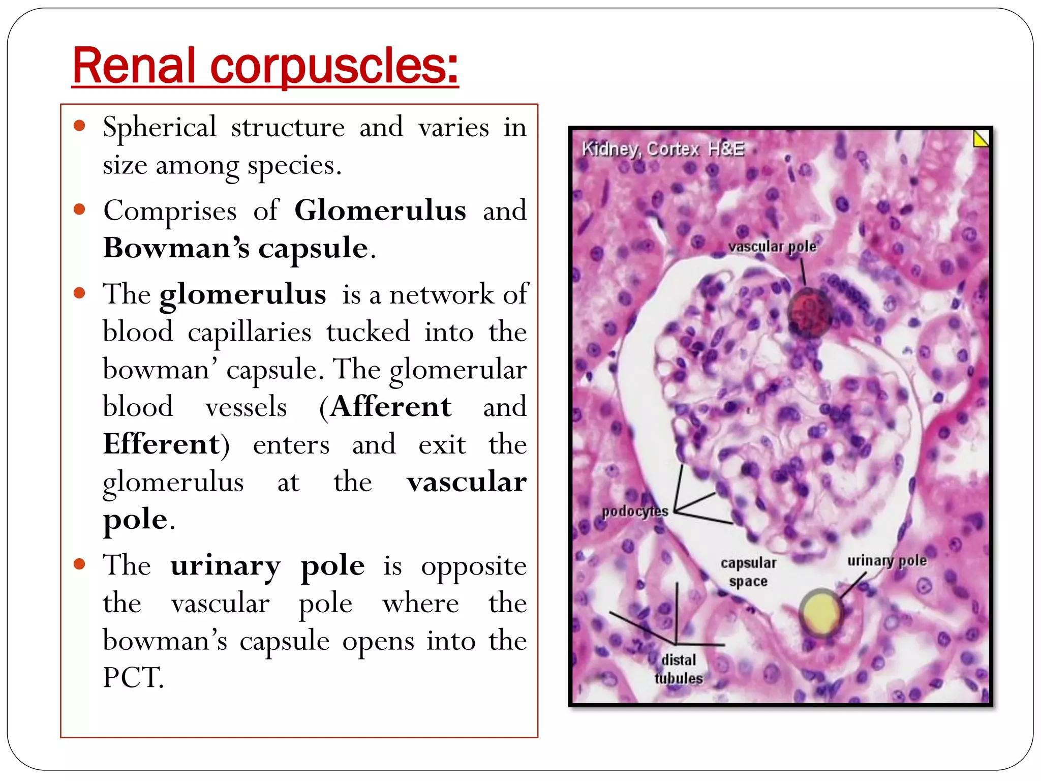
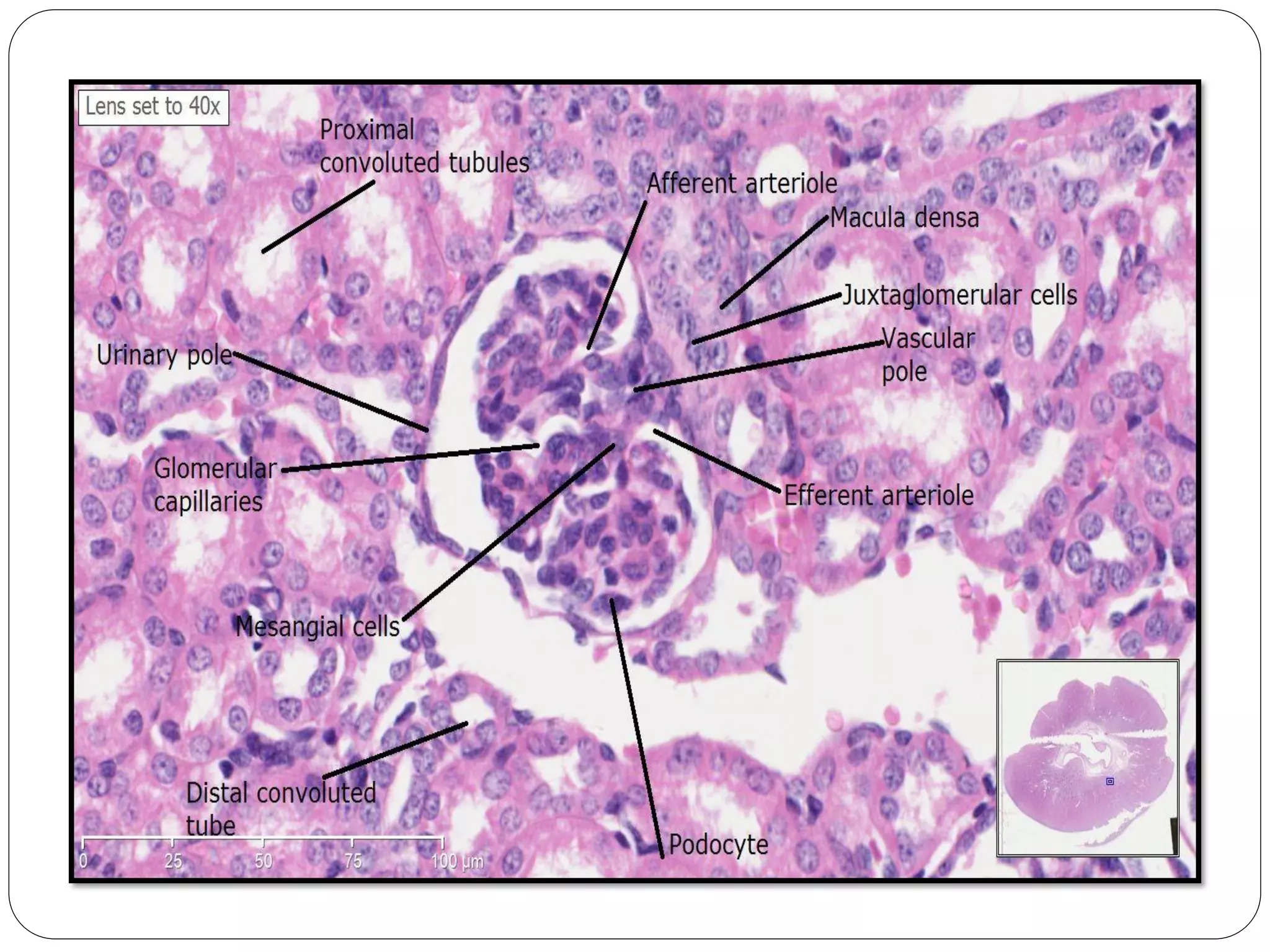
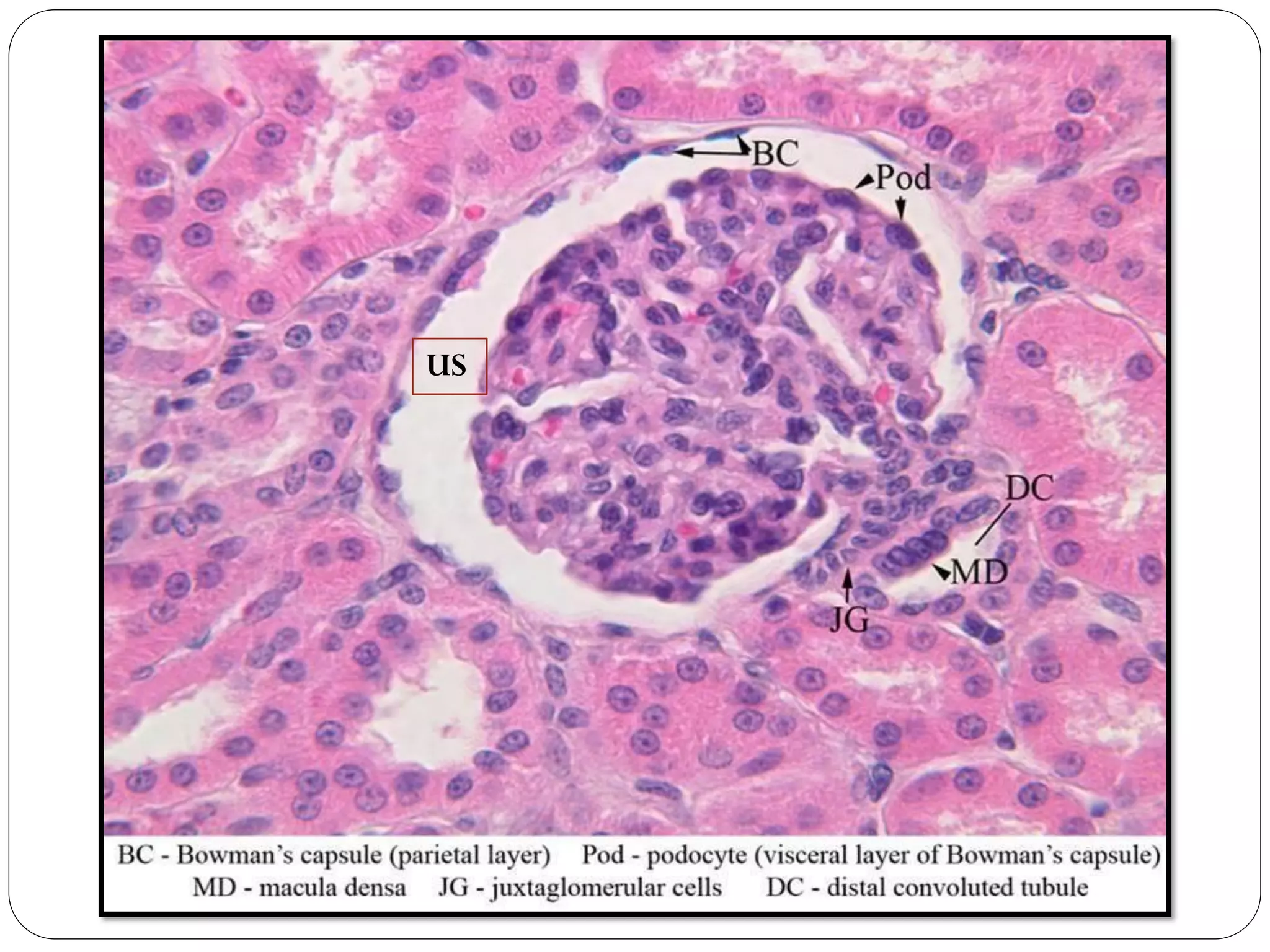
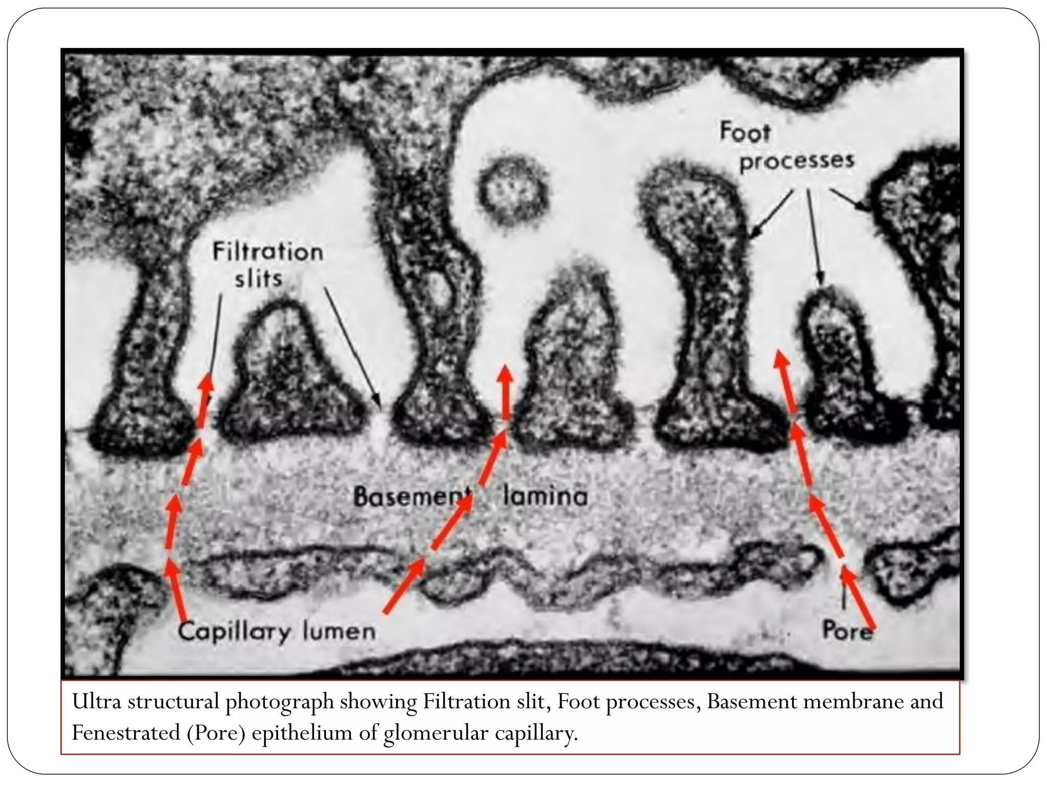
Dr. Rajesh Ranjan
Assistant Professor
Deptt. ofVeterinary Anatomy
C.V.Sc. & A.H., Rewa
HISTOLOGY OF URINARY SYSTEM
 The main organs of this system are:
Kidneys
Ureters
Urinary bladder
Urethra
KIDNEY
Comprises of
Capsule Parenchyma
Cortex Medulla
Cortical labyrinth Medullary rays
Outer Medulla Inner Medulla
Outer stripes Inner stripes
 Capsule is made up of collagen fibers, some smooth muscle
fibers and blood capillaries.
 The Parenchyma consists of millions of nephrons, branches of
renal arteries, veins, lymphatics and nerves.
 The Nephrons are the structural and functional unit of kidney.
 Nephrons can be classified
 On the basis of location of their glomeruli:
 Superficial (near the capsule)
 Mid cortical (near the medulla/Juxtamedullary)
 On the basis of the length of the loop of henle:
 Short looped- generally have superficial or mid cortical glomeruli
and the tubules extend only into the outer medulla before it reflects
back into the cortex.
 Long looped- have juxtamedullary glomeruli and tubules extend into
the inner medulla before reflecting back into the cortex.
Nephrons comprises of:
1. Renal corpuscles
 Glomerulus
 Glomerular capillaries
 Mesangiam
 Bowman’s capsule
 Parietal layer
 Visceral layer
2. Proximal tubules
 Proximal convoluted tubule (PCT)
 Proximal straight tubule (PST)
3. Henle’s loop
 Thin descending portion
 Thin ascending portion
 Thick ascending portion
4. Distal convoluted tubule
5. Connecting segment
6. Collecting duct
 Arcade- initial collecting tubule
 Straight portion-
 Cortical collecting duct
 Outer medullary duct
 Inner medullary duct
Renal Cortex
Cortical labyrinth
PCT, DCT, Connecting
segment, Renal corpuscles,
Thick ascending limb,
Initial collecting tubule
Medullary rays
Cortical collecting ducts,
Cortical thick ascending
limb and Proximal straight
tubule
Renal Medulla (Pyramids)
Outer Medulla Inner Medulla
Outer stripes Inner stripes
Proximal straight tubule,
Thick ascending limb,
Collecting duct
Transition of PST to thin
descending limb of
henle’s loop, Collecting
duct.
Collecting duct, Loop of
Henle’s thin descending
and ascending limb.
Renal corpuscles:
 Spherical structure and varies in
size among species.
 Comprises of Glomerulus and
Bowman’s capsule.
 The glomerulus is a network of
blood capillaries tucked into the
bowman’ capsule. The glomerular
blood vessels (Afferent and
Efferent) enters and exit the
glomerulus at the vascular
pole.
 The urinary pole is opposite
the vascular pole where the
bowman’s capsule opens into the
PCT.
 The glomerular capillaries are
lined by an extremely thin layer
of fenestrated endothelium.
 Beneath the endothelium is the
Glomerular basement membrane
(GBM).
 The GBM is made up of collagen
fibers, heparan sulfate and
glycoproteins.
 Beneath the GBM there is visceral
layer of bowman’s capsule.
•Mesangiam forms the core of
the glomerulus and is
composed of specialized
contractile cells embedded in a
acellular matrix.
•Messangial cells have
elongated, irregular cell
processes, contains bundles of
microfilament made up of
contractile proteins.
• it is responsible for
phagocytosis, production of
mesangial matrix, maintenance
of capillary loops and blood
flow.
 Bowman’s capsule comprises of visceral and parietal layer.
 In between the two layers, Urinary space (US) is present which
continues with the lumen of PCT.
 Visceral layer is lined by Podocytes. They have primary, secondary
and tertiary processes, the smallest of these are called foot
processes or pedicels.
 Narrow space between foot processes are called Filtration slits
which are bridged by the slit diaphragm.
 Parietal layer is lined by simple squamous epithelium that
abruptly changes to simple cuboidal at the urinary pole.
 An additional epithelial cell in the bowman’s capsule is the Peripolar
cell, located at the vascular pole at the junction between the parietal
and visceral layer.
 Its cytoplasm contains dark stained, membrane bound granules.
US
Arrow indicates peripolar cell at the vascular pole between the
junction of parietal and visceral layer of bowman’s capsule.
Ultra structural photograph showing Filtration slit, Foot processes, Basement membrane and
Fenestrated (Pore) epithelium of glomerular capillary.
Proximal Tubules:
 It twists and turns in the cortical labyrinth (PCT) until it enters
the medullary rays where it becomes straight and called as PST.
 Lining epithelium is simple cuboidal with brush border.
 The brush border appearance is due to numerous microvilli on
the apical cell membrane of cuboidal cells.
 Nucleus is single, spherical and situated in the middle to basal
part of the cell.
 The cuboidal cells have eosinophilic granular cytoplasm.
 Cell boundaries are not distinct because of extensive basal and
lateral cell membrane interdigitation with neighbouring cells.
 The lumen is small and uneven.
Loop of Henle:
 The Thin limb has simple squamous epithelium that
gradually changes to low cuboidal at the end of the thin
segment.
 The nuclei protrude into the lumen to a greater degree than do
the nuclei of the endothelial cells of adjacent capillaries.
 The Thick ascending limb has simple cuboidal
epithelium with stuby apical microprojections and
undualating cell borders.
 No brush border.
Distal Convoluted Tubule:
 The lining epithelium is tall
cuboidal without any brush
border (microvilli are
absent).
 Apical cell borders are
simple without undulation.
 Nucleus is located close to
the apical border of the
cell.
 The lumen appears larger
in DCT as compared to
PCT.
Connecting segment:
 It contains a variety of epithelial cell types which includes tall
cuboidal cells, connecting segment cells, intercalated cells
and principal cells.
 The mixture of variety of cell types with different cell
heights, staining density and shape produces an irregular
appearance in light microscopy.
 Principal cells are low cuboidal cells with smooth apical
surface and few short apical micro projections.
 Intercalated cells are epithelial cells traditionally
associated with the regulation of acid-base homeostasis.
Collecting ducts:
 Connecting segments merge with the initial collecting tubule in
the cortical labyrinth and these merge with the cortical
collecting duct in the medullary rays.
 The lining epithelium is simple cuboidal.
 Collecting ducts can be recognised by clear lateral
demarcation between neighbouring epithelial cells.
 These collecting ducts extend towards the renal papillae and the
renal calyx.
 The collecting ducts at the tip of the medullary pyramids are
called ducts of Bellini.
Juxtaglomerular Apparatus:
 Located at the vascular pole of the glomerulus.
 Includes
 Macula densa
 Extraglomerular mesangial cells/ Polkissen cells/ Lacis cells
 Juxtaglomerular cells (JG cells)
 Macula densa is a patch of specialized epithelial cells in the thick
ascending limb where it passes between the afferent and efferent
arterioles. Cells are tall and narrow and the intercellular space is
dilated. It regulates the glomerular blood pressure and renin-
angiotensin system.
 Lacis cells are found between the macula densa and the two
arterioles.
 JG cells are found in the afferent arterioles and are specialized
smooth muscle cells.
Ureter:
 The lumen of ureter is stellate shaped and the wall comprises of
following layers from inside outwards:
 Tunica mucosa- the lining epithelium is transitional
epithelium. The epithelium is 3-5 cell layers thick. Beneath the
epithelium is lamina propria. The lamina muscularis is absent,
hence the lamina propria intermingles with the underlying tunica
submucosa to form propria-submucosa. It consists of loose
connective tissue.
 Tunica muscularis- upper 2/3rd of the ureter is made of two
layers of smooth muscle cells.
 Inner longitudinal and outer circular layer
 Lower 1/3rd of the ureter has a third outer longitudinal layer (inner
longitudinal, middle circular and outer longitudinal).
 Tunica serosa/ adventitia- made of fibrous connective tissue.
Transitional epithelium
lumen
Urinary Bladder:
 The histological details of urinary bladder is same as that of the
ureter except that:
 The lamina muscularis is present as small isolated bundles of
smooth muscles.
 The tunica muscularis is thick and the muscles are collectively
called as Detrusor muscle.
 Rest of the features are same.
Urethra:
 The histological details include the basic four layers:
 Tunica mucosa: The epithelium is transitional but
changes to stratified squamous at the external urethral
orifice.
 Tunica submucosa: It is a connective tissue layer and has
cavernous spaces that are typical of erectile tissue.
 Tunica muscularis: It has inner and outer longitudinal
and a middle circular layer of smooth muscles as in bladder
but towards the external urethral orifice, it acquires an
external layer of skeletal muscle called Urethralis
muscle.
 Tunica serosa/ adventitia: is a fibrous layer.
Assignment:
 A well labelled pictorial representation of
Histology of different organs and parts of the
Urinary system in practical note book.
Thanks!

Histology-of-urinary-system.pdf

  • 1.
    Dr. Rajesh Ranjan AssistantProfessor Deptt. ofVeterinary Anatomy C.V.Sc. & A.H., Rewa HISTOLOGY OF URINARY SYSTEM
  • 2.
     The mainorgans of this system are: Kidneys Ureters Urinary bladder Urethra
  • 3.
    KIDNEY Comprises of Capsule Parenchyma CortexMedulla Cortical labyrinth Medullary rays Outer Medulla Inner Medulla Outer stripes Inner stripes
  • 5.
     Capsule ismade up of collagen fibers, some smooth muscle fibers and blood capillaries.  The Parenchyma consists of millions of nephrons, branches of renal arteries, veins, lymphatics and nerves.  The Nephrons are the structural and functional unit of kidney.  Nephrons can be classified  On the basis of location of their glomeruli:  Superficial (near the capsule)  Mid cortical (near the medulla/Juxtamedullary)  On the basis of the length of the loop of henle:  Short looped- generally have superficial or mid cortical glomeruli and the tubules extend only into the outer medulla before it reflects back into the cortex.  Long looped- have juxtamedullary glomeruli and tubules extend into the inner medulla before reflecting back into the cortex.
  • 7.
    Nephrons comprises of: 1.Renal corpuscles  Glomerulus  Glomerular capillaries  Mesangiam  Bowman’s capsule  Parietal layer  Visceral layer 2. Proximal tubules  Proximal convoluted tubule (PCT)  Proximal straight tubule (PST)
  • 8.
    3. Henle’s loop Thin descending portion  Thin ascending portion  Thick ascending portion 4. Distal convoluted tubule 5. Connecting segment 6. Collecting duct  Arcade- initial collecting tubule  Straight portion-  Cortical collecting duct  Outer medullary duct  Inner medullary duct
  • 9.
    Renal Cortex Cortical labyrinth PCT,DCT, Connecting segment, Renal corpuscles, Thick ascending limb, Initial collecting tubule Medullary rays Cortical collecting ducts, Cortical thick ascending limb and Proximal straight tubule
  • 10.
    Renal Medulla (Pyramids) OuterMedulla Inner Medulla Outer stripes Inner stripes Proximal straight tubule, Thick ascending limb, Collecting duct Transition of PST to thin descending limb of henle’s loop, Collecting duct. Collecting duct, Loop of Henle’s thin descending and ascending limb.
  • 11.
    Renal corpuscles:  Sphericalstructure and varies in size among species.  Comprises of Glomerulus and Bowman’s capsule.  The glomerulus is a network of blood capillaries tucked into the bowman’ capsule. The glomerular blood vessels (Afferent and Efferent) enters and exit the glomerulus at the vascular pole.  The urinary pole is opposite the vascular pole where the bowman’s capsule opens into the PCT.
  • 12.
     The glomerularcapillaries are lined by an extremely thin layer of fenestrated endothelium.  Beneath the endothelium is the Glomerular basement membrane (GBM).  The GBM is made up of collagen fibers, heparan sulfate and glycoproteins.  Beneath the GBM there is visceral layer of bowman’s capsule. •Mesangiam forms the core of the glomerulus and is composed of specialized contractile cells embedded in a acellular matrix. •Messangial cells have elongated, irregular cell processes, contains bundles of microfilament made up of contractile proteins. • it is responsible for phagocytosis, production of mesangial matrix, maintenance of capillary loops and blood flow.
  • 14.
     Bowman’s capsulecomprises of visceral and parietal layer.  In between the two layers, Urinary space (US) is present which continues with the lumen of PCT.  Visceral layer is lined by Podocytes. They have primary, secondary and tertiary processes, the smallest of these are called foot processes or pedicels.  Narrow space between foot processes are called Filtration slits which are bridged by the slit diaphragm.  Parietal layer is lined by simple squamous epithelium that abruptly changes to simple cuboidal at the urinary pole.  An additional epithelial cell in the bowman’s capsule is the Peripolar cell, located at the vascular pole at the junction between the parietal and visceral layer.  Its cytoplasm contains dark stained, membrane bound granules.
  • 15.
  • 16.
    Arrow indicates peripolarcell at the vascular pole between the junction of parietal and visceral layer of bowman’s capsule.
  • 17.
    Ultra structural photographshowing Filtration slit, Foot processes, Basement membrane and Fenestrated (Pore) epithelium of glomerular capillary.
  • 18.
    Proximal Tubules:  Ittwists and turns in the cortical labyrinth (PCT) until it enters the medullary rays where it becomes straight and called as PST.  Lining epithelium is simple cuboidal with brush border.  The brush border appearance is due to numerous microvilli on the apical cell membrane of cuboidal cells.  Nucleus is single, spherical and situated in the middle to basal part of the cell.  The cuboidal cells have eosinophilic granular cytoplasm.  Cell boundaries are not distinct because of extensive basal and lateral cell membrane interdigitation with neighbouring cells.  The lumen is small and uneven.
  • 20.
    Loop of Henle: The Thin limb has simple squamous epithelium that gradually changes to low cuboidal at the end of the thin segment.  The nuclei protrude into the lumen to a greater degree than do the nuclei of the endothelial cells of adjacent capillaries.  The Thick ascending limb has simple cuboidal epithelium with stuby apical microprojections and undualating cell borders.  No brush border.
  • 22.
    Distal Convoluted Tubule: The lining epithelium is tall cuboidal without any brush border (microvilli are absent).  Apical cell borders are simple without undulation.  Nucleus is located close to the apical border of the cell.  The lumen appears larger in DCT as compared to PCT.
  • 23.
    Connecting segment:  Itcontains a variety of epithelial cell types which includes tall cuboidal cells, connecting segment cells, intercalated cells and principal cells.  The mixture of variety of cell types with different cell heights, staining density and shape produces an irregular appearance in light microscopy.  Principal cells are low cuboidal cells with smooth apical surface and few short apical micro projections.  Intercalated cells are epithelial cells traditionally associated with the regulation of acid-base homeostasis.
  • 24.
    Collecting ducts:  Connectingsegments merge with the initial collecting tubule in the cortical labyrinth and these merge with the cortical collecting duct in the medullary rays.  The lining epithelium is simple cuboidal.  Collecting ducts can be recognised by clear lateral demarcation between neighbouring epithelial cells.  These collecting ducts extend towards the renal papillae and the renal calyx.  The collecting ducts at the tip of the medullary pyramids are called ducts of Bellini.
  • 26.
    Juxtaglomerular Apparatus:  Locatedat the vascular pole of the glomerulus.  Includes  Macula densa  Extraglomerular mesangial cells/ Polkissen cells/ Lacis cells  Juxtaglomerular cells (JG cells)  Macula densa is a patch of specialized epithelial cells in the thick ascending limb where it passes between the afferent and efferent arterioles. Cells are tall and narrow and the intercellular space is dilated. It regulates the glomerular blood pressure and renin- angiotensin system.  Lacis cells are found between the macula densa and the two arterioles.  JG cells are found in the afferent arterioles and are specialized smooth muscle cells.
  • 28.
    Ureter:  The lumenof ureter is stellate shaped and the wall comprises of following layers from inside outwards:  Tunica mucosa- the lining epithelium is transitional epithelium. The epithelium is 3-5 cell layers thick. Beneath the epithelium is lamina propria. The lamina muscularis is absent, hence the lamina propria intermingles with the underlying tunica submucosa to form propria-submucosa. It consists of loose connective tissue.  Tunica muscularis- upper 2/3rd of the ureter is made of two layers of smooth muscle cells.  Inner longitudinal and outer circular layer  Lower 1/3rd of the ureter has a third outer longitudinal layer (inner longitudinal, middle circular and outer longitudinal).  Tunica serosa/ adventitia- made of fibrous connective tissue.
  • 29.
  • 30.
    Urinary Bladder:  Thehistological details of urinary bladder is same as that of the ureter except that:  The lamina muscularis is present as small isolated bundles of smooth muscles.  The tunica muscularis is thick and the muscles are collectively called as Detrusor muscle.  Rest of the features are same.
  • 32.
    Urethra:  The histologicaldetails include the basic four layers:  Tunica mucosa: The epithelium is transitional but changes to stratified squamous at the external urethral orifice.  Tunica submucosa: It is a connective tissue layer and has cavernous spaces that are typical of erectile tissue.  Tunica muscularis: It has inner and outer longitudinal and a middle circular layer of smooth muscles as in bladder but towards the external urethral orifice, it acquires an external layer of skeletal muscle called Urethralis muscle.  Tunica serosa/ adventitia: is a fibrous layer.
  • 34.
    Assignment:  A welllabelled pictorial representation of Histology of different organs and parts of the Urinary system in practical note book.
  • 35.