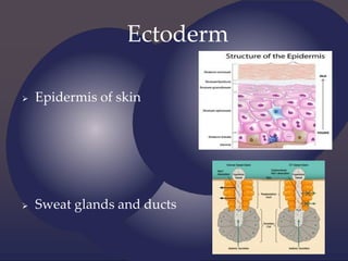
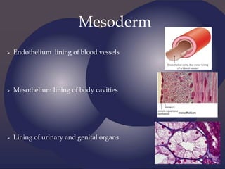
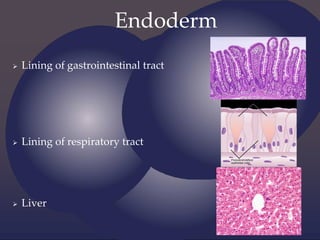
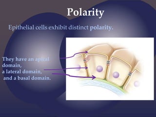
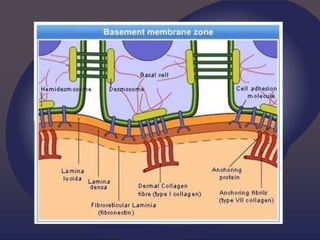
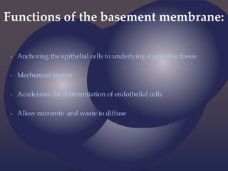
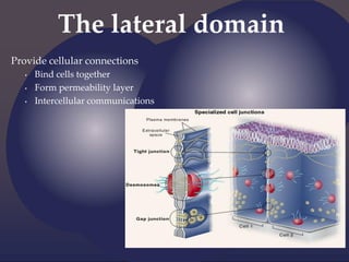
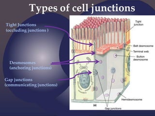
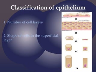
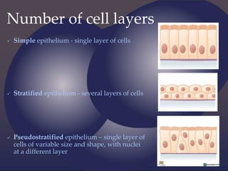
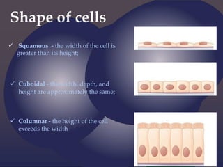
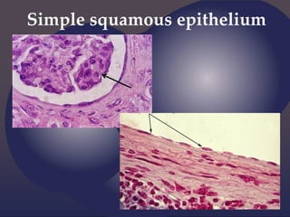
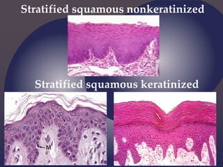
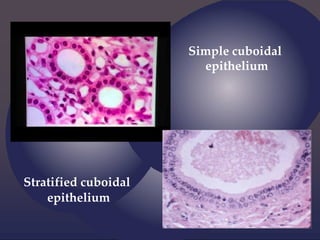
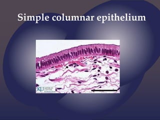
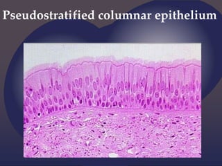
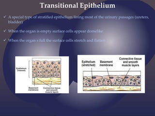
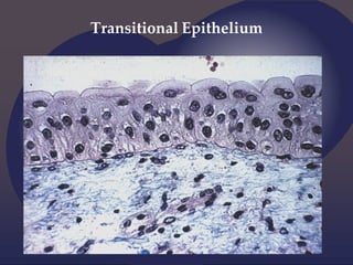
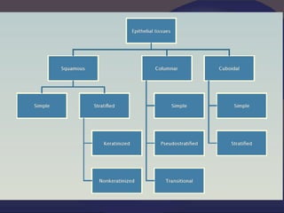
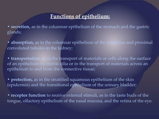
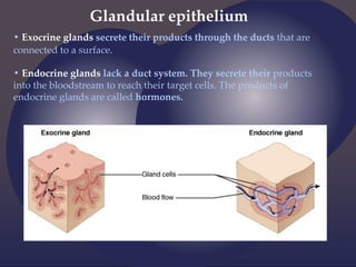
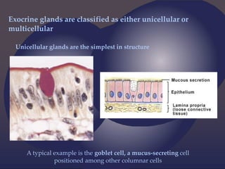
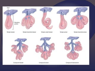
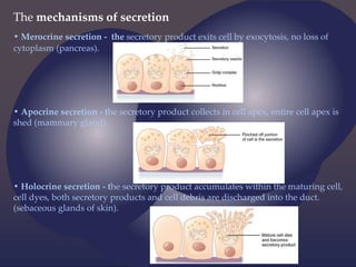
SEACOM PHARMACY COLLEGE
Presented by
Souvick Maity
B.Pharm
1st Year;1st Semester
Reg.No- 233830210071
Roll No- 38305923071
{ detailed
note on
EPITHELIAl
TISSUE
Histology = the study of tissues
Tissue = a complex of cells
that have a common function
The Four Primary Tissue Types:
Epithelium (epithelial tissue) covers body
surfaces, lines body cavities, and forms glands.
Connective tissue underlies or supports the other
three basic tissues, both structurally and functionally.
Muscle tissue is made up of contractile cells and
is responsible for movement.
Nerve tissue receives, transmits, and integrates
information from outside and inside the body to
control the activities of the body.
General features of Epithelium:
1. Can be derivate of all three germ layers
2. Cells show polarity
3. Little intercellular material
4. Cells are laying close to each other
5. Tend to form junctions
6. Rest on a basement membrane
7. Lack vessels
8. Line surfaces and cavities or form glands
9. Mitotically active
Derivation
Ectoderm
Mesoderm
Endoderm
Ectoderm
 Epidermis of skin
 Sweat glands and ducts
Mesoderm
 Endothelium lining of blood vessels
 Mesothelium lining of body cavities
 Lining of urinary and genital organs
Endoderm
 Lining of gastrointestinal tract
 Lining of respiratory tract
 Liver
Polarity
Epithelial cells exhibit distinct polarity.
They have an apical
domain,
a lateral domain,
and a basal domain.
The apical domain
• Part of the epithelial cell that looks towards the
lumen or the surface
• Can have:
- stereocilia
- microvilli
- cilia
Microvilli
• Fingerlike cytoplasmic projections on the apical surface
• Internal structure of microvilli contain a core of actin filaments
• Increase the surface of absorption (small intestine)
Stereocilia
• Unusually long, immotile microvilli
• In sense organs (hair cells) serve as mechanoreceptors
• In male reproductive system (epididymis) have absorptive
function
Cilia
• capable of moving fluid and particles along the epithelial
surface (bronchial tree, oviducts)
• present in sperm cell as flagella (forward movement)
The basal domain
• A part of the cell faced towards the basement membrane
Basement membrane
• a thin, fibrous, extracellular matrix that separates the epithelial cells
from underlying connective tissue
Basement membrane
Basal lamina: Reticular lamina
- lamina lucida
- lamina densa
Basal lamina
 Collagen type IV
 Heparan sulfate
 Fibronectine and
laminine
Reticular lamina
Collagen type III (reticular fibers)
Proteoglycans
Functions of the basement membrane:
• Anchoring the epithelial cells to underlying connective tissue
• Mechanical barrier
• Accelerates the differentiation of endothelial cells
• Allow nutrients and waste to diffuse
The lateral domain
Provide cellular connections
• Bind cells together
• Form permeability layer
• Intercellular communications
Types of cell junctions
Tight Junctions
(occluding junctions )
Desmosomes
(anchoring junctions)
Gap junctions
(communicating junctions)
Tight junctions
(occluding junctions)
 Plasma membranes of the
neighboring cells are tightly sealed
with each other
3 types of proteins:
- occludins
- claudins
- junctional adhesion molecules
 Functions as a permeability
barrier
(ex. makes us waterproof)
Anchoring junctions
Desmosomes Hemidesmosomes
 Attach cytoskeleton of neighboring cells or to the extracellular matrix
- Intracellular anchor proteins
- Transmembrane adhesion proteins
 Do not prevent the flow of substances around cells
Hemidesmosome
Gap junction
(communicating junction or nexus)
 Direct connection of the
cytoplasm of two cells
allows various molecules
and ions to pass between
cells
 Channels composed of
transmembrane proteins –
connexins
 Connexon – assembly of six
connexin proteins creating a
gap between plasma
membranes of two cells
Junctional Complexes
Examples:
 Tight junctions
- in the intestines – helps to keep the intestinal bacteria and
toxins out of the bloodstream
 Desmosomes
- skin – gives the ability to stretch but keeps cells together
 Gap junctions
- cardiac muscle – allows to spread action potential for
contraction of cardiomyocytes
Mitotic activity
 Most epithelial cells have a life span less than that of the whole
organism
 The replacement of cells produced by mitotic activity of adult
stem cells
- The stratified squamous
epithelium of skin is
replaced in a period of
approximately 28 days.
Classification of epithelium
1. Number of cell layers
2. Shape of cells in the superficial
layer
Number of cell layers
 Simple epithelium - single layer of cells
 Stratified epithelium - several layers of cells
 Pseudostratified epithelium – single layer of
cells of variable size and shape, with nuclei
at a different layer
Shape of cells
 Squamous - the width of the cell is
greater than its height;
 Cuboidal - the width, depth, and
height are approximately the same;
 Columnar - the height of the cell
exceeds the width
Simple squamous epithelium
Stratified squamous nonkeratinized
Stratified squamous keratinized
Simple cuboidal
epithelium
Stratified cuboidal
epithelium
Simple columnar epithelium
Pseudostratified columnar epithelium
Transitional Epithelium
 A special type of stratified epithelium lining most of the urinary passages (ureters,
bladder)
 When the organ is empty surface cells appear domelike
 When the organ s full the surface cells stretch and flatten
Transitional Epithelium
Functions of epithelium:
• secretion, as in the columnar epithelium of the stomach and the gastric
glands;
• absorption, as in the columnar epithelium of the intestines and proximal
convoluted tubules in the kidney;
• transportation, as in the transport of materials or cells along the surface
of an epithelium by motile cilia or in the transport of materials across an
epithelium to and from the connective tissue;
• protection, as in the stratified squamous epithelium of the skin
(epidermis) and the transitional epithelium of the urinary bladder;
• receptor function to receive external stimuli, as in the taste buds of the
tongue, olfactory epithelium of the nasal mucosa, and the retina of the eye.
Glandular epithelium
• Exocrine glands secrete their products through the ducts that are
connected to a surface.
• Endocrine glands lack a duct system. They secrete their products
into the bloodstream to reach their target cells. The products of
endocrine glands are called hormones.
Exocrine glands are classified as either unicellular or
multicellular
Unicellular glands are the simplest in structure
A typical example is the goblet cell, a mucus-secreting cell
positioned among other columnar cells
 Multicellular glands are composed of more than one cell.
 Can be classified according to the arrangement of the secretory cells
(parenchyma) and the branching of the duct elements.
Ducts:
 if the duct is unbranched, the
gland is called simple;
 if the duct is branched, it is called
compound.
Secretary portion:
if the secretory portion is shaped like
a tube, the gland is tubular;
 if it is shaped like a flask, the gland
is alveolar or acinar;
if the tube ends in a saclike
dilation, the gland is tubuloalveolar.
The mechanisms of secretion
• Merocrine secretion - the secretory product exits cell by exocytosis, no loss of
cytoplasm (pancreas).
• Apocrine secretion - the secretory product collects in cell apex, entire cell apex is
shed (mammary gland).
• Holocrine secretion - the secretory product accumulates within the maturing cell,
cell dyes, both secretory products and cell debris are discharged into the duct.
(sebaceous glands of skin).
Type of secretory product:
 Mucous – thick secretion rich in glycoproteins (sublingual gland)
 Serous – thin, watery secretion containing proteins (parotid gland)
 Seromucous – mixed secretion of intermediate thickness (sebaceous
gland)
Thank you for attention

HUMAN ANATOMY AND PHYSIOLOGY.PPT.pdf.pdf

  • 1.
    SEACOM PHARMACY COLLEGE Presentedby Souvick Maity B.Pharm 1st Year;1st Semester Reg.No- 233830210071 Roll No- 38305923071
  • 2.
  • 3.
    Histology = thestudy of tissues Tissue = a complex of cells that have a common function
  • 4.
    The Four PrimaryTissue Types:
  • 5.
    Epithelium (epithelial tissue)covers body surfaces, lines body cavities, and forms glands. Connective tissue underlies or supports the other three basic tissues, both structurally and functionally. Muscle tissue is made up of contractile cells and is responsible for movement. Nerve tissue receives, transmits, and integrates information from outside and inside the body to control the activities of the body.
  • 6.
    General features ofEpithelium: 1. Can be derivate of all three germ layers 2. Cells show polarity 3. Little intercellular material 4. Cells are laying close to each other 5. Tend to form junctions 6. Rest on a basement membrane 7. Lack vessels 8. Line surfaces and cavities or form glands 9. Mitotically active
  • 7.
  • 8.
    Ectoderm  Epidermis ofskin  Sweat glands and ducts
  • 9.
    Mesoderm  Endothelium liningof blood vessels  Mesothelium lining of body cavities  Lining of urinary and genital organs
  • 10.
    Endoderm  Lining ofgastrointestinal tract  Lining of respiratory tract  Liver
  • 11.
    Polarity Epithelial cells exhibitdistinct polarity. They have an apical domain, a lateral domain, and a basal domain.
  • 12.
    The apical domain •Part of the epithelial cell that looks towards the lumen or the surface • Can have: - stereocilia - microvilli - cilia
  • 13.
    Microvilli • Fingerlike cytoplasmicprojections on the apical surface • Internal structure of microvilli contain a core of actin filaments • Increase the surface of absorption (small intestine)
  • 14.
    Stereocilia • Unusually long,immotile microvilli • In sense organs (hair cells) serve as mechanoreceptors • In male reproductive system (epididymis) have absorptive function
  • 15.
    Cilia • capable ofmoving fluid and particles along the epithelial surface (bronchial tree, oviducts) • present in sperm cell as flagella (forward movement)
  • 16.
    The basal domain •A part of the cell faced towards the basement membrane
  • 17.
    Basement membrane • athin, fibrous, extracellular matrix that separates the epithelial cells from underlying connective tissue
  • 18.
    Basement membrane Basal lamina:Reticular lamina - lamina lucida - lamina densa
  • 19.
    Basal lamina  Collagentype IV  Heparan sulfate  Fibronectine and laminine Reticular lamina Collagen type III (reticular fibers) Proteoglycans
  • 21.
    Functions of thebasement membrane: • Anchoring the epithelial cells to underlying connective tissue • Mechanical barrier • Accelerates the differentiation of endothelial cells • Allow nutrients and waste to diffuse
  • 22.
    The lateral domain Providecellular connections • Bind cells together • Form permeability layer • Intercellular communications
  • 23.
    Types of celljunctions Tight Junctions (occluding junctions ) Desmosomes (anchoring junctions) Gap junctions (communicating junctions)
  • 24.
    Tight junctions (occluding junctions) Plasma membranes of the neighboring cells are tightly sealed with each other 3 types of proteins: - occludins - claudins - junctional adhesion molecules  Functions as a permeability barrier (ex. makes us waterproof)
  • 25.
    Anchoring junctions Desmosomes Hemidesmosomes Attach cytoskeleton of neighboring cells or to the extracellular matrix - Intracellular anchor proteins - Transmembrane adhesion proteins  Do not prevent the flow of substances around cells
  • 27.
  • 28.
    Gap junction (communicating junctionor nexus)  Direct connection of the cytoplasm of two cells allows various molecules and ions to pass between cells  Channels composed of transmembrane proteins – connexins  Connexon – assembly of six connexin proteins creating a gap between plasma membranes of two cells
  • 29.
  • 30.
    Examples:  Tight junctions -in the intestines – helps to keep the intestinal bacteria and toxins out of the bloodstream  Desmosomes - skin – gives the ability to stretch but keeps cells together  Gap junctions - cardiac muscle – allows to spread action potential for contraction of cardiomyocytes
  • 31.
    Mitotic activity  Mostepithelial cells have a life span less than that of the whole organism  The replacement of cells produced by mitotic activity of adult stem cells - The stratified squamous epithelium of skin is replaced in a period of approximately 28 days.
  • 32.
    Classification of epithelium 1.Number of cell layers 2. Shape of cells in the superficial layer
  • 33.
    Number of celllayers  Simple epithelium - single layer of cells  Stratified epithelium - several layers of cells  Pseudostratified epithelium – single layer of cells of variable size and shape, with nuclei at a different layer
  • 34.
    Shape of cells Squamous - the width of the cell is greater than its height;  Cuboidal - the width, depth, and height are approximately the same;  Columnar - the height of the cell exceeds the width
  • 35.
  • 36.
  • 37.
  • 38.
  • 39.
  • 40.
    Transitional Epithelium  Aspecial type of stratified epithelium lining most of the urinary passages (ureters, bladder)  When the organ is empty surface cells appear domelike  When the organ s full the surface cells stretch and flatten
  • 41.
  • 43.
    Functions of epithelium: •secretion, as in the columnar epithelium of the stomach and the gastric glands; • absorption, as in the columnar epithelium of the intestines and proximal convoluted tubules in the kidney; • transportation, as in the transport of materials or cells along the surface of an epithelium by motile cilia or in the transport of materials across an epithelium to and from the connective tissue; • protection, as in the stratified squamous epithelium of the skin (epidermis) and the transitional epithelium of the urinary bladder; • receptor function to receive external stimuli, as in the taste buds of the tongue, olfactory epithelium of the nasal mucosa, and the retina of the eye.
  • 44.
    Glandular epithelium • Exocrineglands secrete their products through the ducts that are connected to a surface. • Endocrine glands lack a duct system. They secrete their products into the bloodstream to reach their target cells. The products of endocrine glands are called hormones.
  • 45.
    Exocrine glands areclassified as either unicellular or multicellular Unicellular glands are the simplest in structure A typical example is the goblet cell, a mucus-secreting cell positioned among other columnar cells
  • 47.
     Multicellular glandsare composed of more than one cell.  Can be classified according to the arrangement of the secretory cells (parenchyma) and the branching of the duct elements. Ducts:  if the duct is unbranched, the gland is called simple;  if the duct is branched, it is called compound. Secretary portion: if the secretory portion is shaped like a tube, the gland is tubular;  if it is shaped like a flask, the gland is alveolar or acinar; if the tube ends in a saclike dilation, the gland is tubuloalveolar.
  • 49.
    The mechanisms ofsecretion • Merocrine secretion - the secretory product exits cell by exocytosis, no loss of cytoplasm (pancreas). • Apocrine secretion - the secretory product collects in cell apex, entire cell apex is shed (mammary gland). • Holocrine secretion - the secretory product accumulates within the maturing cell, cell dyes, both secretory products and cell debris are discharged into the duct. (sebaceous glands of skin).
  • 50.
    Type of secretoryproduct:  Mucous – thick secretion rich in glycoproteins (sublingual gland)  Serous – thin, watery secretion containing proteins (parotid gland)  Seromucous – mixed secretion of intermediate thickness (sebaceous gland)
  • 51.
    Thank you forattention